2416
Reduced Noise and Motion Artifacts for MUSE Reconstruction using Deep Learning-based Phase Correction1GE HealthCare, Menlo Park, CA, United States, 2GE HealthCare, Houston, TX, United States, 3GE HealthCare, Boston, MA, United States
Synopsis
Keywords: Diffusion Reconstruction, Diffusion/other diffusion imaging techniques
Motivation: Filter-based phase estimation requires tuning and is subject to the tradeoff between signal bias and vulnerability against phase inhomogeneity. DL-based phase correction has been shown to effectively remove both high- and low-frequency phase while minimizing signal bias.
Goal(s): To evaluate a DL-based phase correction method that improves the robustness of motion-induced phase estimation and its impact on noise and motion artifacts in MUSE reconstruction.
Approach: Volunteer brain and abdomen data were acquired with a MUSE sequence and reconstruction was performed offline.
Results: Compared to filter-based phase estimation, DL-based phase correction results in reduced noise and motion artifacts in MUSE reconstructed images.
Impact: MUSE enables high resolution DWI over a large FOV with reduced geometric distortion, but is very sensitive to shot-to-shot differences in motion-induced phase. DL-based phase correction can improve robustness in MUSE reconstruction, especially in anatomical regions with significant motion.
Introduction
Compared to single-shot echo planar imaging, multi-shot DWI (MUSE) has been shown to enable higher resolution DWI over a larger FOV with reduced geometric distortion.1 However, shot-to-shot differences in motion-induced phase can result in significant aliasing artifacts and signal cancellation. A common method to estimate this motion-induced phase is by reconstructing each shot using conventional parallel imaging and using the phase of a low-pass filtered image. In general, this method works well when the shot number is low and the motion induced phase is linearly smooth. However, when there is significant motion, the non-linear or high-frequency phase cannot be resolved and MUSE reconstruction becomes suboptimal. In this study, we aim to evaluate a deep-learning based phase correction method that improves the robustness of motion-induced phase estimation and ultimately multi-shot DWI image quality.Methods
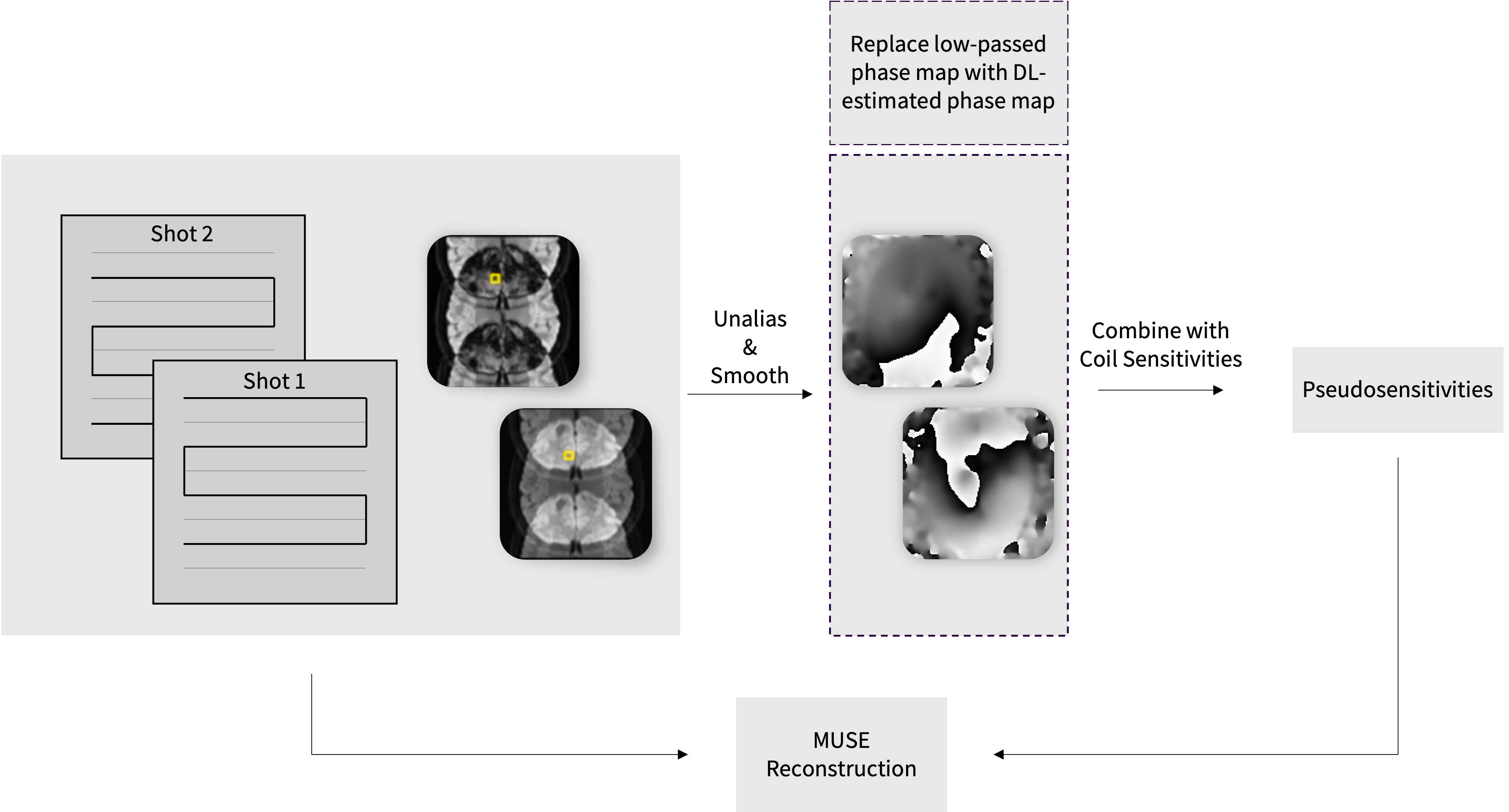
Volunteer scans were performed with IRB approval and written informed consent on 3T scanners (GE HealthCare, SIGNA™ Premier and SIGNA™ Architect, Waukesha, WI) using product MUSE sequence. The axial brain scan was acquired with 5 shots, whereas the sagittal spinal cord and axial abdomen scans were acquired with 2 shots. Specific imaging parameters are located in the figure captions. The deep learning-based phase correction (DLPC) model was trained using a database of over 10,000 images with various SNR levels and background phases as described by Wang et al.2 Figure 1 shows a schematic of MUSE reconstruction and where in the pipeline the low-pass filter for shot-to-shot phase estimation would be replaced by our proposed DLPC method to generate high resolution and high SNR phase maps. Compatibility of DLPC with the product denoising and deringing feature AIR™ Recon DL (ARDL)3 was also assessed to further improve in-plane resolution and SNR of the final MUSE image. Image reconstruction was performed offline to generate three sets of images from the same dataset: (1) original non-DL image with filter-based phase estimation, (2) image with DLPC phase estimation, (3) image with DLPC phase estimation with ARDL. Postprocessing was performed with READY View (GE HealthCare, Waukesha, WI).Results and Discussion
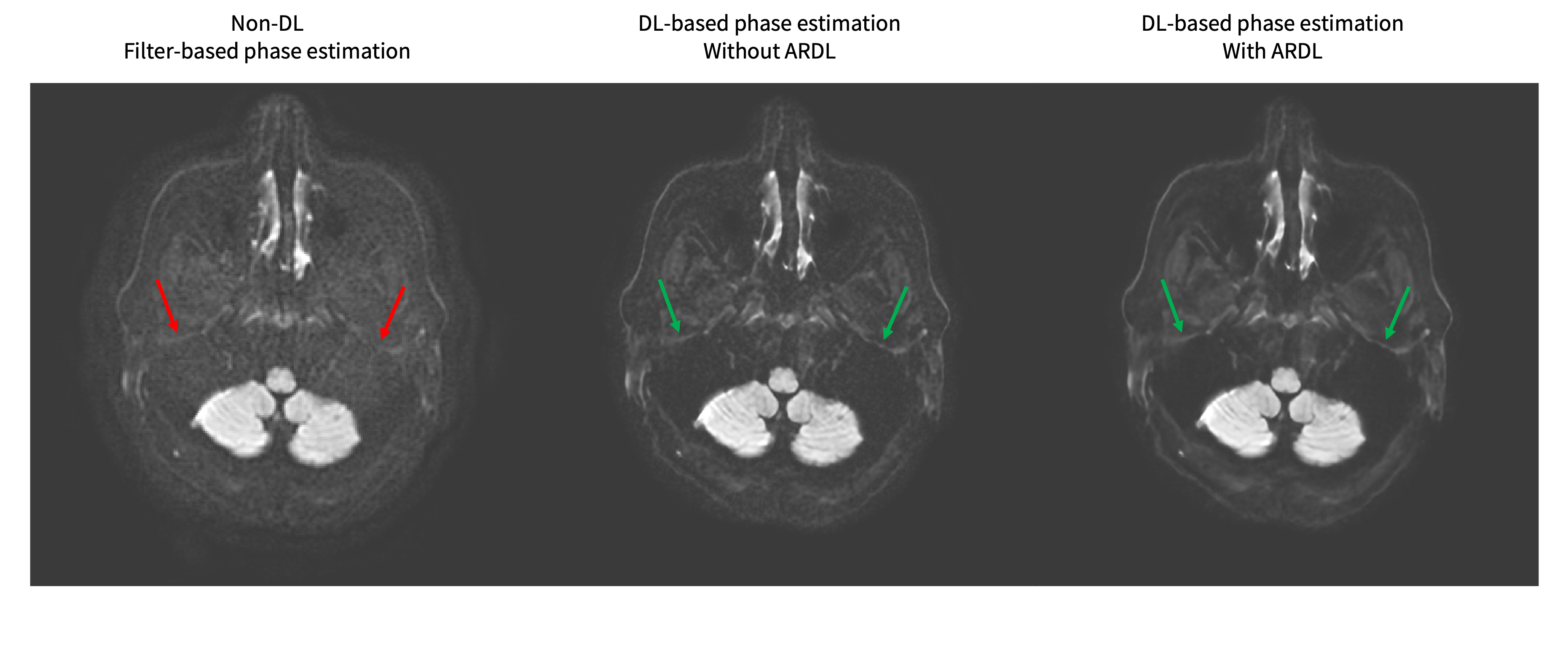
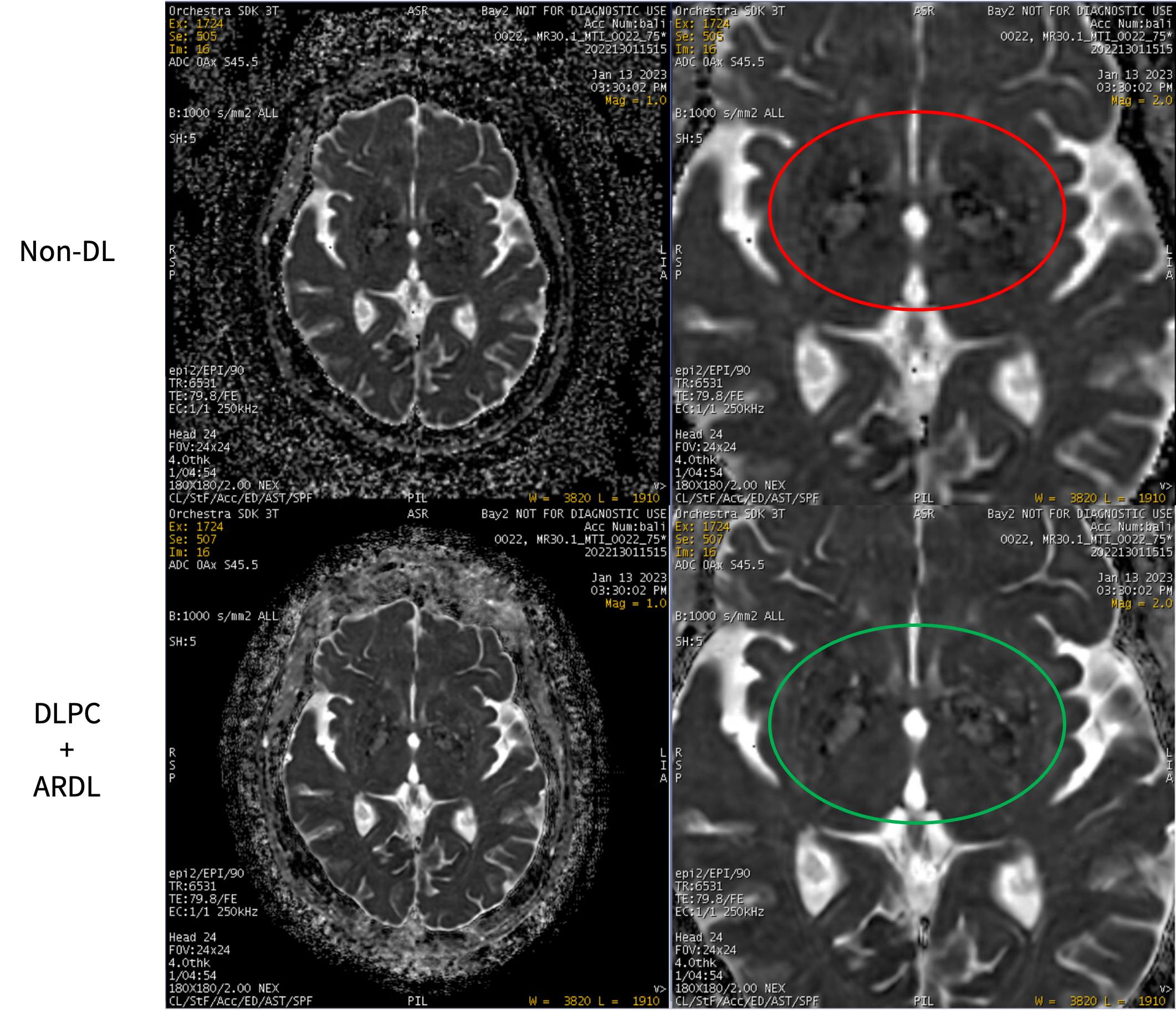
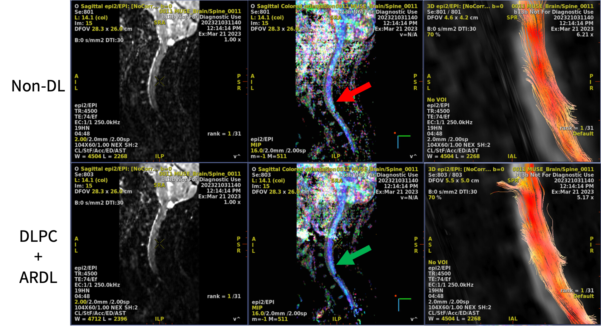
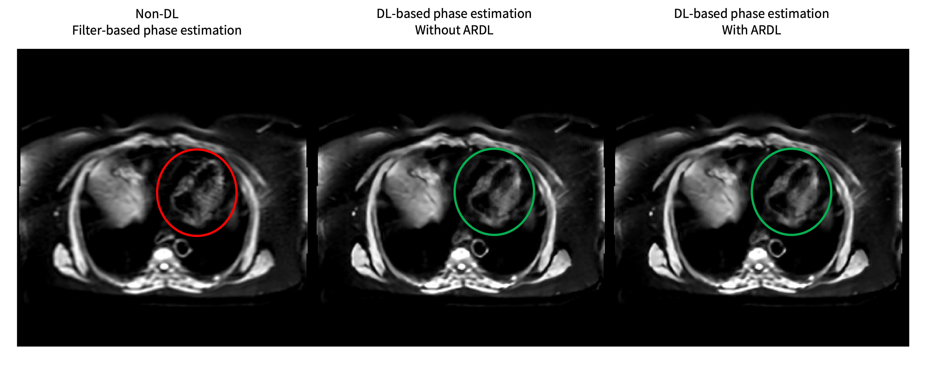
Comparisons between non-DL filter-based phase estimation, DLPC phase estimation, and DLPC phase estimation with ARDL for a MUSE brain scan are shown in Figure 2. The DLPC phase estimation significantly cleans up the noise amplification compared to filter-based phase estimation, resulting in better visualization of structures such as the edges of the temporal lobes (indicated with green arrows). Combining DLPC with ARDL further increases the SNR of the final MUSE image. The impact of DLPC and ARDL propagates downstream to postprocessing as well. Figure 3 shows cleaner ADC maps with DLPC and ARDL; Figure 4 depicts colored fractional anisotropy (FA) maps with higher SNR and fiber tracts with increased density (Figure 4). DLPC also improves robustness in MUSE reconstruction in anatomical regions with significant motion. Unlike DLPC, filter-based phase estimation often cannot capture such high frequency phase changes due to motion. The abdomen MUSE scan in Figure 5 is one example of this scenario, where the low pass filter-based phase estimation fails to capture the high frequency phase changes from the motion of the heart, leading to signal cancellation in the final image. DLPC adequately corrects for both low and high frequency phase changes and recovers artifact-free signal in the myocardial tissue of a beating heart.Conclusion
Low pass filtering is commonly used in multi-shot DWI reconstruction. However, the assumption that the shot-to-shot motion induced phase maps are smooth is often violated due to motion and susceptibility differences in tissue boundaries. The proposed DLPC method addresses this challenge by capturing these non-linear or high frequency phase changes, improving robustness in MUSE reconstruction with reduced noise amplification and increased signal recovery. The improved MUSE image quality with higher SNR and tissue conspicuity also affects the downstream postprocessing applications such as ADC map, FA map, and fiber tract generation.Acknowledgements
No acknowledgement found.References
- Chen NK, Guidon A, Chang HC, Song AW. A robust multi-shot scan strategy for high-resolution diffusion weighted MRI enabled by multiplexed sensitivity-encoding (MUSE). Neuroimage. 2013;72:41-7.
- Wang X, Litwiller D, Guidon A, Lan PS, Sprenger T. Robust complex signal averaging for diffusion weighted imaging. In: Proceedings of the 31st Annual Meeting of the International Society for Magnetic Resonance in Medicine (ISMRM). Toronto, Canada; 2023; #3963.
- Lebel RM. Performance characterization of a novel deep learning-based MR image reconstruction pipeline. arXiv preprint arXiv:2008.06559. 2020.
Figures