2413
Prospective gradient nonlinearity correction for diffusion MRI: uncover lost sensitivity to non-Gaussian diffusion and tissue microstructure1Technology and Innovation Center, GE Healthcare, Niskayuna, NY, United States, 2University of Michigan, Ann Arbor, MI, United States, 3Mayo Clinic, Rochester, MN, United States
Synopsis
Keywords: Microstructure, Diffusion/other diffusion imaging techniques
Motivation: Time-dependent diffusion MRI, which is sensitive to non-Gaussian diffusion, reveals tissue microstructures and has been shown to improve cancer imaging and neuroimaging. However, gradient nonlinearity results in subject position-dependent bias for non-Gaussian diffusion characterization. Correction methods are needed.
Goal(s): To reduce the effect of gradient nonlinearity on 2D time-dependent diffusion MRI.
Approach: Slice-by-slice scaling of diffusion encoding gradients was applied to compensate for gradient nonlinearity.
Results: Uncorrected $$$\frac{ADC(60Hz)}{ADC(0Hz)}$$$ of a non-Gaussian diffusion phantom showed errors in off-center slices, where the actual diffusion gradient amplitude was reduced compared to prescribed values. The errors were reduced by prospectively increasing the prescribed diffusion gradient amplitude.
Impact: MR physicists, neuroimaging scientists, and radiologists, who are interested in microstructure imaging by probing time-dependent, non-Gaussian diffusion, will benefit from increased robustness to gradient nonlinearity and subject position, especially when using high-performance gradient systems that may have increased nonlinearity.
Introduction
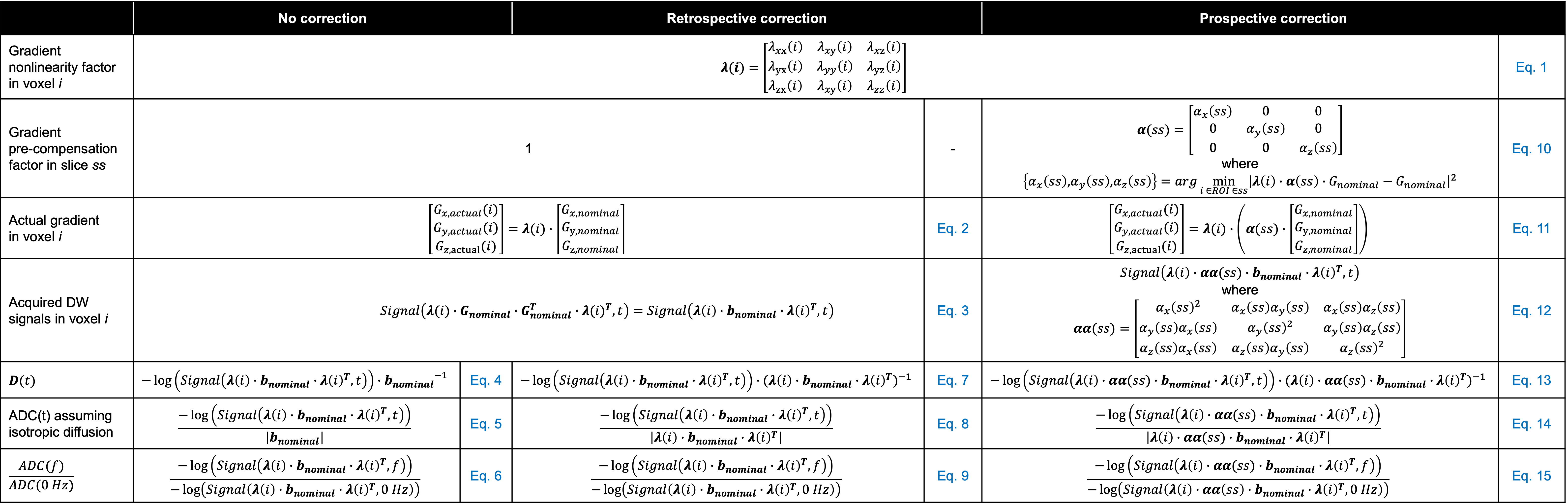
Diffusion MRI-based microstructure imaging techniques have been shown to improve clinical diagnosis and treatment evaluation of cancer1,2, as well as advance our understanding of neurological disorders and diseases3. For example, time-dependent diffusion MRI has shown to disentangle intracellular water signal from extracellular water signal and quantify cell size4 and exchange rate5. Despite its promise, the accuracy and reproducibility of microstructure imaging are reduced by the nonlinearity of gradient fields. Gradient nonlinearity results in spatially varying b-values and introduces subject position-dependent variation of diffusion measurements6 and microstructure characteristics7. Retrospective gradient nonlinearity correction has been implemented by calculating the actual b-value and tensor, and applying them in the parametric estimation8. These methods improve the accuracy of apparent diffusion coefficient (ADC)9,10. However, they cannot recover reduced sensitivity to non-Gaussian diffusion if the diffusion weighted (DW) signals are acquired at a decreased b-value.We propose prospective compensation for gradient nonlinearity in two-dimensional diffusion MRI. The systematic bias of gradient nonlinearity on time-dependent diffusivity, which is sensitive to non-Gaussian diffusion, was analyzed and the prospective compensation was applied to reduce the bias.
Theory
The actual, applied gradient $$$G_{x/y/z,actual}(i)$$$ deviates from the prescribed, nominal gradient $$$G_{x/y/z,nominal}(i)$$$, characterized by scale factors λ(i) due to the nonlinearity11 in the voxel i (Figure 1, Eqs.1-2). Thus, the acquired DW signals are weighted by both nominal b-value and λ(i) (Eq. 3). Without correction, diffusion tensor and ADC(t) at diffusion time t are calculated with reference to the nominal b-value (Eqs. 4-6). Retrospective gradient nonlinearity correction applies an estimated λ(i) to the ADC calculation8 (Eqs. 7-9).We propose to prospectively scale diffusion encoding gradients with factors αx/y/z(i). Ideally, αx/y/z(i) would be chosen such that the L2 norm of α(i)·λ(i)·bnominal-bnominal is close to 0 for each voxel. In 2D diffusion MRI, αx/y/z(ss) of slice sscan be determined to achieve an optimal value over a certain region-of-interest (ROI) (Figure 1, Eq. 10). Thus, the acquired DW signals are weighted by α(ss)·λ(i) (Eqs. 11-12) and the calculated ADC (Eqs. 13-15) more accurately reflects the tissue property at the prescribed, nominal b-value.
Methods
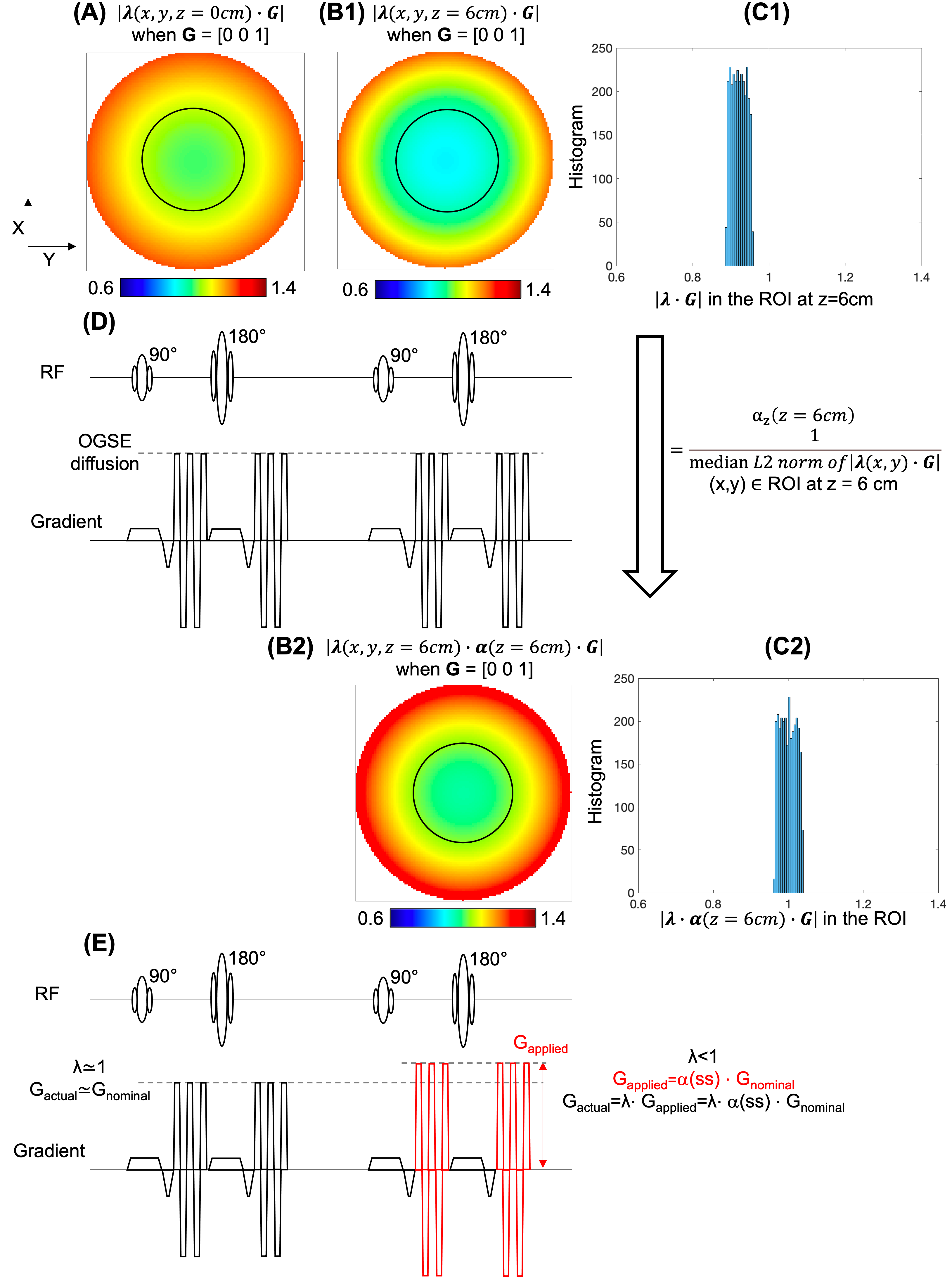
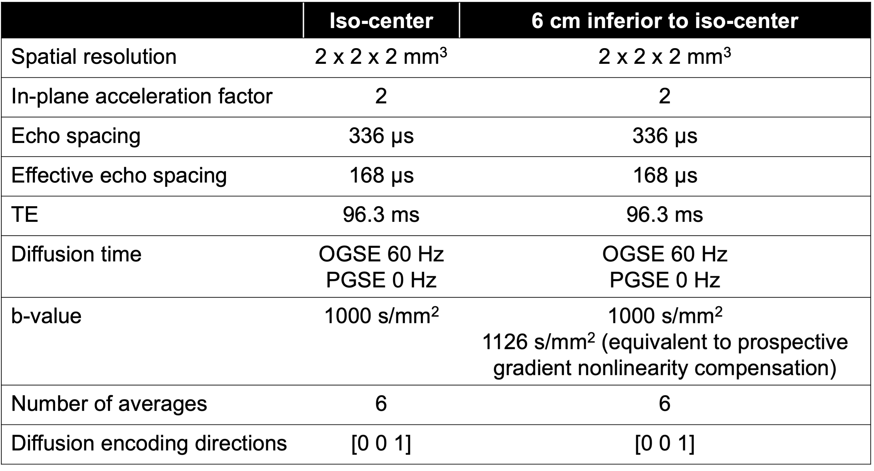
Figure 2 shows the estimated Z gradient field of a small-bore MAGNUS12 gradient coil at z=0 cm and 6 cm. As an example, the pre-compensation scaling factor αz(ss) was calculated as the inverse of the median L2 norm of λ(i) in a circular ROI with 6 cm radius, and αy(ss) and αx(ss) were set to 0.A cylindrical non-Gaussian diffusion phantom with ~1.6 cm radius, consisting of water and lipid nanoparticle vesicles13-16, was scanned in a 3.0T MANGUS MRI system12 (GE Healthcare, USA) at 300 mT/m and 750 T/m/s, and a 32-channel phased-array receive RF coils (Nova Medical, USA). Oscillating gradient spin echo diffusion encoding17 with a frequency of 60 Hz (short diffusion time) and pulsed gradient spin echo (approximating 0 Hz) at b-value of 1000 s/mm2 were acquired. The scan was performed at two positions, i.e., iso-center and 6 cm off-center in inferior direction. At the 6 cm off-center position, two datasets were acquired with and without gradient scaling for diffusion encoding gradient. Imaging protocols are shown in Figure 3. ADC(0 Hz), ADC(60 Hz) and $$$\frac{ADC(60 Hz)}{ADC(0 Hz)}$$$, which reflects time-dependent diffusion that is sensitive to tumor microstructure17, were calculated.
Results
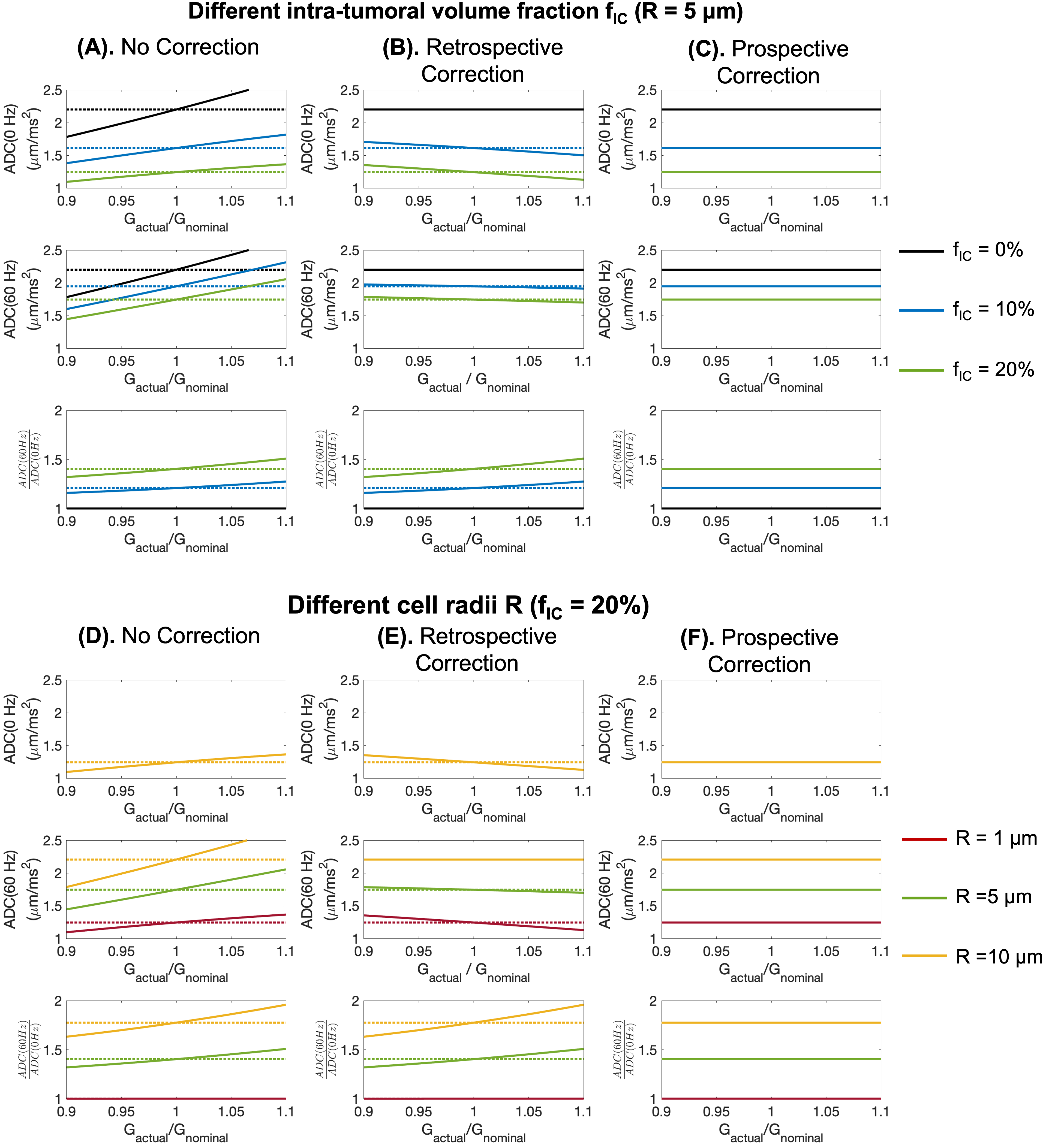
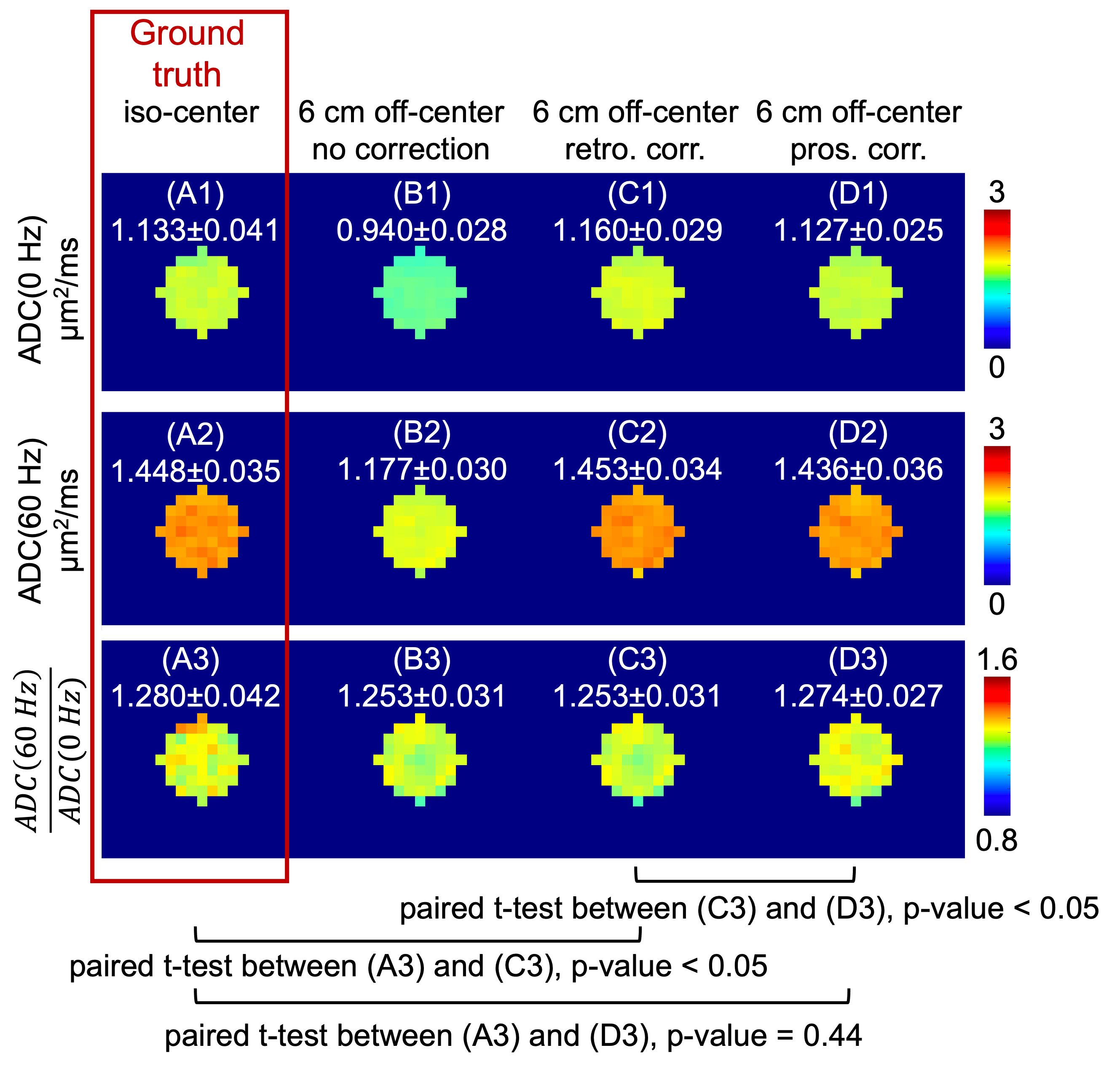
Simulations in Figure 4 show the effect of gradient nonlinearity on ADC(0 Hz), ADC(60 Hz) and $$$\frac{ADC(60 Hz)}{ADC(0 Hz)}$$$ of tumor with different intra-tumoral volume fractions and cell radii, modeled following previous studies17. Due to gradient nonlinearity (Figures 4A and 4D), all three measurements deviated from true values (dashed lines). The errors of ADC(0 Hz) and ADC(60 Hz) were reduced using retrospective correction (Figures 4B and 4E), whereas the error of $$$\frac{ADC(60 Hz)}{ADC(0 Hz)}$$$ remained unchanged. The prospective correction (Figures 4C and 4F) corrected for errors of all three measurements.In the phantom acquisitions without correction, the mean ADC(0 Hz), ADC(60 Hz) and $$$\frac{ADC(60 Hz)}{ADC(0 Hz)}$$$ at 6 cm off-center (Figures 5B) were significantly different from the measurements at iso-center (Figures 5A). The retrospective correction (Figures 5C) reduced the error on ADC(0 Hz) and ADC(60 Hz), but not on $$$\frac{ADC(60 Hz)}{ADC(0 Hz)}$$$. The prospective correction (Figures 5D) reduced errors on all three measurements.
Discussions and Conclusions
Our preliminary study demonstrated that gradient nonlinearity affects the sensitivity of time-dependent diffusion measurements for non-Gaussian diffusion, which can be corrected prospectively by applying slice-by-slice scaling of the diffusion encoding gradients in 2D imaging. Emerging small-bore, head-only gradient coils tend to have higher gradient nonlinearity12,18,19. Because an important application of these high-performance is to advance microstructure imaging, specifically by probing non-Gaussian diffusion, the effect of gradient nonlinearity requires careful consideration.Acknowledgements
Research reported in this publication was supported by the National Institute of Biomedical Imaging and Bioengineering of the National Institutes of Health (NIH) under award Number U01EB034313, and the National Cancer Institute of the NIH under award Number R01CA190299. The content is solely the responsibility of the authors and does not necessarily represent the official views of the NIH.References
1. Zhu A, Shih R, Huang RY, DeMarco JK, Bhushan C, Morris HD, Kohls G, Yeo DTB, Marinelli L, Mitra J, Hood M, Ho VB, Foo TKF. Revealing tumor microstructure with oscillating diffusion encoding MRI in pre-surgical and post-treatment glioma patients. Magnetic Resonance in Medicine 2023;90(5):1789-1801.
2. Wu D, Jiang K, Li H, Zhang Z, Ba R, Zhang Y, Hsu Y-C, Sun Y, Zhang Y-D. Time-Dependent Diffusion MRI for Quantitative Microstructural Mapping of Prostate Cancer. Radiology;0(0):211180.
3. Lee H-H, Papaioannou A, Kim S-L, Novikov DS, Fieremans E. A time-dependent diffusion MRI signature of axon caliber variations and beading. Communications biology 2020;3(1):1-13.
4. Jiang X, Li H, Xie J, McKinley ET, Zhao P, Gore JC, Xu J. In vivo imaging of cancer cell size and cellularity using temporal diffusion spectroscopy. Magnetic Resonance in Medicine 2017;78(1):156-164.
5. Chakwizira A, Zhu A, Foo T, Westin C-F, Szczepankiewicz F, Nilsson M. Diffusion MRI with free gradient waveforms on a high-performance gradient system: Probing restriction and exchange in the human brain. NeuroImage 2023:120409.
6. Malyarenko DI, Newitt DC, Amouzandeh G, Wilmes LJ, Tan ET, Marinelli L, Devaraj A, Peeters JM, Giri S, Vom Endt A, Hylton NM, Partridge SC, Chenevert TL. Retrospective Correction of ADC for Gradient Nonlinearity Errors in Multicenter Breast DWI Trials: ACRIN6698 Multiplatform Feasibility Study. Tomography 2020;6(2):86-92.
7. Dai E, Zhu A, Yang GK, Quah K, Tan ET, Fiveland E, Foo TKF, McNab JA. Frequency-dependent diffusion kurtosis imaging in the human brain using an oscillating gradient spin echo sequence and a high-performance head-only gradient. NeuroImage 2023;279:120328.
8. Tan ET, Marinelli L, Slavens ZW, King KF, Hardy CJ. Improved correction for gradient nonlinearity effects in diffusion‐weighted imaging. Journal of Magnetic Resonance Imaging 2013;38(2):448-453.
9. Malyarenko DI, Newitt D, J. Wilmes L, Tudorica A, Helmer KG, Arlinghaus LR, Jacobs MA, Jajamovich G, Taouli B, Yankeelov TE, Huang W, Chenevert TL. Demonstration of nonlinearity bias in the measurement of the apparent diffusion coefficient in multicenter trials. Magnetic Resonance in Medicine 2016;75(3):1312-1323.
10. Tao AT, Shu Y, Tan ET, Trzasko JD, Tao S, Reid RD, Weavers PT, Huston III J, Bernstein MA. Improving apparent diffusion coefficient accuracy on a compact 3T MRI scanner using gradient nonlinearity correction. Journal of Magnetic Resonance Imaging 2018;48(6):1498-1507.
11. Bammer R, Markl M, Barnett A, Acar B, Alley MT, Pelc NJ, Glover GH, Moseley ME. Analysis and generalized correction of the effect of spatial gradient field distortions in diffusion-weighted imaging. Magnetic Resonance in Medicine 2003;50(3):560-569.
12. Foo TK, Tan ET, Vermilyea ME, Hua Y, Fiveland EW, Piel JE, Park K, Ricci J, Thompson PS, Graziani D. Highly efficient head‐only magnetic field insert gradient coil for achieving simultaneous high gradient amplitude and slew rate at 3.0 T (MAGNUS) for brain microstructure imaging. Magnetic resonance in medicine 2020;83(6):2356-2369.
13. Solomon E, Lemberskiy G, Baete S, Hu K, Malyarenko D, Swanson S, Shukla-Dave A, Russek SE, Zan E, Kim SG. Time-dependent diffusivity and kurtosis in phantoms and patients with head and neck cancer. Magnetic Resonance in Medicine 2023;89(2):522-535.
14. Malyarenko DI, Swanson SD, Konar AS, LoCastro E, Paudyal R, Liu MZ, Jambawalikar SR, Schwartz LH, Shukla-Dave A, Chenevert TL. Multicenter Repeatability Study of a Novel Quantitative Diffusion Kurtosis Imaging Phantom. Tomography 2019;5(1):36-43.
15. Malyarenko D, Chenevert TL. Practical correction of gradient nonlinearity bias for mean diffusion kurtosis model parameters.16. Swanson SD, Malyarenko DI, Chenevert TL. Nanoscopic materials for quantitative water exchange phantoms.
17. Zhu A, Shih R, Huang RY, DeMarco JK, Bhushan C, Morris HD, Kohls G, Yeo DTB, Marinelli L, Mitra J, Hood M, Ho VB, Foo TKF. Revealing tumor microstructure with oscillating diffusion encoding MRI in pre-surgical and post-treatment glioma patients. Magn Reson Med 2023;90(5):1789-1801.
18. Weiger M, Overweg J, Rösler MB, Froidevaux R, Hennel F, Wilm BJ, Penn A, Sturzenegger U, Schuth W, Mathlener M. A high‐performance gradient insert for rapid and short‐T2 imaging at full duty cycle. Magnetic resonance in medicine 2018;79(6):3256-3266.
19. Davids M, Dietz P, Ruyters G, Roesler M, Klein V, Guérin B, Feinberg DA, Wald LL. Peripheral nerve stimulation informed design of a high-performance asymmetric head gradient coil. Magnetic Resonance in Medicine 2023;90(2):784-801.
Figures