2411
A Docker-based Ensemble Pipeline for Tractogram with High-throughput, Reproducible and Comprehensive Mapping of White Matter Fibers (DEPTH)1Department of Radiology, Children's Hospital of Philadelphia, Philadelphia, PA, United States, 2School of Software, Dalian University of Technology, Dalian, China, 3Department of Bioengineering, University of Pennsylvania, Philadelphia, PA, United States, 4Department of Radiology, University of Pennsylvania, Philadelphia, PA, United States
Synopsis
Keywords: Diffusion Software, Software Tools, High-throughput, Docker, Reproducibility, Autism Spectrum Disorder, Brain Connectivity
Motivation: The complex whole brain connectome is underlined by white matter (WM) fibers featuring both long and short-range connections. Most existing protocols are tailored to trace long-range fibers, incapable of comprehensively tracing all fibers including both long and short-range fibers.
Goal(s): To achieve comprehensive mapping of whole-brain WM fibers in a unified framework including specifically high-fidelity tractogram of short-range fibers.
Approach: We developed DEPTH, a Docker-containerized pipeline extending advanced short-range fiber tracing.
Results: DEPTH offers high-throughput, reproducible and comprehensive tractogram of both long and short-range WM fibers. The Docker-based containerization ensures cross-platform usability and versatility without complex setup and configuration.
Impact: High-throughput, reproducible and comprehensive whole-brain white matter fibers across multisite datasets could now be traced by our software.
Introduction
Information transmission within the human brain occurs through a complex distributed network, arising from interactions between both nearby regions known as “short-range” and projections from non-adjacent areas referred to as “long-range” or distant connections1-3. Existing protocols designed for the tracing of a particular category of tracts4-7 are inadequate for the comprehensive mapping of whole brain white matter fibers. To bridge this gap, we extend our prior STTAR8 protocol to form a unified white matter fiber tracing pipeline, DEPTH.Methods
Docker-base Encapsulation (Figure 1): DEPTH is containerized using Docker9-11, packaging the extended STTAR8 protocol and other necessary dependencies such as ANTs12, FreeSurfer13, FSL14 and DIPY15 to form an all-in-one platform. It integrates preprocessing, tracing and clustering of high-throughput and reproducible white matter tracts, compatible with most operating systems.Overview of DEPTH protocol by extending STTAR (Figure 2): Raw data is organized as BIDS16 and preprocessed by the dMRI and anatomical preprocessing workflows (a, b). STTAR tracing is conducted by seeding from the whole brain (c), short/long-range fibers that terminate in adjacent/non-adjacent regions are extracted (d) followed by HDBSCAN17 for delineating clusters (e). To delineate well-defined deep tracts, the traced whole brain tractogram is registered to a reference streamline atlas18 and pruned using RecoBundles19 implemented in DIPY15 (f). Details of each step are explained below.
Data used in this study include ten subjects from each dataset (in total forty) aged 22-25 years from Human Connectome Project (HCP), 22-25 years from Autism Brain Imaging Data Exchange (ABIDE), 9-10 years from Adolescent Brain Cognitive Development (ABCD) and 5-20 years from Healthy Brain Network (HBN).
dMRI and anatomical preprocessing: Raw diffusion weighted images (DWI) are first denoised using Patch2Self20 and corrected for B0 susceptibility-induced distortion using FSL’s topup. Eddy current and motion correction are jointly carried out by FSL’s eddy.
The T1w images are first corrected for intensity nonuniformity with ANTs12 and skull-stripped with mri_synthstrip21 in FreeSurfer13. Brain parcellation is performed on the fused T1w by mri_synthseg22 in Freesurfer13. The parcellated labels are then transferred to DWI space by structural-to-diffusion coregistration. FSL’s FLIRT is run as initialization, followed by FSL’s epi_reg as refinement.
Extended STTAR tracing: The whole brain mask is used as seed ROI. FDT (FMRIB’s Diffusion Toolbox) bedpostx23-25 is applied for local fiber orientation estimation with two fibers per voxel. Probtrackx23-24 is used for streamline tracing with 10 seeds per voxel and traced in opposite directions from the seed.
ROI-based filtering and clustering: Streamlines that terminate in adjacent regions are extracted as short-range fibers and filtered with NOT ROI and length threshold using the same setting specified in STTAR8. Streamlines connecting non-adjacent regions are extracted as long-range fibers and filtered with the same NOT ROI. HDBSCAN17 is then applied to produce clusters.
Atlas-based deep tract parcellation: The traced whole brain tractogram is registered to the reference atlas18 using streamline-based linear registration26. Deep white matter tracts are parcellated based on their minimum average direct-flip distance with the atlas clusters using RecoBundles19.
Results
Figure 3 demonstrates representative white matter clusters from three HCP subjects identified by DEPTH. Left panel (a) shows short-range clusters from eight pairs of cortical regions traced, right panel (b) shows long-range clusters from two pairs of cortical regions and four well-defined deep tracts of consistent shapes. Figure 4 shows merged centroids and probability maps of clusters from four datasets: HCP, ABCD, HBN and ABIDE, with ten subjects per dataset. Specifically, five centroid streamlines are extracted from each cluster to form a cluster with fifty streamlines across ten subjects. Each cluster is converted to a binary map and averaged to generate the probability map. For example, cluster#1 and cluster#2 connecting cMFG and PrCG show a left-oriented U- and an up-oriented U-shape respectively. In Figure 5, we compare the mean fractional anisotropy (FA) of six clusters from three gyrus pairs between healthy subjects and autism spectrum disorder (ASD) subjects. Ten subjects from HCP and ABIDE of age 22-25 are adopted respectively. We observe significant differences from cluster#2 linking IPG-SPG, cluster#1 and cluster#2 linking cMFG-PrCG and cluster#1 linking SPG-SMG that averaged FAs in healthy subjects are higher than ASD subjects.Discussion and Conclusion
The proposed DEPTH is versatile and reproducible for comprehensive mapping of both long and short-range white matter fibers. It can be potentially used to automatically trace fibers connecting any user-customized ROIs to study the connectivity in specific focused brain regions. Statistics significance shown in Figure 5 indicates the applicability of DEPTH in studying relevant neuropsychiatric disorders. Whole brain white matter fiber atlas can also be established as the foundation for broader neuroscientific and clinical discoveries.Acknowledgements
This study is funded by NIH R01MH092535, R01MH125333, R01EB031284, R01MH129981, R21MH123930 and P50HD105354.References
1. Ouyang, M., Dubois, J., Yu, Q., Mukherjee, P., & Huang, H. (2019). Delineation of early brain development from fetuses to infants with diffusion MRI and beyond. Neuroimage, 185, 836-850.
2. Ouyang, M., Kang, H., Detre, J. A., Roberts, T. P., & Huang, H. (2017). Short-range connections in the developmental connectome during typical and atypical brain maturation. Neuroscience & Biobehavioral Reviews, 83, 109-122.
3. Ouyang, M., Jeon, T., Mishra, V., Du, H., Wang, Y., Peng, Y., & Huang, H. (2016, March). Global and regional cortical connectivity maturation index (CCMI) of developmental human brain with quantification of short-range association tracts. In Medical Imaging 2016: Biomedical Applications in Molecular, Structural, and Functional Imaging (Vol. 9788, pp. 328-334). SPIE.
4. Huang, H., Zhang, J., Jiang, H., Wakana, S., Poetscher, L., Miller, M. I., ... & Mori, S. (2005). DTI tractography based parcellation of white matter: application to the mid-sagittal morphology of corpus callosum. Neuroimage, 26(1), 195-205.
5. Huang, H., Zhang, J., Van Zijl, P. C., & Mori, S. (2004). Analysis of noise effects on DTI‐based tractography using the brute‐force and multi‐ROI approach. Magnetic Resonance in Medicine: An Official Journal of the International Society for Magnetic Resonance in Medicine, 52(3), 559-565.
6. Wakana, S., Caprihan, A., Panzenboeck, M. M., Fallon, J. H., Perry, M., Gollub, R. L., ... & Mori, S. (2007). Reproducibility of quantitative tractography methods applied to cerebral white matter. Neuroimage, 36(3), 630-644.
7. Román, C., Hernández, C., Figueroa, M., Houenou, J., Poupon, C., Mangin, J. F., & Guevara, P. (2022). Superficial white matter bundle atlas based on hierarchical fiber clustering over probabilistic tractography data. NeuroImage, 262, 119550.
8. Zhao, C., Ouyang, M., Yu, Q., Huang, H. (2020). Short-range Tractography with high Throughput and Reproducibility (STTAR) characterized by FDT tracing and HDBSCAN clustering. Proceeding of ISMRM, 857.
9. Gorgolewski, K. J., Alfaro-Almagro, F., Auer, T., Bellec, P., Capotă, M., Chakravarty, M. M., ... & Poldrack, R. A. (2017). BIDS apps: Improving ease of use, accessibility, and reproducibility of neuroimaging data analysis methods. PLoS computational biology, 13(3), e1005209.
10. Cieslak, M., Cook, P. A., He, X., Yeh, F. C., Dhollander, T., Adebimpe, A., ... & Satterthwaite, T. D. (2021). QSIPrep: an integrative platform for preprocessing and reconstructing diffusion MRI data. Nature methods, 18(7), 775-778.
11. Esteban, O., Markiewicz, C. J., Blair, R. W., Moodie, C. A., Isik, A. I., Erramuzpe, A., ... & Gorgolewski, K. J. (2019). fMRIPrep: a robust preprocessing pipeline for functional MRI. Nature methods, 16(1), 111-116.
12. Avants, B. B., Epstein, C. L., Grossman, M., & Gee, J. C. (2008). Symmetric diffeomorphic image registration with cross-correlation: evaluating automated labeling of elderly and neurodegenerative brain. Medical image analysis, 12(1), 26-41.
13. Fischl, B. (2012). FreeSurfer. Neuroimage, 62(2), 774-781.
14. Andersson, J. L., & Sotiropoulos, S. N. (2016). An integrated approach to correction for off-resonance effects and subject movement in diffusion MR imaging. Neuroimage, 125, 1063-1078.
15. Garyfallidis, E., Brett, M., Amirbekian, B., Rokem, A., Van Der Walt, S., Descoteaux, M., ... & Dipy Contributors. (2014). Dipy, a library for the analysis of diffusion MRI data. Frontiers in neuroinformatics, 8.
16. Gorgolewski, K. J., Auer, T., Calhoun, V. D., Craddock, R. C., Das, S., Duff, E. P., ... & Poldrack, R. A. (2016). The brain imaging data structure, a format for organizing and describing outputs of neuroimaging experiments. Scientific data, 3(1), 1-9.
17. McInnes, L., Healy, J., & Astels, S. (2017). hdbscan: Hierarchical density based clustering. J. Open Source Softw., 2(11), 205.
18. Yeh, F. C., Panesar, S., Fernandes, D., Meola, A., Yoshino, M., Fernandez-Miranda, J. C., ... & Verstynen, T. (2018). Population-averaged atlas of the macroscale human structural connectome and its network topology. Neuroimage, 178, 57-68.
19. Garyfallidis, E., Côté, M. A., Rheault, F., Sidhu, J., Hau, J., Petit, L., ... & Descoteaux, M. (2018). Recognition of white matter bundles using local and global streamline-based registration and clustering. NeuroImage, 170, 283-295.
20. Fadnavis, S., Batson, J., & Garyfallidis, E. (2020). Patch2Self: Denoising Diffusion MRI with Self-Supervised Learning. Advances in Neural Information Processing Systems, 33, 16293-16303.
21. Hoopes, A., Mora, J. S., Dalca, A. V., Fischl, B., & Hoffmann, M. (2022). SynthStrip: Skull-stripping for any brain image. NeuroImage, 260, 119474.
22. Billot, B., Greve, D. N., Puonti, O., Thielscher, A., Van Leemput, K., Fischl, B., ... & Iglesias, J. E. (2023). SynthSeg: Segmentation of brain MRI scans of any contrast and resolution without retraining. Medical image analysis, 86, 102789.
23. Behrens, T. E., Berg, H. J., Jbabdi, S., Rushworth, M. F., & Woolrich, M. W. (2007). Probabilistic diffusion tractography with multiple fibre orientations: What can we gain?. Neuroimage, 34(1), 144-155.
24. Behrens, T. E., Woolrich, M. W., Jenkinson, M., Johansen‐Berg, H., Nunes, R. G., Clare, S., ... & Smith, S. M. (2003). Characterization and propagation of uncertainty in diffusion‐weighted MR imaging. Magnetic Resonance in Medicine: An Official Journal of the International Society for Magnetic Resonance in Medicine, 50(5), 1077-1088.
25. Jbabdi, S., Sotiropoulos, S. N., Savio, A. M., Graña, M., & Behrens, T. E. (2012). Model‐based analysis of multishell diffusion MR data for tractography: How to get over fitting problems. Magnetic resonance in medicine, 68(6), 1846-1855.
26. Garyfallidis, E., Ocegueda, O., Wassermann, D., & Descoteaux, M. (2015). Robust and efficient linear registration of white-matter fascicles in the space of streamlines. NeuroImage, 117, 124-140.
Figures
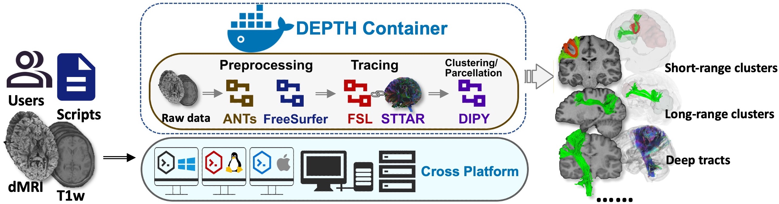
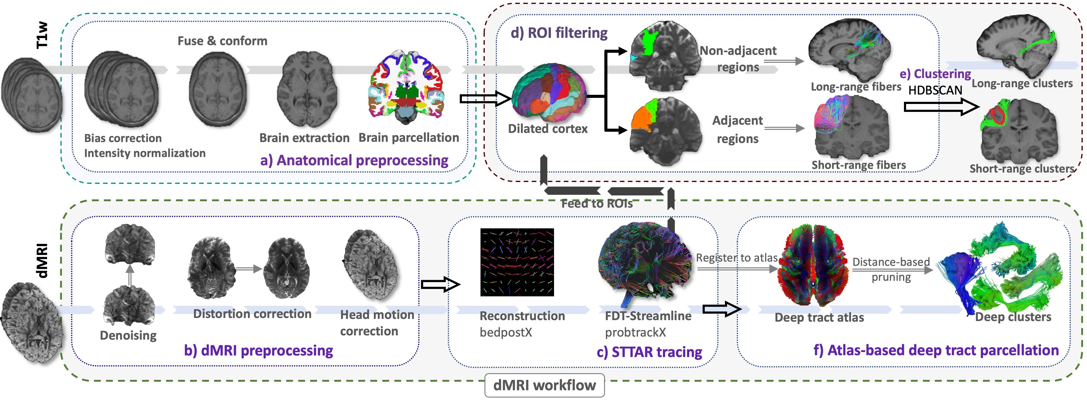
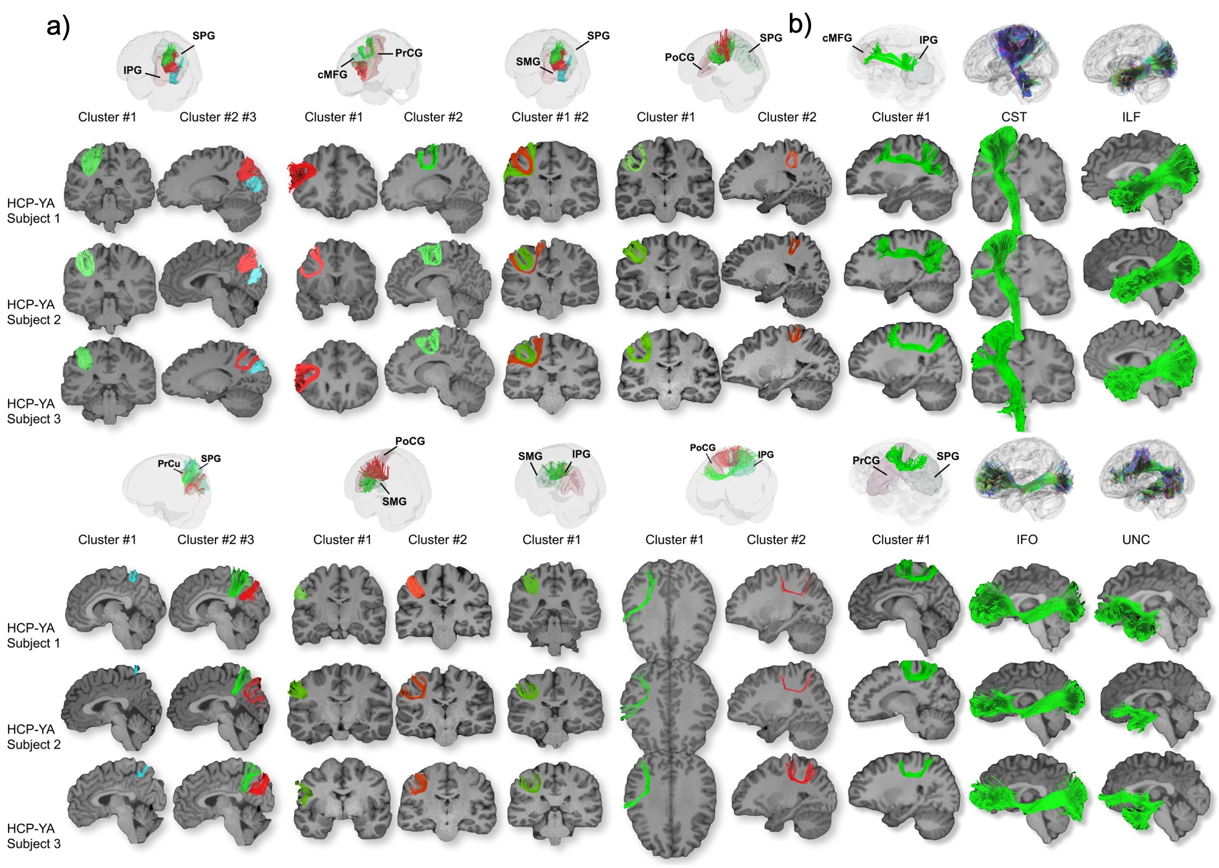
Figure 3. Visual display of representative (a) short-range clusters from eight pairs of gyri and (b) six long-range clusters identified by DEPTH. Abbreviation: IPG=inferior parietal gyrus, SPG=superior parietal gyrus, cMFG= caudal middle frontal gyrus, PrCG=precentral gyrus, SMG=supramarginal gyrus, PoCG=postcentral gyrus, PrCu=precuneus gyrus, CST=corticospinal tract, ILF=inferior longitudinal fasciculus, IFO=inferior longitudinal fasciculus, UNC= Uncinate fasciculus.
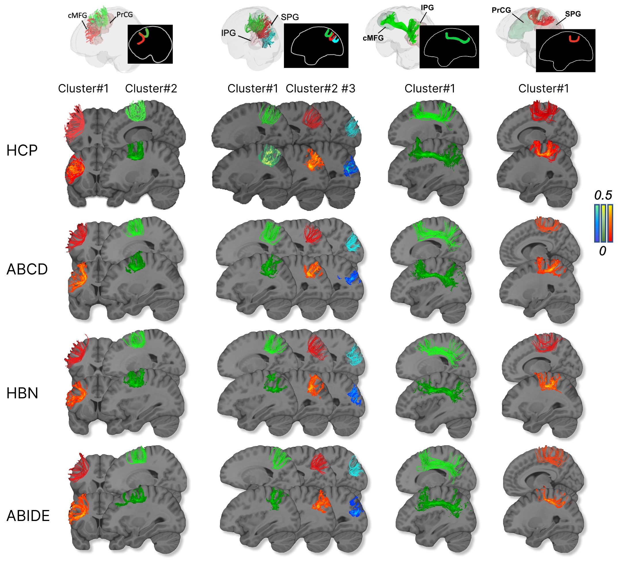
Figure 4. High reproducibility of identified short-range fibers across multiple datasets: HCP, ABCD, HBN and ABIDE. The consistent shape of merged centroids and probabilistic maps indicate that DEPTH can be applied to data of various qualities and scanning protocols. It shows the reproducible trajectories of clusters, with higher intensity representing better reproducibility. Abbreviation: IPG=inferior parietal gyrus, SPG=superior parietal gyrus, cMFG= caudal middle frontal gyrus, PrCG=precentral gyrus.
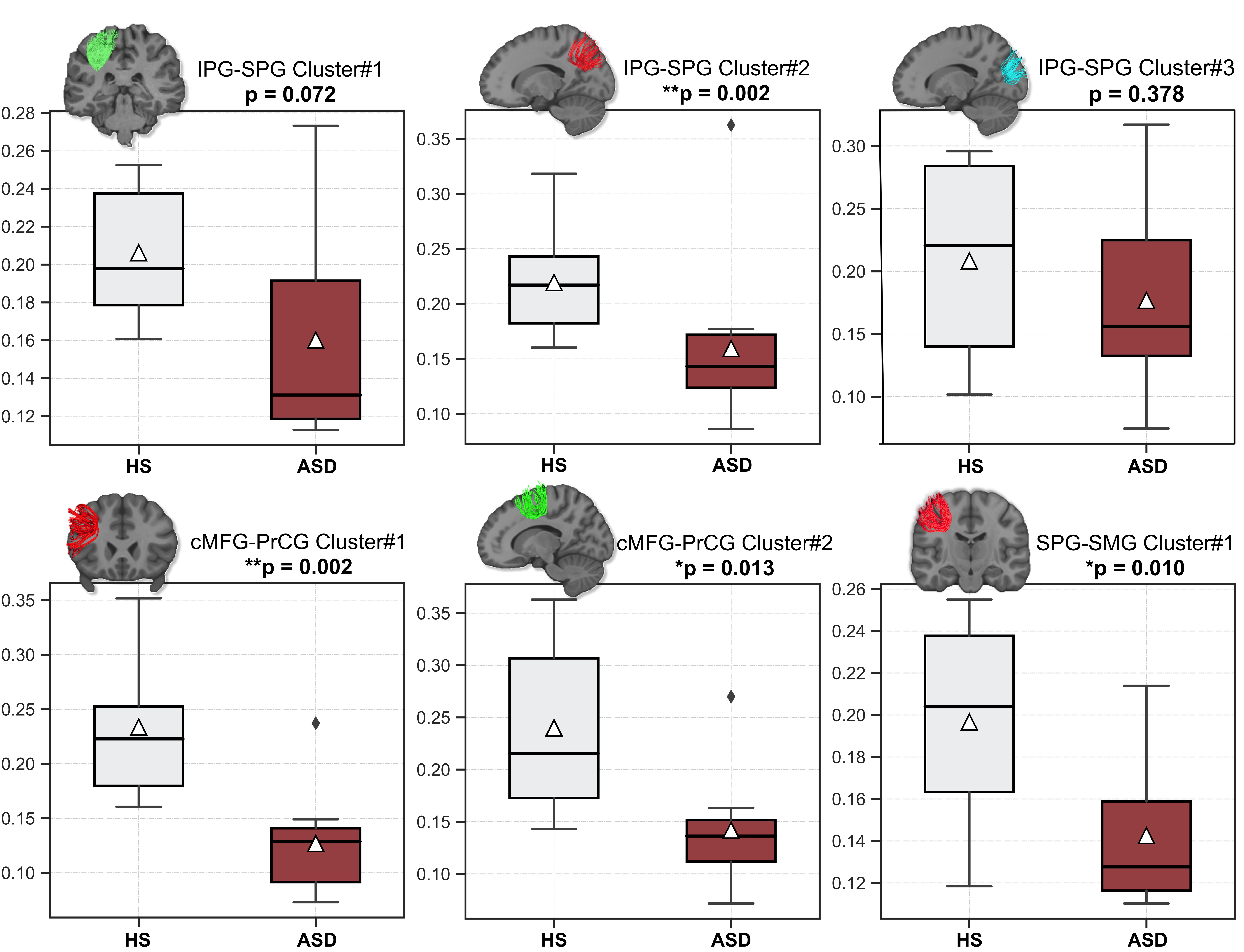
Figure 5. Compared mean FA over six short-range clusters between healthy subjects (HS) and Autism spectrum disorder (ASD) subjects. We observe significant differences on the second cluster linking IPG-SPG, two clusters linking cMFG-PrCG and one cluster linking SPG-SMG. The triangles and lines in the boxes denote mean and medium values. Abbreviation: IPG=inferior parietal gyrus, SPG=superior parietal gyrus, cMFG= caudal middle frontal gyrus, PrCG=precentral gyrus, SMG=supramarginal gyrus.