2409
The Pseudo-Fiber Effect: Fiber Density Impacts on Diffusion Metrics1Mallinckrodt Institute of Radiology, Washington University School of Medicine in St. Louis, St. Louis, MO, United States, 2Department of Mathematics, Washington University in St. Louis, St. Louis, MO, United States
Synopsis
Keywords: Diffusion Modeling, Diffusion/other diffusion imaging techniques
Motivation: To understand how axon density affects degeneracy-related errors in diffusion metric estimation.
Goal(s): Assuming ideal conditions (e.g., noise free and no inter-compartmental exchange), are there correlations between voxel contents and model degeneracies?
Approach: A systematically-generated set of simulated voxels spanning a wide range of microstructural compositions, processed with various diffusion models, and analyzed both comparatively and component-wise.
Results: Based on the individual signal component results, the extra-axonal water is the largest confounding factor. A model's depends largely on its ability to distinguish truly intra-axonal water from constrained extra-axonal water.
Impact: It is important to consider degeneracies when interpreting the results of diffusion modeling lest the associated errors propagate downstream. Our results offer a more robust understanding of the conditions which give rise to these degeneracy-related errors.
Introduction
Model degeneracies whereby two or more microstructural configurations within an image voxel give rise to equivalent solutions when processed are known to occur in the analysis of diffusion-weighted images (DWI) [see, e.g., 1–3]. One example of degeneracy-related phenomena leading to systematic errors in diffusion metrics estimated via microstructural diffusion models is the “parallel fibers problem” [4, 5]. To illustrate this further, imagine a voxel comprising two equally proportioned parallel axon bundles. The ground-truth microstructure in the scenario is described by two anisotropic diffusion tensors X and Y ; however, when this voxel is processed with a diffusion MRI (dMRI) model, X and Y become mathematically indistinguishable from a single anisotropic diffusion tensor Z with a diffusivity equal to the mean of X and Y . A more nuanced example of conditions that produce degenerate solutions—the “pseudo-fiber effect”—occurs due to incomplete or erroneous signal partitioning among modeled microstructural components [6]. Pseudo-fibers, or systematic errors in axonal metrics, are hypothesized to arise due to the spurious inclusion of extra-axonal signals within the employed microstructural model's axonal term(s).Methods
To investigate how the pseudo-fiber effect varies with fiber volume fraction for selected diffusion MRI models, we generated 300 noise-free isotropic (75×75×75-µm) diffusion MRI voxels with unique microstructural configurations were generated via simDRIFT, a Python package for massively parallel simulations of water self-diffusion in biophysically relevant tissue features [15]. The in silico phantom formed by these voxels comprises a broad range of biophysically meaningful microstructural conditions. We compared the results from this mosaic produced by diffusion basis spectrum imaging (DBSI, [7–9]) and the white matter tract integrity imaging (WMTI, [10, 11]) interpretation of the diffusion kurtosis imaging (DKI, [12–14]) model, two tensor-based models with differing algorithmic approaches. The test mosaic, shown in Figure 1, contains voxels with four fiber crossing angles, fifteen fiber fractions, and five cell fractions. Each voxel contains two fiber bundles with equal volume fractions and radii (1.0 µm) but distinct diffusivities (1.0 µm2/ms and 2.0 µm2/ms), hereafter referred to as “fiber 1” and “fiber 2”, respectively. The diffusivity of extra-axonal water was set as 3.0 µm2/ms.Within the simulated voxels, water self-diffusion was simulated via a random walk of 106 randomly initialized “spins” with a time-step of dt = 1×10⁻⁶s and a total diffusion time of ∆=1×10⁻²s. Since the diffusion time used for the simulations is less than the pre-exchange lifetime of axonal water (∼5×10⁻² s, [16]), inter-compartmental exchange is programmatically excluded. Given that dt ≪ ∆ for these simulations, the narrow-pulse-width approximation (i.e., dt ≈ δ, [17]) is employed.
Results
The results for DBSI clearly illustrate two manifestations of the pseudo-fiber effect. First, DBSI fails to distinguish the two fiber bundles when they are parallel, resulting in the inaccurate determination of fiber AD even when the signal consists only of the portion emanating from the axons (the square data points in Figure 2A–B). Second, at volume fractions ≳50%, the constraints imposed on the extra-cellular water by the closely packed fibers cause DBSI to identify this extra-axonal water as axonal. This artificially inflates the measured AD and introduces further errors in fiber fraction estimates, as seen in the uptick in relative error in Figure 3B. The results obtained from the test mosaic by DKI show a similarly apparent increase in erroneously identified axons at higher fiber-packing densities. The role of water in these errors is solidified by comparing the fiber fractions obtained from the signal composed of only fibers to those obtained from the total (i.e., extra-fiber-water included) signal. Moreover, since DKI identifies only the lower-AD fiber of the two present, its fiber fraction estimates are systematically under-valued for even the fiber-only signal. This fiber’s range of measured AD varies with orientation, covering a ±0.2 µm2/ms range from the mean predicted value.Discussion and Conclusions
Both of the examined diffusion models suffer to some extent from a loss of accuracy under a subset of potential biophysical contexts. Separating and processing the signals produced by each component of the imaging voxel has allowed us to identify which sets of conditions produce degenerate results for each model. The factor primarily responsible for the pseudo-fiber effect appears to be the extra-cellular water, as has been suggested by other researchers [6, 18, 19]. From our tests, we propose that this contribution arises from the structure imposed by nearby fiber bundles on the extra-axonal water. The constrained water engenders artificially inflated axonal diffusivity and signal fraction values through incomplete partitioning by the chosen model. To resolve the pseudo-fiber effect, future models will likely need to completely reimagine signal partitioning methods.Acknowledgements
This work was funded in part via support from NIH NINDS R01 NS11691 and R01 NS047592. The authors would like to acknowledge the early contributions of Chunyu Song and Anthony Wu.References
1. O. Jelescu, J. Veraart, E. Fieremans, and D. S. Novikov, “Degeneracy in model parameter estimation for multi-compartmental diffusion in neuronal tissue”, NMR in Biomedicine 29, 33–47 (2015).
2. S. Coelho, J. M. Pozo, S. N. Jespersen, D. K. Jones, and A. F. Frangi, “Resolving degeneracy in diffusion MRI biophysical model parameter estimation using double diffusion encoding”, Magnetic Resonance in Medicine 82, 395–410 (2019).
3. H. Rafipoor, Y.-Q. Zheng, L. Griffanti, S. Jbabdi, and M. Cottaar, “Identifying microstructural changes in diffusion MRI: How to circumvent parameter degeneracy”, NeuroImage 260, 119452 (2022).
4. K. K. Seunarine and D. C. Alexander, “Multiple Fibers: Beyond the Diffusion Tensor”, in Diffusion MRI (Elsevier, 2014), pp. 105–123.
5. K. Schilling, Y. Gao, V. Janve, I. Stepniewska, B. A. Landman, and A. W. Anderson, “Can increased spatial resolution solve the crossing fiber problem for diffusion MRI?”, NMR in Biomedicine 30, 10.1002/nbm.3787 (2017).
6. M. Kumar, R. K. Gupta, K. Nath, R. K. S. Rathore, G. Bayu, R. Trivedi, M. Husain, K. N. Prasad, R. P. Tripathi, and P. A. Narayana, “Can we differentiate true white matter fibers from pseudofibers inside a brain abscess cavity using geometrical diffusion tensor imaging metrics?”, NMR in Biomedicine 21, 581–588 (2007).
7. Y. Wang, Q. Wang, J. P. Haldar, F.-C. Yeh, M. Xie, P. Sun, T.-W. Tu, K. Trinkaus, R. S. Klein, A. H. Cross, and S.-K. Song, “Quantification of increased cellularity during inflammatory demyelination”, Brain 134, 3590–3601 (2011).
8. A. H. Cross and S.-K. Song, “A new imaging modality to non-invasively assess multiple sclerosis pathology”, Journal of Neuroimmunology 304, 81–85 (2017).
9. Z. Ye, S. E. Gary, P. Sun, S. M. Mustafi, G. R. Glenn, F.-C. Yeh, H. Merisaari, C. Song, R. Yang, G.-S. Huang, H.-W. Kao, C.-Y. Lin, Y.-C. Wu, J. H. Jensen, and S.-K. Song, “The impact of edema and fiber crossing on diffusion MRI metrics assessed in an ex vivo nerve phantom: Multi-tensor model vs. diffusion orientation distribution function”, NMR in Biomedicine 34, 10.1002/nbm.4414 (2020).
10. E. Fieremans, J. H. Jensen, and J. A. Helpern, “White matter characterization with diffusional kurtosis imaging”, NeuroImage 58, 177–188 (2011).
11. E. Fieremans, A. Benitez, J. H. Jensen, M. F. Falangola, A. Tabesh, R. L. Deardorff, M. V. S. Spampinato, J. S. Babb, D. S. Novikov, S. H. Ferris, and J. A. Helpern, “Novel white matter tract integrity metrics sensitive to Alzheimer disease progression”, American Journal of Neuroradiology 34, 2105–2112 (2013).
12. J. H. Jensen, J. A. Helpern, A. Ramani, H. Lu, and K. Kaczynski, “Diffusional kurtosis imaging: the quantification of non-Gaussian water diffusion by means of magnetic resonance imaging”, Magnetic Resonance in Medicine 53, 1432–1440 (2005).
13. J. H. Jensen, M. F. Falangola, C. Hu, A. Tabesh, O. Rapalino, C. Lo, and J. A. Helpern, “Preliminary observations of increased diffusional kurtosis in human brain following recent cerebral infarction”, NMR in Biomedicine 24, 452–457 (2010).
14. H. Lu, J. H. Jensen, A. Ramani, and J. A. Helpern, “Three-dimensional characterization of non-Gaussian water diffusion in humans using diffusion kurtosis imaging”, NMR in Biomedicine 19, 236–247 (2006).
15. J. Blum and K. L. Utt, “Simulated diffusion in realistic imaging features of tissue (SimDRIFT)”, Journal of Open Source Software 8, 5621 (2023).
16. X. He, M. E. Raichle, and D. A. Yablonskiy, “Transmembrane dynamics of water exchange in human brain”, Magnetic Resonance in Medicine 67, 562–571 (2011).
17. L. Z. Wang, A. Caprihan, and E. Fukushima, “The narrow-pulse criterion for pulsed-gradient spin-echo diffusion measurements”, Journal of Magnetic Resonance, Series A 117, 209–219 (1995).
18. J. Veraart, E. Fieremans, and D. S. Novikov, “On the scaling behavior of water diffusion in human brain white matter”, NeuroImage 185, 379–387 (2019).
19. G. Rojas-Vite, R. Coronado-Leija, O. Narvaez-Delgado, A. Ramírez-Manzanares, J. L. Marroquín, R. Noguez-Imm, M. L. Aranda, B. Scherrer, J. Larriva-Sahd, and L. Concha, “Histological validation of per-bundle water diffusion metrics within a region of fiber crossing following axonal degeneration”, NeuroImage 201, 116013 (2019).
Figures
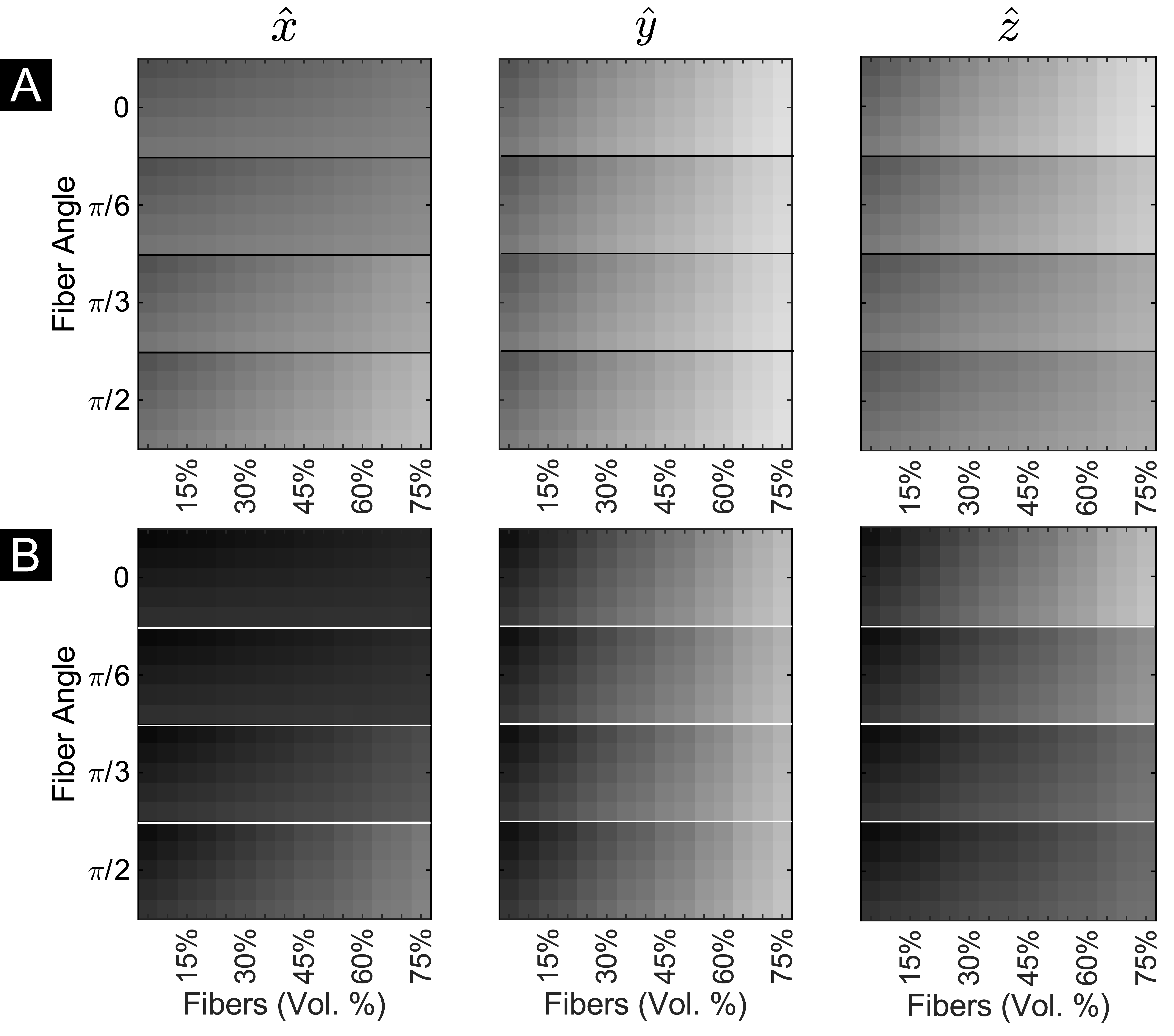
Figure 1: Diffusion-weighted signal obtained for the 20×15 voxel test mosaic along each Cartesian axis. The images shown in (A) were generated using a diffusion gradient strength of b = 400 s/mm2, whereas those shown in (B) were generated with b = 1200 s/mm2. Intensity is logarithmically scaled to the maximum intensity (i.e., the b = 0 image) and plotted with arbitrary units.
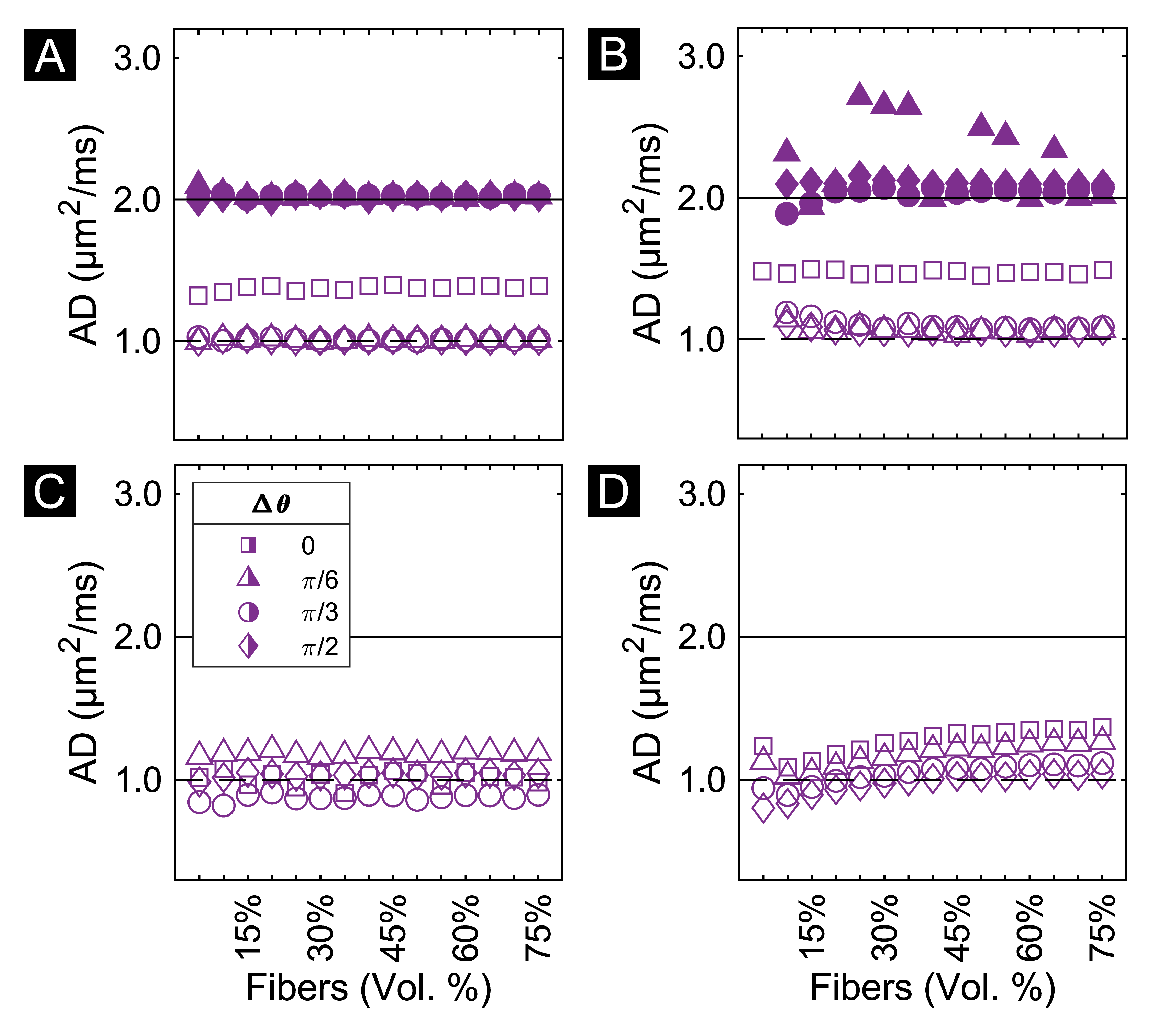
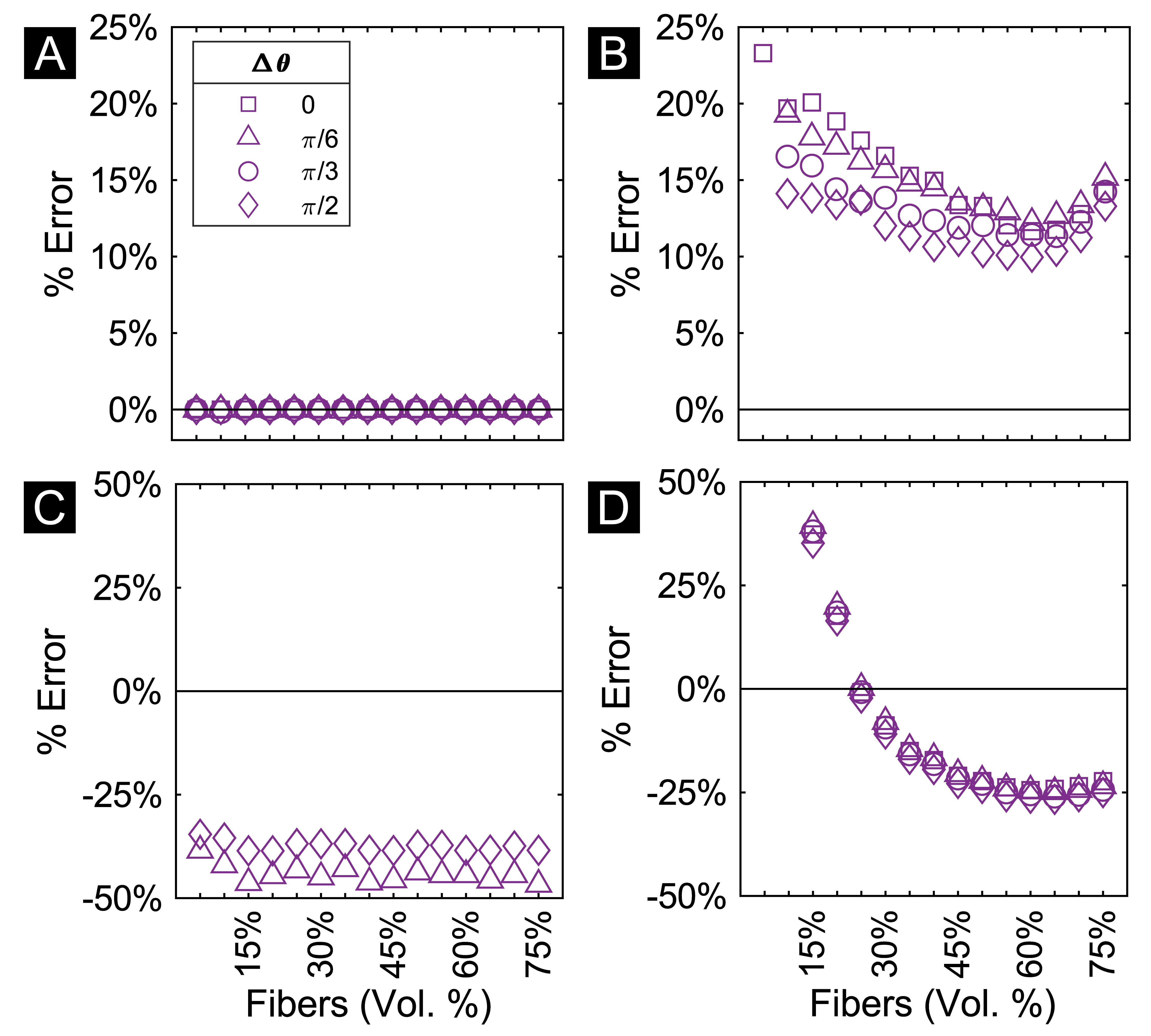
Figure 3: Axonal signal fraction estimates for simulated voxels without cells. The plotted values were calculated as the relative error between the estimated and ground truth values. (A-B) DBSI fiber fraction estimates from the fibers-only signal and the total signal, respectively. (C-D) DKI axonal water fraction estimates obtained using the fibers-only signal and the total signal, respectively.