2407
A GPU-Accelerated Software Toolbox for Simulation and Analysis of Diffusion MRI in Microstructures with a Graphical User Interface1Vanderbilt University Medical Center, Nashville, TN, United States, 2Vanderbilt University, Nashville, TN, United States, 3High School, Knoxville, TN, United States
Synopsis
Keywords: Diffusion Software, Diffusion/other diffusion imaging techniques, software, simulation, analysis
Motivation: Simulation and analysis of diffusion MRI (dMRI) in microstructures requires specialized expertise that limits its wide usage.
Goal(s): To provide an easy-to-use dMRI software package to enable end users to simulate and analyze diffusion in complex media.
Approach: MATI is written using object-oriented programming in two versions (MATLAB and Python) and can be used via a GUI or scripts. It can handle typical diffusion pulse sequences and various microstructures for data fitting, and arbitrary digitalized microstructures for simulating dMRI with GPU acceleration.
Results: A GPU-accelerated dMRI simulation and data fitting toolbox with a GUI has been developed for public use.
Impact: MATI, a GPU-accelerated toolbox for simulating and analyzing dMRI signals in various microstructures with a graphical user interface (GUI), has been developed. This easy-to-use package enables non-expert and expert users to simulate and analyze diffusion MRI signals in complex media.
Introduction
Diffusion MRI (dMRI) continues to be of considerable interest for characterizing tissue microstructure beyond the resolution limits of image voxel sizes to obtain information at cellular or subcellular levels. Several software toolboxes have been developed to simulate dMRI signals from arbitrary tissue structures such as SpinDoctor1 and RMS2, or to fit biophysical signal models to dMRI data to extract microstructural parameters non-invasively, such as FSL, MRTrix3, DSI-Studio, Camino, and Dipy. This work aims to provide an easy-to-use toolbox that provides both capabilities. We developed MATI (Microstructural Analysis of Tissues by Imaging) as a GPU-accelerated diffusion MRI signal simulation and data fitting toolbox with a graphical user-friendly interface (GUI). MATI provides a framework that emphasizes usability, so non-expert end users can perform microstructural dMRI research, and extensibility, so experts can extend the toolbox with new pulse sequences and biophysical models.Methods
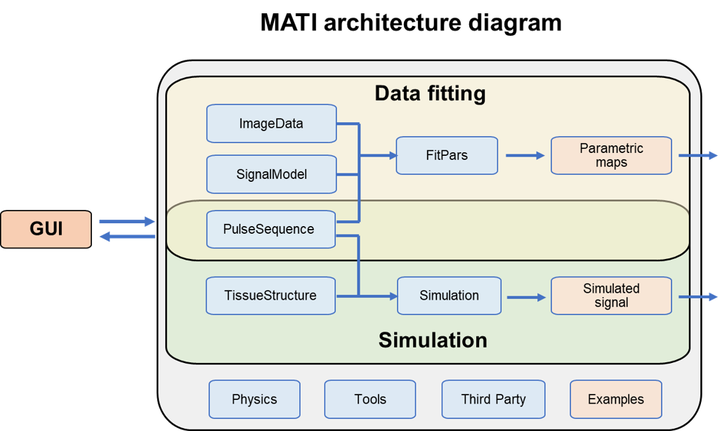
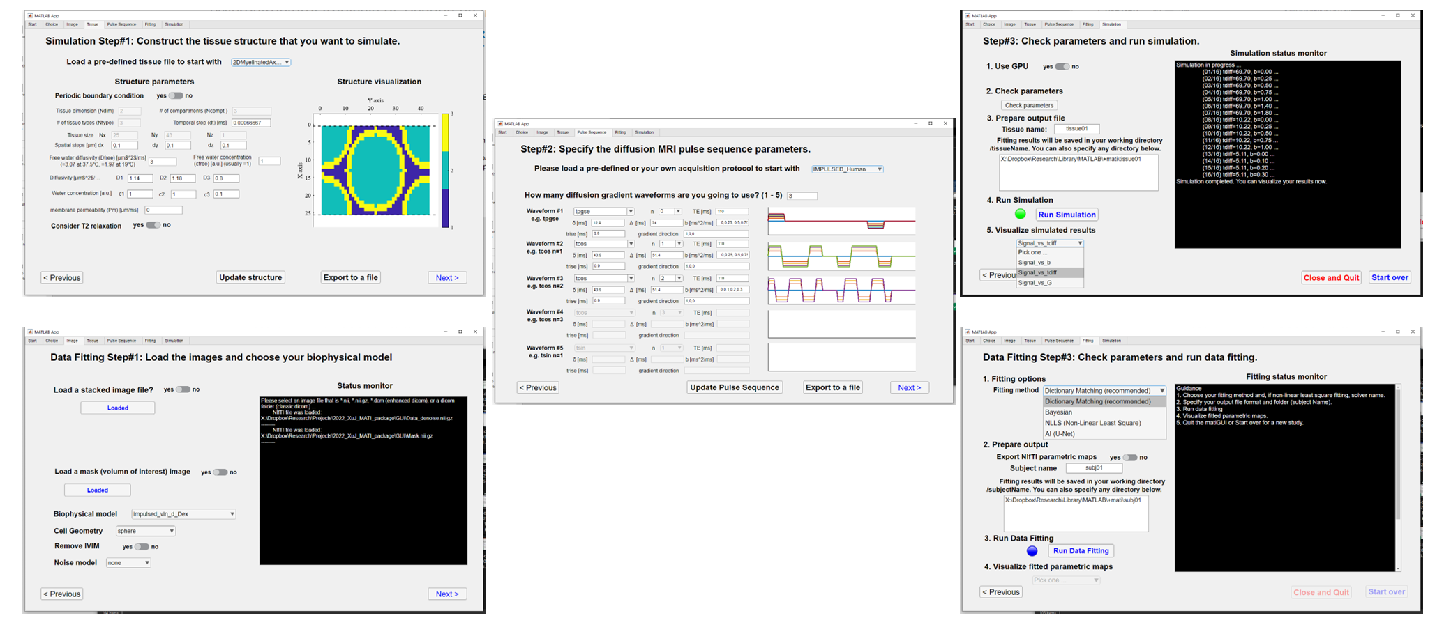
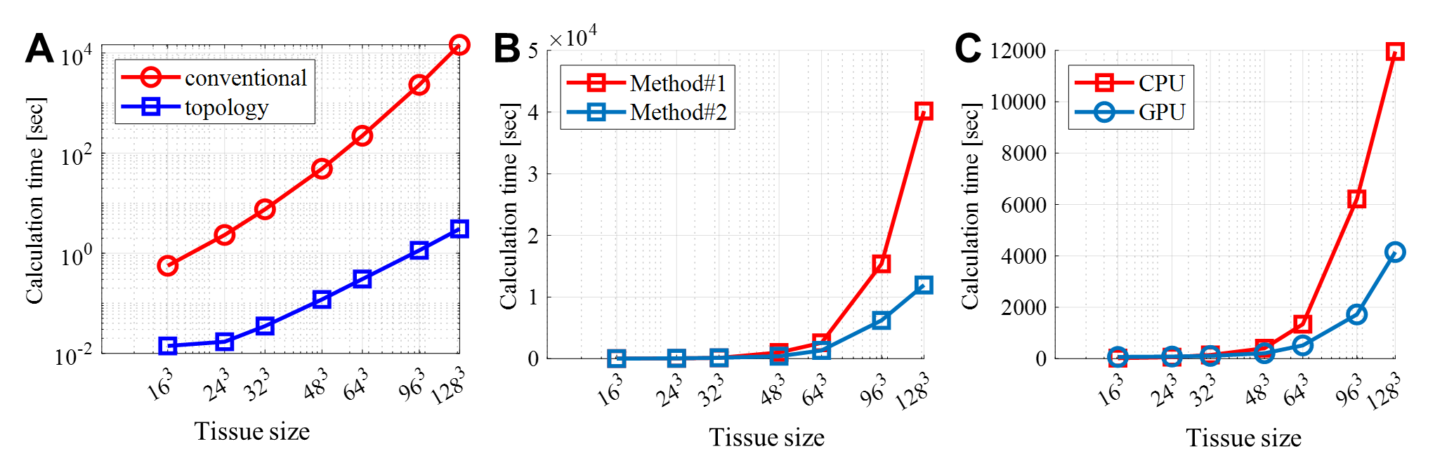
Software architecture: MATI was written with object-oriented programming in two versions (MATLAB and Python). A GUI was provided as a MATLAB App but both versions provide scripts and examples for advanced users. For dMRI simulation, a GPU-accelerated Finite Different (FD) method is used to synthesize dMRI signals with arbitrary digitalized structures with arbitrary diffusion gradient waveforms, which in turn provides great flexibility to investigate a wide selection of diffusion MRI problems. For data fitting, different dMRI biophysical signal models can be fit to dMRI data loaded from DICOM or NIfTI files to extract microstructural information such as cell size, cell density, intra- and extra-cellular diffusivities, and transcytolemmal water exchange rate constant. The software architecture is shown in Figure 1 and sample screenshots are shown in Figure 2.Performance: Three improvements were made to accelerate simulations: (1) Instead of calculating with loops, a topology method is used to calculate a sparse FD matrix in order to use matrix multiplication, which can be readily parallelized. (2) A more time-efficient approach was used to deal with time-varying FD matrix related to finite gradient durations. (3) GPU support is added. For data fitting, the standard non-linear least square (NLLS) fitting was provided with multiple initial conditions and global optimization algorithms to avoid local minima. However, a dictionary matching method (like that used in MR fingerprinting3) and the Bayesian method4 are recommended because their full vectorization is much faster and more reliable than conventional NLLS.
Output: MATLAB .mat files that contain all intermediate and final simulated results and fitted parametric maps in the NIfTI format can be saved in any folder specified by the user. Some third-party and in-house image visualization tools are provided.
Results
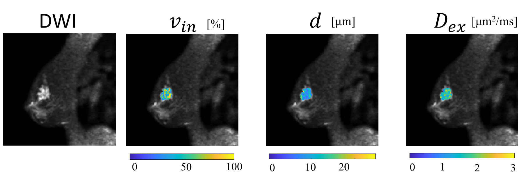
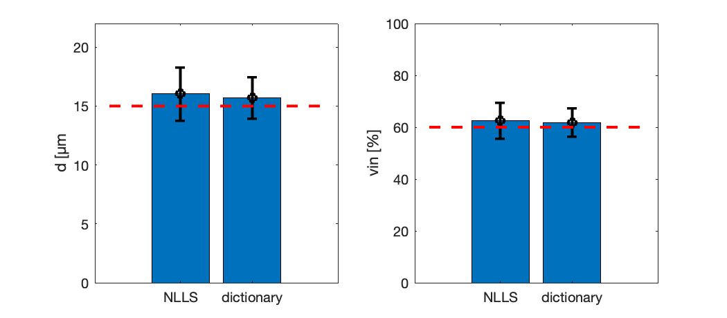
Figure 3 demonstrates examples of how FD simulations are significantly accelerated compared with the standard FD simulation5. The CPU used was an Intel Xeon E5-2643 @3.30 GHz whereas the GPU was an NVIDIA GeForce GTX TITAN X. An OGSE sequence with 25 Hz and TE = 110 ms was analyzed. For a digitalized tissue with a size of 128^3, all three improvements together achieved two orders of magnitude of acceleration. Figure 4 shows representative parametric maps ( intracellular volume fraction, mean cell size, and extracellular diffusivity) overlaid on a diffusion-weighted image from a breast cancer patient. Figure 5 shows the comparison of different fitting strategies for the IMPULSED MRI. Fittings were performed 100 times on simulated data with different noise at a SNR=50. The means and standard deviations represent the accuracy and precision of the fitting methods. The dictionary matching method is superior in both compared with the standard NLLS method.Discussion and Conclusion
A GPU-accelerated microstructural diffusion MRI signal simulation and data fitting toolbox with a graphical user-friendly interface has been developed, which is suitable for a broad range of end users such as clinicians. MATI may provide a valuable tool to the diffusion MRI field and assist the interpretation of more pre-clinical and clinical dMRI studies.Acknowledgements
No acknowledgement found.References
1. J. R. Li et al., SpinDoctor: A MATLAB toolbox for diffusion MRI simulation. Neuroimage 202, 116120 (2019).
2. H. H. Lee, E. Fieremans, D. S. Novikov, Realistic Microstructure Simulator (RMS): Monte Carlo simulations of diffusion in three-dimensional cell segmentations of microscopy images. J Neurosci Methods 350, 109018 (2021).
3. D. Ma et al., Magnetic resonance fingerprinting. Nature 495, 187-192 (2013).
4. X. Jiang, J. Xu, J. C. Gore, Quantitative temporal diffusion spectroscopy as an early imaging biomarker of radiation therapeutic response in gliomas: A preclinical proof of concept. Adv Radiat Oncol 4, 367-376 (2019).
5. J. Xu, M. D. Does, J. C. Gore, Numerical study of water diffusion in biological tissues using an improved finite difference method. Phys Med Biol 52, N111-126 (2007).
Figures