2405
Comparison of water exchange measurements between filter-exchange and diffusion time-dependent kurtosis imaging in human brain1Department of Physical Medicine and Rehabilitation of the Affiliated Sir Run Run Shaw Hospital, School of Medicine, Zhejiang University, Hangzhou, China, 2MR Application Predevelopment, Siemens Healthcare GmbH, Erlangen, Germany, 3MR Research Collaboration Team, Siemens Healthineers Ltd., Shanghai, China, 4Interdisciplinary Institute of Neuroscience and Technology, School of Medicine, Zhejiang university, Hangzhou, China, 5MOE Frontier Science Center for Brain Science and Brain-machine Integration, School of Brain Science and Brain Medicine, Zhejiang university, Hangzhou, China
Synopsis
Keywords: DWI/DTI/DKI, Brain, Trans-membrane water exchange; FEXI; time-dependent DKI; in vivo
Motivation: Trans-membrane water exchange rate has been measured by several MRI methods but reported with largely variant results.
Goal(s): To explore whether Filter-exchange imaging (FEXI) and time-dependent DKI are comparable for water exchange measurements on the same subjects in human brain.
Approach: Eight healthy volunteers underwent FEXI and DKI(t) acquisitions on a 3T scanner. ROI-based analysis was performed to determine correlations between FEXI-derived AXR and DKI(t)-derived 1/τex.
Results: A significant correlation between AXR and 1/τex was found only in axial direction in white matter. This correlation should be interpreted cautiously because structural disorder has non-negligible effects on D(t) and K(t) in Kärger model.
Impact: While a significant correlation was observed between AXR and 1/τex in the axial direction, this study suggests cautious use of DKI(t) for water exchange measurements due to potential deviations from the Kärger model's constant diffusivity assumption in the human brain.
Introduction
Trans-membrane water exchange is essential for homeostasis, and its measurement can help characterize cell function in brain diseases. Various magnetic resonance imaging (MRI) methods have been used to estimate trans-membrane water exchange in neuronal tissues. It is important to evaluate whether these methods could lead to comparable water exchange rates. In ex vivo cells, there is good agreement between water exchange measurements using constant-gradient single diffusion-encoding and filter-exchange imaging (FEXI)1. A comparison of diffusion time-dependent kurtosis imaging (DKI(t)) and FEXI in chemically fixed mouse brain showed that they are comparable to water exchange processes in white matter (WM)2. However, water diffusion properties and membrane permeability were affected by fixation in ex vivo tissues3. There still lacks such comparisons in vivo, especially in human brain. This study investigated whether FEXI and DKI(t) are comparable for water exchange measurements in human brain.Methods
Eight healthy volunteers (age 22±2 years) underwent MRI with a 3T MRI scanner (MAGNETOM Prisma, Siemens Healthcare, Erlangen, Germany). All Subjects signed informed consent before study and after Ethics Committee approval of the First Affiliated Hospital, Zhejiang University. The MRI protocol included 3D SPACE T2-weighted images (1.0×1.0×1.0mm3), DKI(t) and FEXI. DKI(t) data were acquired using a diffusion-weighted stimulated-echo sequence, with resolution=3.0×3.0×4.0mm3, TR/TE=3000/52ms, slices=12, b-values=1000 and 2000s/mm2, δ=15ms, and nine diffusion times (33, 45, 60, 80, 100, 150, 200, 250, 350, 450ms) in 30 directions. Additionally, b=0s/mm² images were acquired with five repetitions. FEXI was performed with bf=830s/mm2 in filter block, two b-values in detection block (bd=100s/mm2 with three repetitions and 1300s/mm2 with six repetitions), resolution=4.0×4.0×4.0mm3, and slices=12. The directions of bf and bd were kept consistent and along 20 directions. Three mixing times (tm) were used: 25, 200, and 400ms. As equilibrium data for fitting, FEXI was acquired with bf=0s/mm2 and =25ms. FEXI and DKI(t) data pre-processing, diffusivity and kurtosis fitting were completed in TORTOISE4 and DESIGNER5 toolboxes. AXR was estimated as in the previous study6, and exchange time (τex) was estimated based on the Kärger model7. Region of interest (ROI)-based comparison between 1/τex and AXR was conducted, using the Jülich and MNI atlases for extraction of WM tracts and gray matter (GM) ROIs. Pearson correlation analyses between the 1/τex and AXR of each ROI were performed in GraphPad Prism to evaluate whether FEXI and DKI(t) were comparable for water exchange measurements.Results
Figure 1(a) shows that MD and MK from DKI(t) data in polyvinylpyrrolidone (35%) phantom demonstrated no time-dependent changes. In human WM and GM, diffusivity exhibited a significant decrease according to diffusion time, represented by simple linear regression (Figure 2(b) and (c)). The mean rate of diffusivity reduction across all participants is shown in Figure 2(d); its uncertainty was estimated via bootstrapping. Although the diffusivity over long diffusion times does not reach a constant tortuosity asymptote, the smaller rate of diffusivity reduction occurred for diffusion times exceeding 100 ms. Thus, we fitted the Kärger model to data beyond 100 ms, with the goal of satisfying the Kärger model's assumption of constant diffusivity over time. Figure 2(a) and (b) show the fitting results for MK, AK and RK in WM, and MK in GM using the Kärger model with K∞=0 and K∞>0, respectively. Furthermore, the MD, AD, and RD-derived AXR in WM and MD-derived AXR in GM were estimated from FEXI data. Good fit was evident, even in the WM tract (acoustic tract) and GM ROI (caudate) with few voxels (Figure 3). Finally, ROI-based correlation analysis revealed a significant correlation between AD-derived AXR and AK-derived 1/τex in WM (Figure 4).Discussion
We observed significant diffusivity time-dependence over a long diffusion time, consistent with an in vivo study involving mouse hippocampus8 and a study involving human WM9, but contradicts other reports in mouse or rat brain2,10 (Table 1). The decreases in diffusivity and kurtosis over a long diffusion time suggest that the influence of microstructure is nontrivial11. It is difficult to separate the microstructural involvement from exchange effects, hindering direct comparison between them. However, we observed a correlation between FEXI AD-derived AXR and DKI(t) AK-derived exchange time (diffusion time > 100 ms) in human WM. This finding emphasizes the need for a more sophisticated model describing diffusion time-dependence in in vivo tissues, with specific water-exchange signatures.Conclusion
We identified a correlation between AD-derived AXR and AK-derived exchange time in human WM. However, this correlation should be interpreted cautiously because structural disorder has non-negligible effects on D(t) and K(t) in Kärger model. These results suggest that the need of a more sophisticated model describing diffusion time-dependence in in vivo tissues, with specific water-exchange signatures.Acknowledgements
This work is supported in part by the National Natural Science Foundation of China (NSFC) (Grant Nos. 82111530201, 82222032, 82172050), the STI2030-Major Projects Q22 of China (Grant No. 2022ZD0206000).References
1. Tian X, Li H, Jiang X, et al. Evaluation and comparison of diffusion MR methods for measuring apparent transcytolemmal water exchange rate constant. Journal of Magnetic Resonance. 2017;275:29-37.
2. Li C, Fieremans E, Novikov DS, et al. Measuring water exchange on a preclinical MRI system using filter exchange and diffusion time dependent kurtosis imaging. 2022;(November):1-15.
3. Thelwall PE, Shepherd TM, Stanisz GJ, et al. Effects of temperature and aldehyde fixation on tissue water diffusion properties, studied in an erythrocyte ghost tissue model. Magn Reson Med. 2006;56(2):282-289.
4. Pierpaoli C, Walker L, Irfanoglu MO, et al. TORTOISE: an integrated software package for processing of diffusion MRI data. ISMRM 18th Annual Meeting. 2010.
5. Ades-Aron B, Veraart J, Kochunov P, et al. Evaluation of the accuracy and precision of the diffusion parameter EStImation with Gibbs and NoisE removal pipeline. Neuroimage. 2018;183:532-543.
6. Li Z, Pang Z, Cheng J, et al. The direction-dependence of apparent water exchange rate in human white matter. Neuroimage. 2022;247.
7. Fieremans E, Novikov DS, Jensen JH, et al. Monte Carlo study of a two-compartment exchange model of diffusion. NMR Biomed. 2010;23(7):711-724.
8. Mougel E, Valette J, Palombo M. Investigating Exchange, Structural Disorder and Restriction in Gray Matter via Water and Metabolites Diffusivity and Kurtosis Time-Dependence. arXiv. 2023
9. Fieremans E, Burcaw LM, Lee HH, et al. In vivo observation and biophysical interpretation of time-dependent diffusion in human white matter. Neuroimage. 2016;129:414-427.
10. Jelescu IO, de Skowronski A, Geffroy F, et al. Neurite Exchange Imaging ((NEXI): A minimal model of diffusion in gray matter with inter-compartment water exchange. Neuroimage. 2022;256(January):119277.
11. Lee HH, Papaioannou A, Novikov DS, et al. In vivo observation and biophysical interpretation of time-dependent diffusion in human cortical gray matter. Neuroimage. 2020;222(14):1-5.
Figures
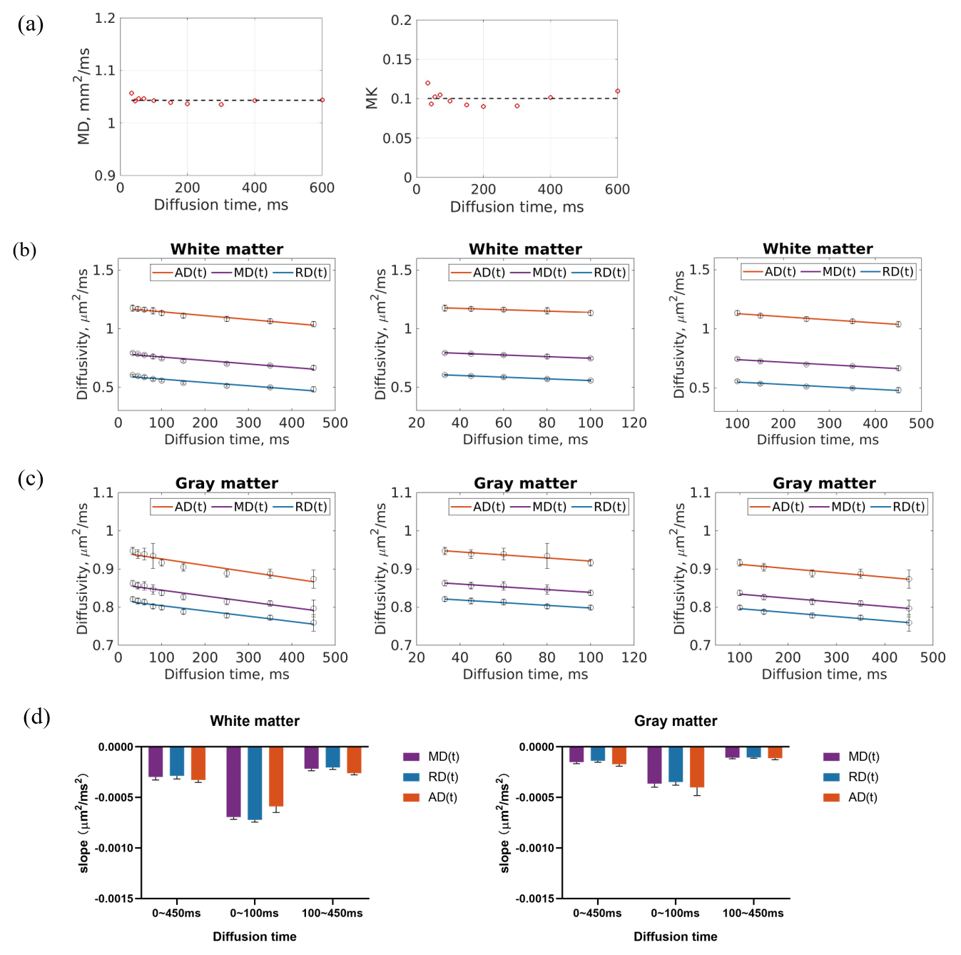
Figure 1: (a) MD and MK estimated from DKI(t) data in polyvinylpyrrolidone (35%) phantom. (b-c) Averaged diffusivity (MD, AD, RD) across all participants according to diffusion time in WM and GM, respectively, fitted via simple linear regression to evaluate time-dependence. (d) Statistical results. Bar represents rate of linear regression in panels b-c; error bar represents uncertainty in rate estimated via bootstrapping. Smaller uncertainty indicates significant time-dependence of diffusivity.
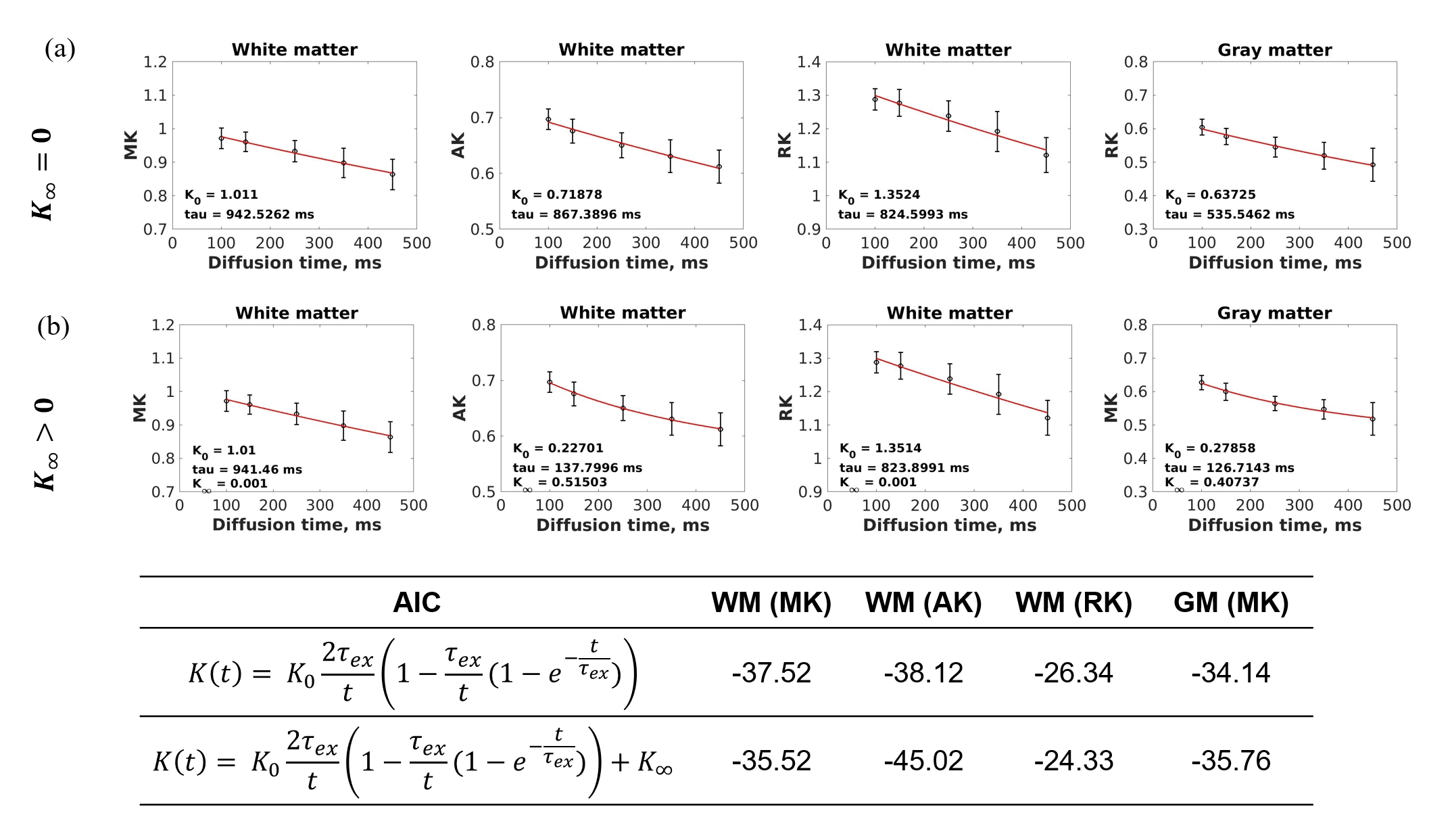
Figure 2: Fitted Kärger models with (a) K∞=0 and (b) K∞>0 for kurtosis at diffusion times exceeding 100 ms. And the AIC (Akaike information criterion) values for the two different Kärger models.
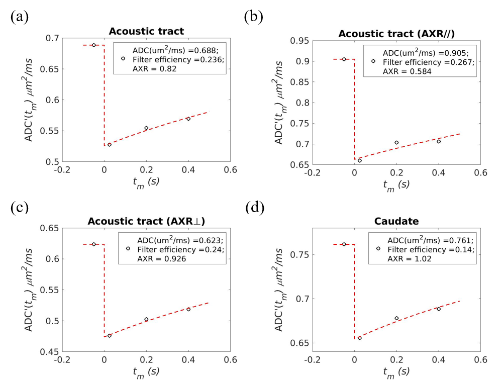
Figure 3: Representative AXR fitting curves from one participant’s WM ROI (acoustic, ~10 voxels) and GM ROI (caudate, ~40 voxels). Good fit was evident, even in ROIs with a relatively limited number of voxels across all WM tracts and GM ROIs, respectively. AXR// refers to AXR values derived from the FEXI dataset, where the gradient direction is parallel to the principal WM tract orientation. AXR⊥ refers to AXR values derived from the FEXI dataset, where the gradient direction is perpendicular to the principal orientation of WM tracts.
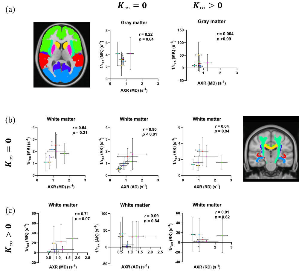
Figure 4: Scatterplot of AXR and 1/τex, and statistical results of ROI-based correlation analysis. (a) No correlation between MD-derived AXR and MK-derived 1/τex in GM ROIs, using Kärger model with K∞=0 or K∞>0. (b) Significant correlation between AD-derived AXR and AK-derived 1/τex, using Kärger model with K∞=0 in WM ROIs. The Error bars represent standard deviations of AXR or 1/τex across all participants.
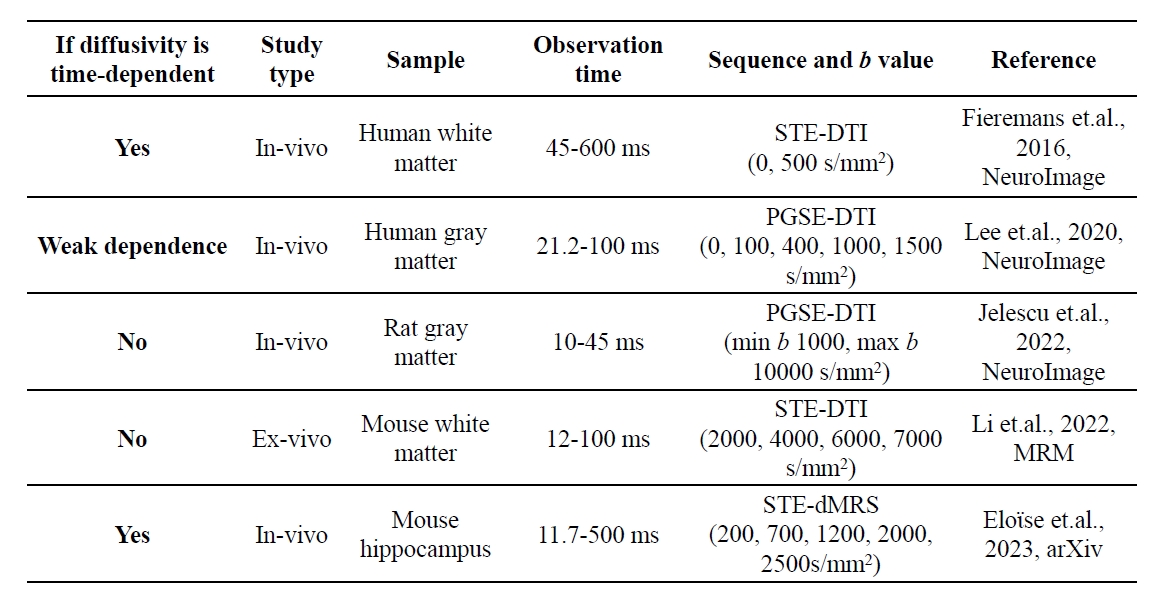
Table 1: Diffusivity time-dependence and methods reported in the literature.