2404
Investigating the influence of post-mortem interval on diffusion anisotropy of whole human brains1Department of Systems Neurosciences, University Medical Center Hamburg-Eppendorf, Hamburg, Germany, 2MR Physics Group, Max Planck Institute for Human Development, Berlin, Germany, 3Department of Neurophysics, Max Planck Institute for Human Cognitive and Brain Sciences, Leipzig, Germany, 4Institute of Molecular and Cellular Cognition, University Medical Center Hamburg-Eppendorf, Hamburg, Germany, 5Department of Neurosurgery, University Medical Center Hamburg-Eppendorf, Hamburg, Germany, 6Department of Legal Medicine, University Medical Center Hamburg-Eppendorf, Hamburg, Germany
Synopsis
Keywords: DWI/DTI/DKI, Ex-Vivo Applications, Post-mortem interval, sample size, fixation, human brain
Motivation: The ISMRM-Diffusion-Study-Group recommends a post-mortem interval (PMI) under six hours to avoid degeneration in ex-vivo tissue for validation of microstructure parameters estimated using preclinical MRI. Fractional anisotropy (FA) deviation from the in-vivo value serves as a quality indicator.[1]
Goal(s): Investigating the influence of PMI and tissue size on FA.
Approach: Five human whole-brains (PMI 15-24h) and a temporal-lobe (TL) specimen (PMI 2h) were examined with diffusion MRI (dMRI) before and after fixation.
Results: The FA of the unfixed whole-brain samples didn’t show differences to the in-vivo values, but between unfixed and fixed states. The FA of the TL specimen was unaffected during fixation.
Impact: For the PMIs examined here, myelin decomposition may not significantly affect FA from dMRI of unfixed post-mortem specimens. However, it can affect whole-brain samples during immersion fixation - an effect that may be mitigated by using smaller samples.
Introduction
Diffusion MRI (dMRI) of fixed ex-vivo brain samples is often used as an intermittent modality to validate dMRI-based microstructure parameters against their histological gold standard counterpart[3]. The ISMRM Diffusion-Study-Group suggests a post-mortem interval (PMI) under 6 hours for accurate preclinical MRI and histological assessments, see also Fig. 1a).[1] They recommend that post-fixation fractional anisotropy (FA) values should mirror in-vivo conditions.[1] This rule is based on the observation by Shepherd et al’s[2] showing that FA values were decreased by 38% in rat spinal cord white matter within the first 6 hours and by 52% within 24 hours, Fig. 1 a). We hypothesize that the effect of PMI on FA in human brains is smaller than on rodents because of the larger brain size. This study tests the influence of PMI on the FA of fixed human brains.Methods
Specimens:Three types of specimens (details in Table 1) were utilized in this study: 1) five whole human brains (referred to as brain 1 to 5) and post-mortem intervals (PMIs) ranging from 15 to 24 hours, 2) two living whole human brains and 3) a temporal-lobe specimen (TL-specimen) with a PMI of 2 hours.
Nomenclature:
In-vivo dMRI from a patient with drug-resistent temporal-lobe epilepsy (in-vivo brain 2) and a healthy brain (in-vivo brain 1).
Unfixed dMRI: for (1) in-situ post-mortem brains (brains 1 to 4) and (2) the freshly excised TL-specimen of the epilepsy patient stored in glucose. All specimens were stored within the PMI at room temperature (21°C).[4]
Ex-vivo dMRI: for (1) all whole brains and (2) TL-specimen, both in fixed state. All specimens were fixated in 4% paraformaldehyde. It is expected that the brains are fixed after 20 days[5] and the TL-specimen after 12 hours (assuming fixative’s diffusion rate ~1mm/h[6]).
Details are available in Table 1 a).
Data acquisition and analysis:
All measurements were performed on a 3 T PRISMA fit MRI (Siemens Healthcare, Erlangen, Germany). The choice of the best b-values for the diffusion measurements were investigated previously for whole human brains[7] and the TL[8]. Acquisition details are listed in Table 1 b). Each dataset was pre-processed and analyzed with the diffusion tensor model (DTI) implemented in the ACID toolbox[9] using SPM 12[10] in Matlab R2021b[11]. Details on the pre-processing of the dMRI data can be found in [7] for the brains and [8] for the TL-specimen. The corpus callosum (CC) and white matter TL were segmented using ITK-snap[12].
Results
The post-mortem unfixed FA value was close to the in-vivo FA for the CC specimen (an average difference of 1%; min. 0.029% for PMI 21h and max. -3.32 % for PMI 18h) whereas it showed a large difference for the TL-specimen (17.7%). In comparison to the in-vivo FA values, the FA values of the CC segments were on average -25.21 ± 4.40% smaller after fixation (min. -19.29 % for PMI 24h and max. -29.70% for PMI 18h) whereas the FA values of the TL-specimen increased (+14.81%).Over the fixation period, the FA values within the CC decreased (on average 24% from unfixed to fixed), whereas the FA value of the TL-specimen stayed more or less the same (2.9% difference from unfixed to fixed), see Fig. 4a). The coefficient of variation (COV) (Fig. 4b) increased for all brains over the fixation time, whereas the COV of the TL-specimen was almost stable across the fixation time (Δ=5.7% from unfixed to fixed).
Discussion & Conclusion
We found that the PMI of human brain tissue affects the difference between in-vivo and fixed ex-vivo FA values. For the whole-brain specimens, this difference was almost negligible between the in-vivo and unfixed post-mortem FA value and became pronounced after fixation. For the TL-specimen, no substantial changes in FA were observed during fixation but a large difference was observed between in-vivo and unfixed post-mortem FA values. The latter FA-difference for the TL-specimen can be explained by partial-volume effects present in the lower-resolution in-vivo dMRI data.Two possible reasons for the opposite behavior between TL and whole-brain specimens during fixation are (1) the difference in PMI (whole-brain specimens >15 hours vs. TL-specimen: 2 hours) and (2) the smaller size of the TL-specimen allows for faster immersion-fixation, i.e. less time remains for additional autolysis processes during immersion-fixation.
Ultimately, our results imply that for the PMIs examined here, myelin decomposition[13], mainly driven by lamellar separation[13-15] (Fig. 1b)), may not significantly affect FA from dMRI of unfixed post-mortem specimens. However, it can affect whole-brain samples during immersion fixation - an effect that may be attenuated by using smaller samples.
Acknowledgements
This work was supported by the German Research Foundation (DFG Priority Program 2041 "Computational Connectomics”, [MO 2397/5-1;MO 2397/5-2], by the Emmy Noether Stipend: MO 2397/4-1; MO 2397/4-2) and by the BMBF (01EW1711A and B) in the framework of ERA-NET NEURON and the Forschungszentrums Medizintechnik Hamburg (fmthh; grant 01fmthh2017).References
[1] Kurt G Schilling et al. (2023), Recommendations and guidelines from the ISMRM Diffusion Study Group for preclinical diffusion MRI: Part 2 -- Ex vivo imaging, arXiv, 2209.13371 Not published yet
[2] Sheperd et al.
(2009), Postmortem interval alters the water relaxation
and diffusion properties of rat nervous tissue - Implications for MRI studies
of human autopsy samples, NeuroImage, doi: 10.1016/j.neuroimage.2008.09.054
[3] Mollink J. et al. (2017), Evaluating fibre orientation dispersion in white matter: Comparison of diffusion MRI, histology and polarized light imaging, Neuroimage, doi: 10.1016/j.neuroimage.2017.06.001
[4] Berger, C. et al. (2022), Post mortem brain temperature and its influence on quantitative MRI of the brain, Magma (New York, N.Y.), doi: 10.1007/s10334-021-00971-8
[5] Dawe, et al. (2009), Postmortem MRI of Human Brain Hemispheres: T2 Relaxation Times during Formaldehyde Fixation, Magnetic Resonance in Medicine 61 (4); 810-18
[6] Fox et al. (1985), Formaldehyde fixation, The Journal of Histochemistry and Cytochemistry Vol. 33, No. 8, pp. 845-853, doi: 10.1177/33.8.3894502
[7] Lüthi et al. (2023), Longitudinal assessment of MD and FA in four whole human brains using the DTI model, in: Proc. Intl. Soc. Mag. Reson. Med. 31.
[8] Fritz, F. J. et al. (2023), Diffusion and relaxometry study of an excised temporal lobe from a drug-resistance epilepsy patient using in vivo and ex vivo MRI, in: Proc. Intl. Soc. Mag. Reson. Med. 31.
[9] Fricke, B. et al. (2022), ACID - an open-source, bids compatible software for brain and spinal cord dMRI: reprocessing, DTI/DKI, biophysical modelling, in: Proc. Intl. Soc. Mag. Reson. Med. 30.
[10] Friston, K. (2007), CHAPTER 2 - Statistical parametric mapping, in: FRISTON, K., ASHBURNER, J., KIEBEL, S., NICHOLS, T., PENNY, W. (Eds.), Statistical Parametric Mapping. Academic Press, London, pp. 10–31. doi: 10.1016/B978-012372560-8/50002-4
[11] MATLAB. (2021). 9.11.0.1837725 (R2021b) Update 2. Natick, Massachusetts: The MathWorks Inc.
[12] Paul A. Yushkevich et al. (2006), User-guided 3D active contour segmentation of anatomical structures: Significantly improved efficiency and reliability. Neuroimage 1;31(3):1116-28.
[13] Krassner M. et al. (2023), Postmortem changes in brain cell structure: a review, Free Neuropathology 4:10, doi https://doi.org/10.17879/freeneuropathology-2023-4790
[14] Shibayama et al. (1976), The Postmortem Changes of Pyramidal Neurons in the Hippocampus of Rats, Psychiatry and Clinical Neurosciences, doi: 10.1111/j.1440-1819.1976.tb00113.x
[15] Choe et al. (1995), Postmortem metabolic and morphologic alterations of the dog brain thalamus with use of in vivo 1H magnetic resonance spectroscopy and electron microscopy. Investigative radiology. doi : https://doi.org/10.1097/00004424-199505000-00001
Figures
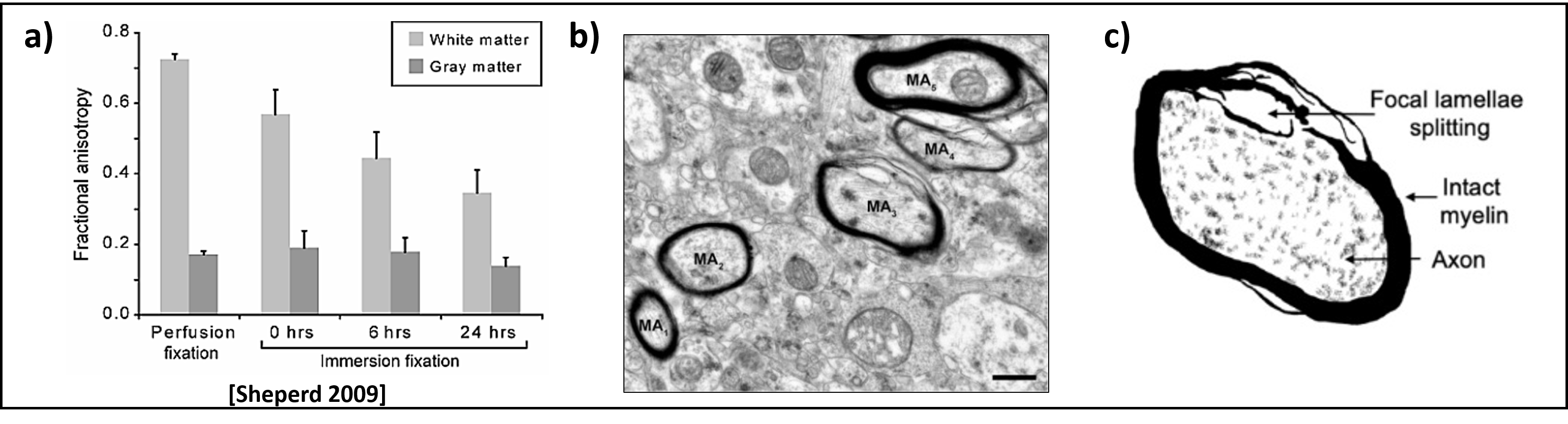
Figure 1:
Figure 1a) Decrease in FA over increasing PMI in white matter in the rat spinal cord, from Sheperd et al.[2]
Figure 1b) Electron Microscopy image (EM) of the prefrontal cortex of the postmortem human brain with good preservation, from Glausier et al.[16]
Figure 1c) shows the myelin lamellae splitting of the myelinated axon MA3 from Figure b) and is taken from Krassner et al.[13]
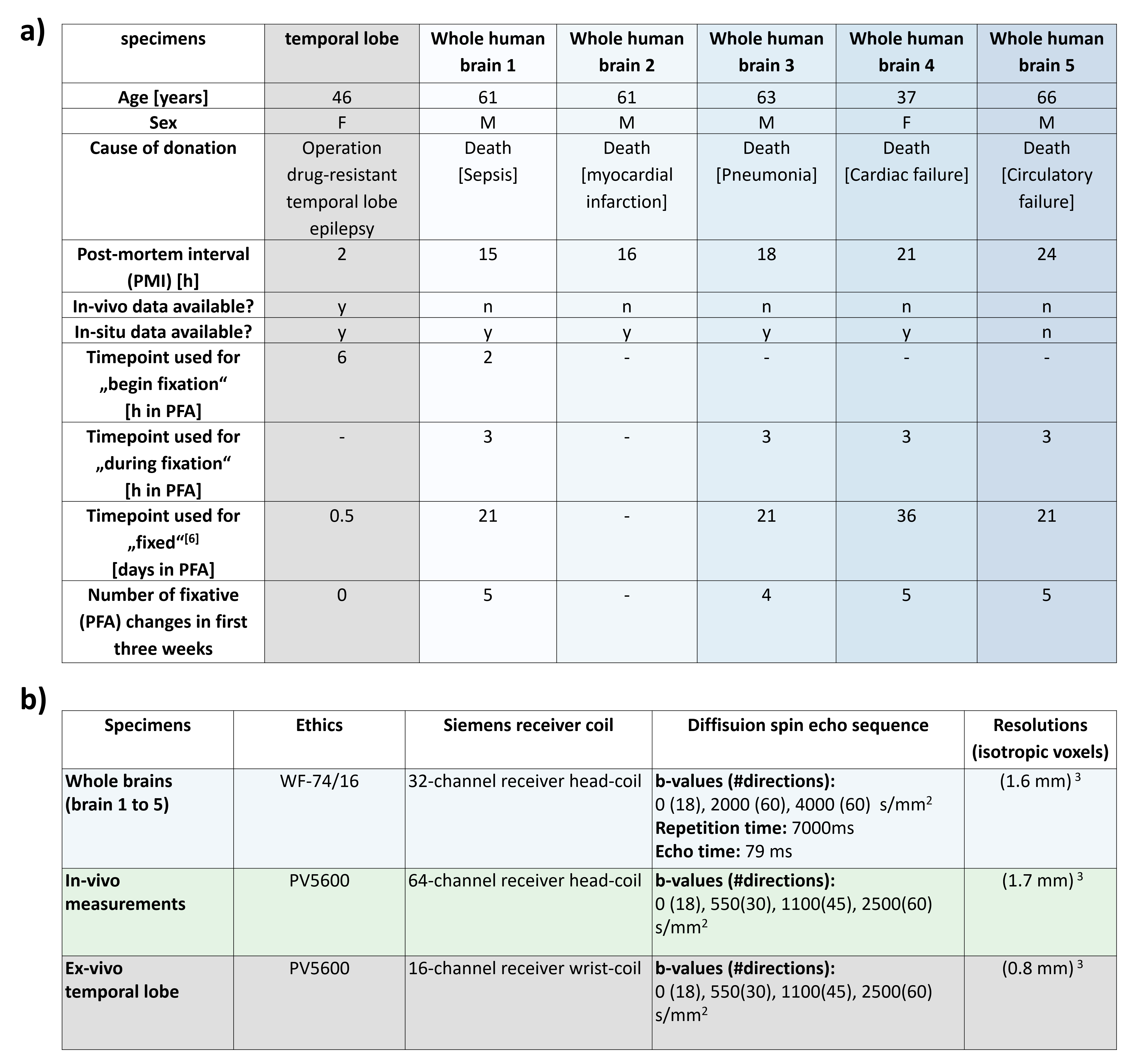
Table 1:
Table 1a) Detailed information on ex-vivo samples examined: one temporal lobe specimen and five whole human brains. Data includes age, gender, cause of death or donation, and details about the measurement times in different fixation steps.
Table 1b) Protocol Details: Information on the protocols used for the diffusion measurements with their respective ethics, along with corresponding resolution.
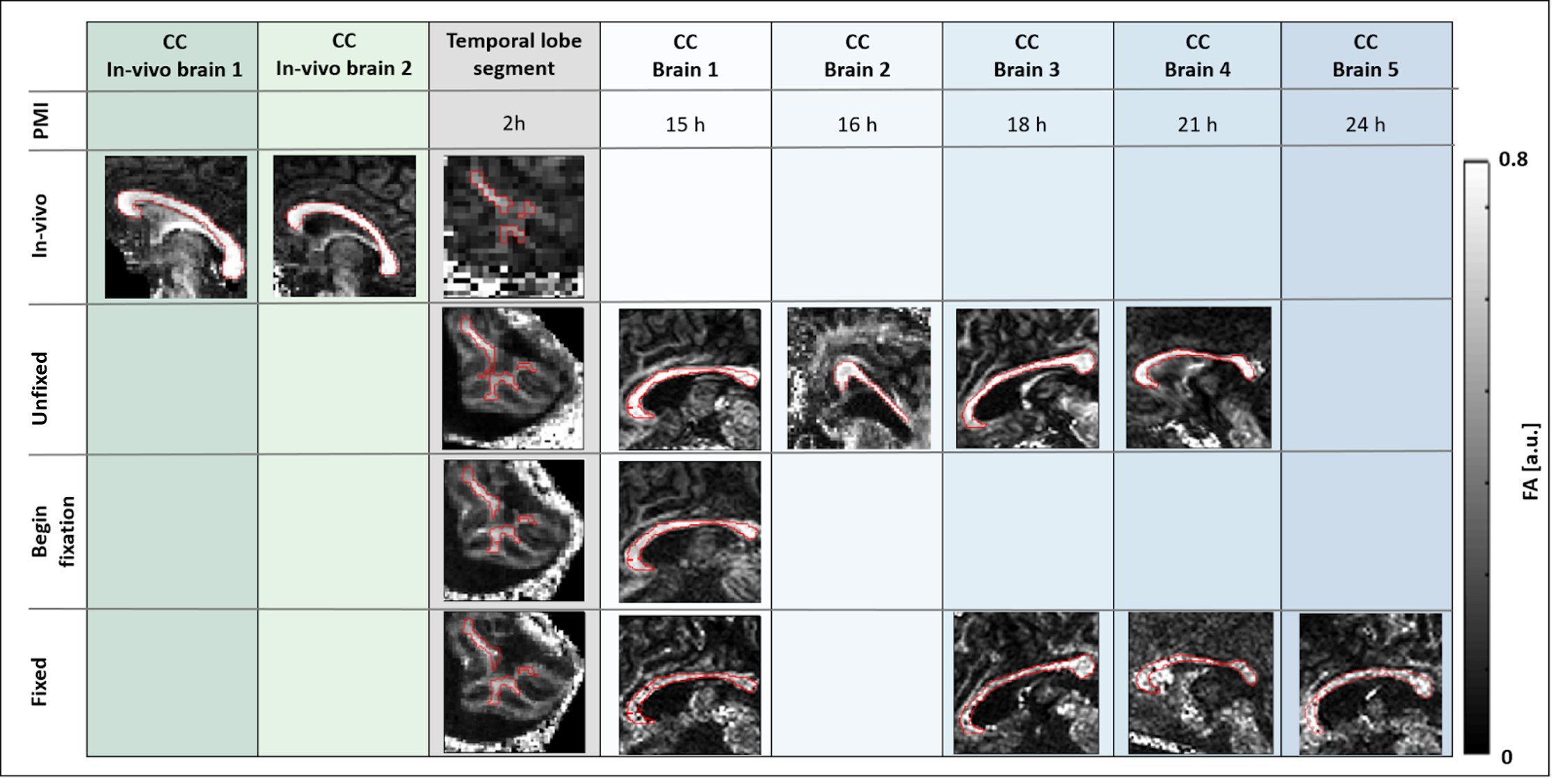
Figure 2:
Segmentation of samples (ascending PMI in columns) at key fixation times (rows). In-vivo brain 2 is the source of the temporal lobe (TL) segment. Brain 1-4 unfixed refers to a deceased state but still inside the skull, at the end of PMI. TL-specimen unfixed means after excision and immersed in glucose. Begin fixation means 3h in fixative for Brain 1 and 6h for TL-segment. Fixed mean >20 days (brains 1-5) and 0.5 days (TL segment) stored in PFA.
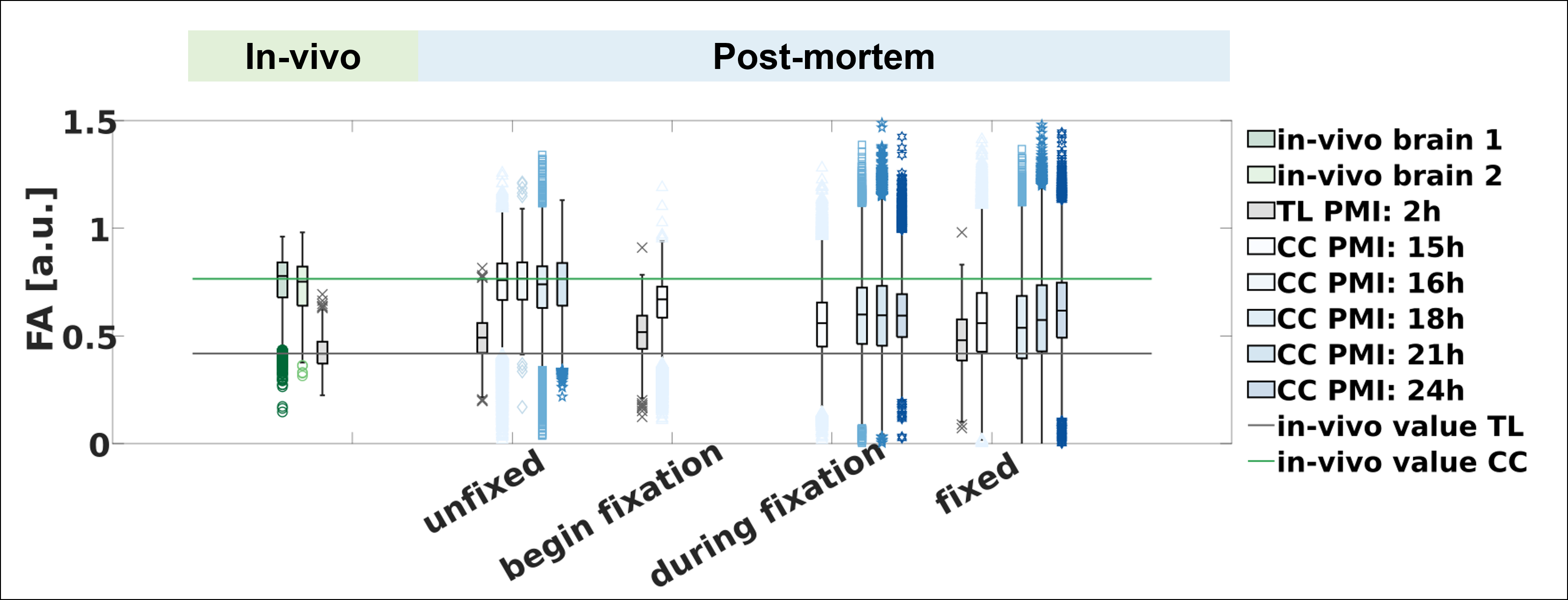
Figure 3:
Change of fractional anisotropy (FA) during fixation as a function of post-mortem interval (PMI) and comparison with its in-vivo value.
Depicted are boxplots of the voxels in the corpus callosum (CC) segment of the whole brains 1-5 and the temporal lobe (TL) specimen at different post-mortem times for the diffusion parameter FA. Note that the in-vivo CC FA values are from a different subject than the other CC FA values.
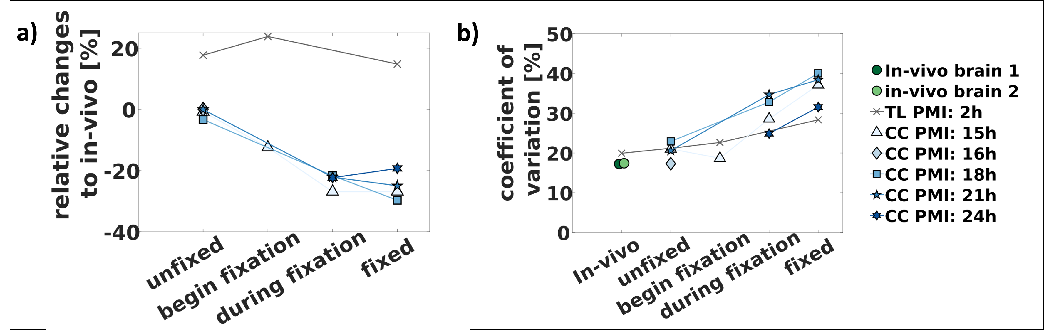
Figure 4:
Change of fractional anisotropy (FA) and its coefficient of variation (COV) during fixation as a function of post-mortem interval (PMI).
Figure 4a) depicts the relative changes of the median FA between brain 1-5 and the TL specimen with respect to the respective in-vivo value.
Figure 4b) depicts the coefficient of variation for the individual samples from Figure 4.