2396
Lactate and total choline show potential prognostic values in diffuse intrinsic pontine glioma1Diagnostic Imaging, St. Jude Children's Research Hospital, Memphis, TN, United States, 2Biomedical Engineering, University of Memphis, Memphis, TN, United States, 3Biomedical Engineering, University of Alabama, Birmingham, AL, United States
Synopsis
Keywords: Tumors (Pre-Treatment), Cancer, metabolism, spectroscopy
Motivation: Metabolic mapping in Diffuse intrinsic pontine glioma (DIPG) may indicate prognosis and potential therapeutic targets.
Goal(s): We intended to identify metabolic ratios that indicated dysregulated metabolic pathways using 1H-MRS and determine whether these metabolites are associated with overall survival length.
Approach: We performed CSI in patients with DIPG prior to radiation therapy. Then we assessed the predictive value of a metabolite ratio for patient overall survival.
Results: Lactate and total choline levels were found to be significantly associated with patient overall survival, making them potential prognostic predictors of overall survival in DIPG.
Impact: This study shows that more aggressive DIPGs are metabolically distinct from less aggressive tumors and that the metabolic differences can predict the overall survival of patients.
Introduction
Diffuse Intrinsic Pontine Glioma (DIPG) is a highly aggressive pediatric cancer with a poor prognosis and median survival of 9 months after diagnosis1.Treatment primarily consists of radiation therapy (RT) and chemotherapy, although the latter has proven largely ineffective1, and the cells’ diffusivity as well as its pontine location precludes surgical resection as a treatment option2. DIPG is characterized by metabolic flux in glial cells including upregulated glycolysis intermediates due to increased dependence on glucose, upregulated tricarboxylic acid (TCA) cycle intermediates, and increased glutamine uptake.2 Magnetic resonance spectroscopy (MRS) has been used recently to measure metabolite levels in glioma as markers of specific gene mutations and predict tumor grades3,4. We proposed that MRS-measured metabolites acquired prior to the patients receiving radiation therapy may inform patient prognosis.Of particular interest were metabolites indicating glial health or high cellular proliferation. Elevated choline-containing compound (Cho) are noted on spectral analysis due to association with cellular membrane, while peaks representing metabolites such as N-acetylaspartate (NAA) are decreased and are used as biomarkers of neuronal density. Other metabolites of interest include lactate (Lac) and pyruvate (Pyr) as markers of glycolysis; Glx (glutamine + glutamate) for neurotransmission; lipid 1.3 and lipid 0.9 showing fatty acid levels; citrate (Cit) a TCA cycle intermediate; myoinositol (Ins) and glycine (Gly) both as neural health markers; 2-hydroxyglutarate (2HG), a marker for IDH mutation; taurine, correlated with apoptosis in glioma and a marker of osmoregularity; and creatine (Cr), an indicator of cellular energetics and internal reference.
Materials and Methods
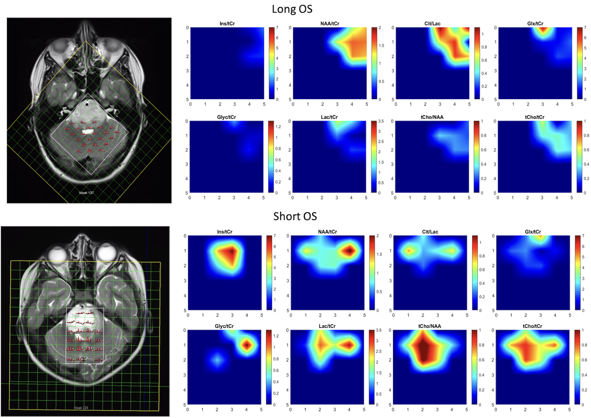
Patients enrolled in the study (n=48) were diagnosed with DIPG, diffuse midline glioma (DMG), or brainstem glioma (BSG). Multiplanar, multisequence imaging including sagittal and axial T1, T2, FLAIR, diffusion, and CSI (TR/TE=1700/135 ms, FOV= 160x160 mm2, ROI matrix size = 6x6 or 6x4, voxel dimensions = 10x10x15 mm3) was acquired (Figure 1). Tumor masks were auto-segmented from conventional imaging using Cascaded Anisotropic Convolutional Neural Networks5 and then manually adjusted by a radiologist if necessary. For the purposes of analysis, a CSI voxel was defined as tumor when it contained ≥80% tumor mask voxels in the conventional imaging. CSI data was processed using LCModel6, a program used for in vivo spectral analysis that established individual metabolite values; 36 spectra were acquired from most patients, with a few single-voxel scans.Metabolic ratios were calculated with Cramer-Rao Lower Bound (CRLB) for each metabolite in a voxel set at a threshold of ≤30% to be considered in the analysis. We assessed the predictive value of a metabolite ratio for patient OS using a novel quantitative method. For a given metabolite ratio of interest, starting at the 10th percentile, metabolite ratio threshold was incremented in steps up to the 90th percentile. The threshold divided the population into two groups, low and high metabolite ratios, for the evaluation of Kaplan-Meier curves and log-rank tests. The metabolite ratio at which the log-rank test achieved significance (p < 0.05) was determined as the threshold value, with the median OS length calculated for the two cohorts. A significant log-rank test would imply that the median OS of the two cohorts are significantly different.
Results and Discussion
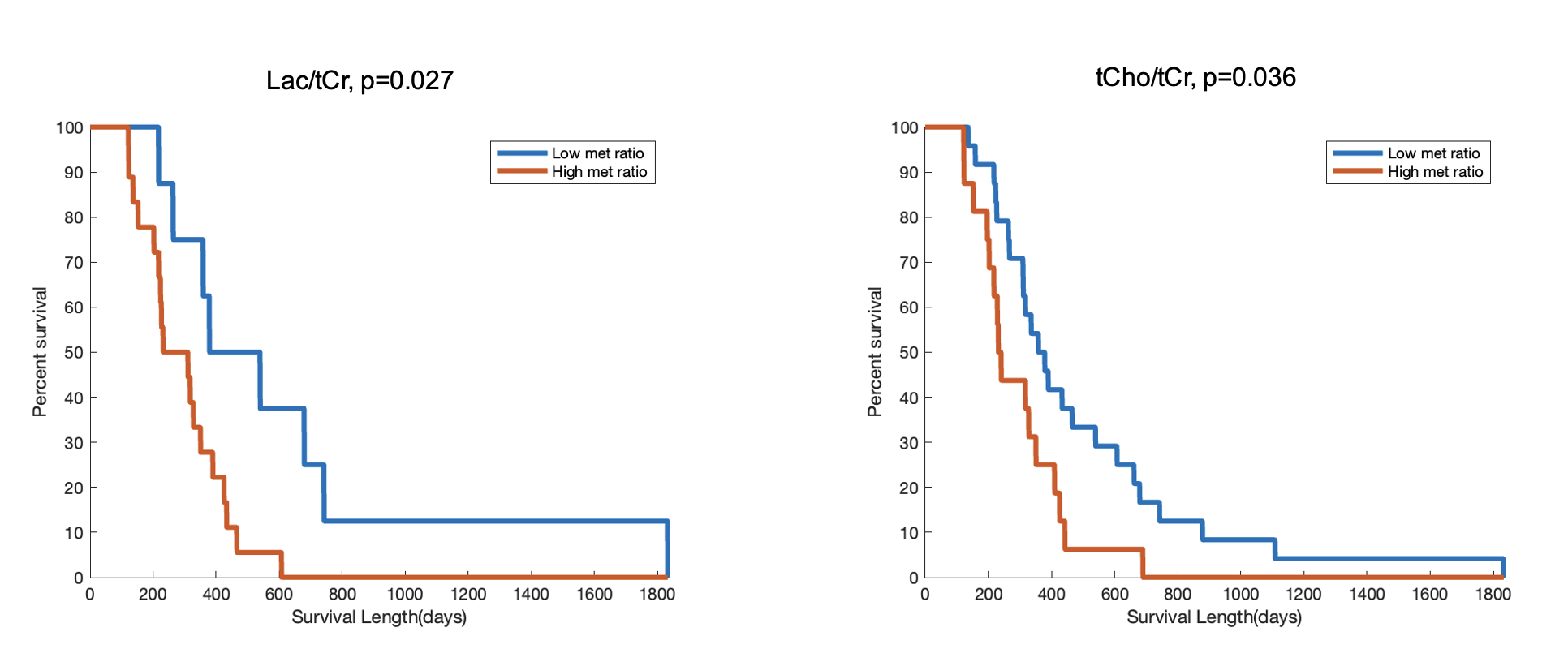
Our results suggest that among the 15 metabolites investigated, only Lac/tCr and tCho/tCr were predictive of OS in this DIPG cohort (Figure 2). More precisely, a Lac/tCr metabolite ratio below 0.76 (53rd percentile) or tCho/tCr ratio between 0.44 – 0.49 (60th percentile) in a DIPG subject denotes longer OS. For a Lac/tCr metabolite ratio threshold of 0.76, patients with lower ratio have a median OS of 460 days, while patients with higher ratio have a median OS of 272 days. Similarly, for a tCho/tCr metabolite ratio threshold of 0.49, the median OS for the lower and higher metabolite ratio groups is 369 days and 237 days, respectively. Other metabolite ratios were found to be not statistically significant in this analysis. Because of “the Warburg effect”, it’s well-known that increased lactate production is indicative of dysregulated glycolytic pathways.7 Additionally, the Cho/Cr ratio has been shown to be a potential prognostic marker used to evaluate cell proliferative activity.8 These ratios indicate that this cohort shows elevated glycolytic and altered metabolic pathways as expected in DIPG patients.Conclusion
We found that tumoral levels of lactate and total choline indicate patient overall survival in patients with brainstem glioma. Further, at certain levels, these ratios are promising prognostic markers in patients with brainstem glioma. Future work should include identifying clinical intervention of these dysregulated pathways.Acknowledgements
No acknowledgement found.References
1. Pellot, Joel E., De Jesus, Orlando. Diffuse Intrinsic Pontine Glioma. [Updated 2022 Aug 8]. In: StatPearls [Internet]. Treasure Island (FL): StatPearls Publishing; 2022 Jan
2. Park, Jiyoon, and Chan Chung. “Epigenetic and Metabolic Changes in Diffuse Intrinsic Pontine Glioma.” Brain tumor research and treatment vol. 11,2 (2023): 86-93. doi:10.14791/btrt.2023.0011
3. Iwahashi, Hirofumi et al. “2-Hydroxyglutarate magnetic resonance spectroscopy in adult brainstem glioma.” Journal of neurosurgery vol. 139,2 355-362. 27 Jan. 2023, doi:10.3171/2022.12.JNS221954
4. Rafique, Zunaira et al. “Diagnostic Accuracy of Magnetic Resonance Spectroscopy in Predicting the Grade of Glioma Keeping Histopathology as the Gold Standard.” Cureus vol. 14,2 e22056. 9 Feb. 2022, doi:10.7759/cureus.22056
5. Wang, G., et al. Automatic brain tumor segmentation using cascaded anisotropic convolutional neural networks. in International MICCAI Brainlesion Workshop. 2017. Springer
6. Provencher, S W. “Automatic quantitation of localized in vivo 1H spectra with LCModel.” NMR in biomedicine vol. 14,4 (2001): 260-4. doi:10.1002/nbm.698
7. Pérez-Tomás, Ricardo, and Isabel Pérez-Guillén. “Lactate in the Tumor Microenvironment: An Essential Molecule in Cancer Progression and Treatment.” Cancers vol. 12,11 3244. 3 Nov. 2020, doi:10.3390/cancers12113244
8. Gao, Wenjing et al. “Cho/Cr ratio at MR spectroscopy as a biomarker for cellular proliferation activity and prognosis in glioma: correlation with the expression of minichromosome maintenance protein 2.” Acta radiologica (Stockholm, Sweden : 1987) vol. 60,1 (2019): 106-112. doi:10.1177/0284185118770899
Figures