2392
Detection of Abnormal Brain Perfusion in Children with Global Developmental Delay Using 3D Pseudo-Continuous Arterial Spin Labeling Imaging1The Third Affiliated Hospital of Zhengzhou University, Zhengzhou, China, 2Henan International Joint Laboratory of Neuroimaging, Zhengzhou, China, 3MR Research China, GE Healthcare, Beijing, China
Synopsis
Keywords: Normal Development, Brain, perfusion; global developmental delay; arterial spin labelling; cerebral blood flow
Motivation: Early detection of global developmental delay (GDD) in young children is imperative for effective intervention.
Goal(s): The goal of this study is to determine whether CBF values, measured through 3D-pcASL, can aid in early GDD diagnosis and intervention.
Approach: Twenty-six children with GDD and 13 age- and sex-matched healthy controls underwent 3D-T1 and 3D-pcASL imaging. CBF values in various brain regions were analyzed and compared using statistical tests.
Results: The study found significantly lower CBF values in the right occipital lobe and left basal ganglia in GDD children. CBF values in these regions demonstrated potential as diagnostic markers for GDD.
Impact: This study's findings suggest that 3D-pcASL perfusion imaging can aid in early diagnosis of global developmental delay (GDD) in young children. Detecting GDD through non-invasive imaging may enhance early intervention, ultimately improving developmental outcomes.
Introduction:
Global developmental delay (GDD) in children aged 2-4 years is a concerning condition characterized by significant delays in cognitive, motor, and social development. Early and accurate assessment of regional cerebral blood flow (CBF) can aid in diagnosing and managing GDD effectively. This study evaluates the potential of three-dimensional pseudo-continuous arterial spin labeling (3D-pcASL) perfusion imaging as a diagnostic tool for assessing CBF in children with GDD.Materials and Methods:
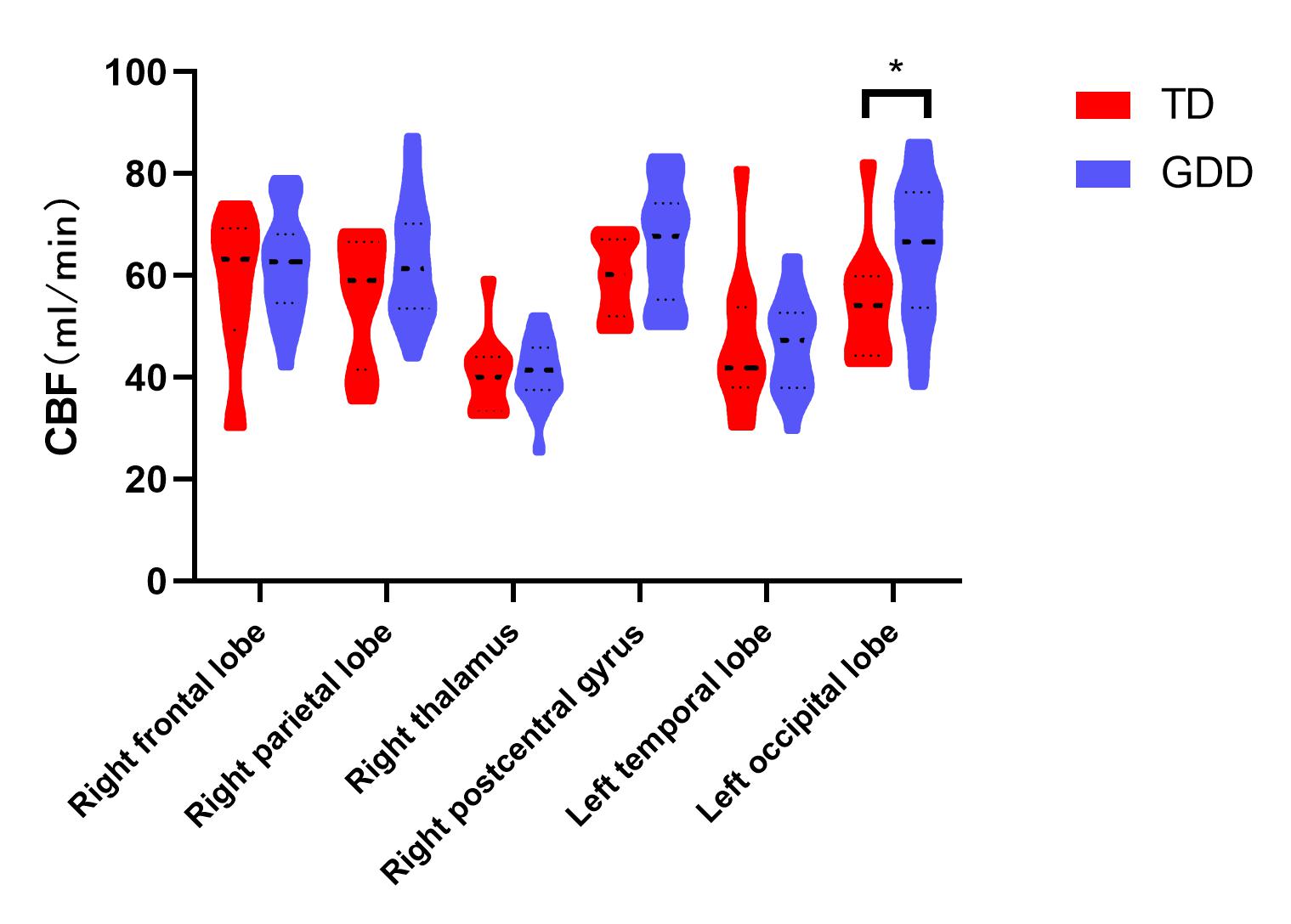
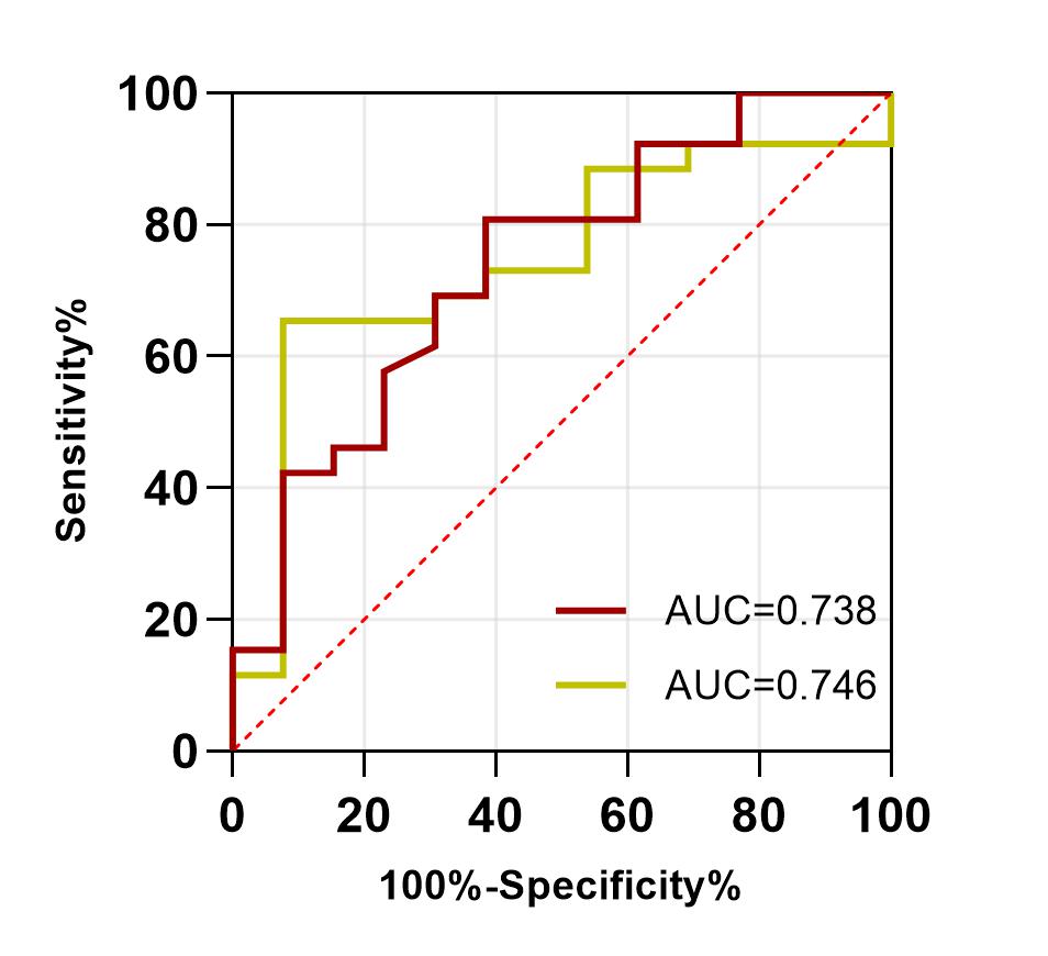
In this study, we examined 26 children aged 2-4 years (GDD group) who were diagnosed with GDD at the Third Affiliated Hospital of Zhengzhou University between March 2022 and October 2023. Additionally, 13 age- and sex-matched typical developmental children(TD group) were included. All participants underwent conventional magnetic resonance imaging (MRI) sequences, specifically 3D-T1 and 3D-pcASL, utilizing a GE 3.0T MRI scanner (SIGNA PIONEER, GE Healthcare, WI, USA). Subsequently, post-processing was performed using the ADW4.7 workstation, and image fusion software was employed to derive CBF perfusion values in various brain regions 1,2. Regions of interest (ROIs), including the frontal lobe, temporal lobe, parietal lobe, occipital lobe, basal ganglia, thalamus, and postcentral gyrus, were selected for analysis(Figure 1).To compare CBF values across different brain regions between children with GDD and healthy children and to analyze CBF characteristics within the GDD group, we expressed normal data as mean ± standard deviation and employed an independent sample t-test for inter-group comparisons. Non-normally distributed data were analyzed using M (Q1, Q2), and the rank sum test was utilized for group comparisons (Figure 2 and 3). Furthermore, we assessed the diagnostic efficacy of CBF values as independent predictors for children aged 2-4 years with GDD by calculating the area under the receiver operating characteristic (ROC) curve (AUC) 3 (Figure 4).
Results:
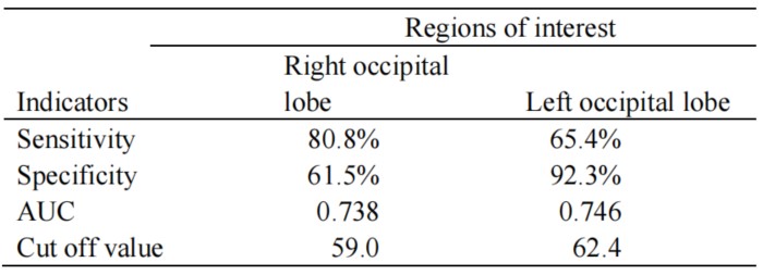
The study results revealed significant differences in CBF values between children with GDD and healthy children, with notably lower CBF values in the right occipital lobe and left basal ganglia in the GDD group (P < 0.05). The ROC curve analysis demonstrated an AUC of 0.738 for the CBF perfusion value in the right occipital lobe, featuring a sensitivity of 80.8%, specificity of 61.5%, and a threshold of 59. For the left occipital lobe, the AUC of CBF was 0.746, with a sensitivity of 65.4%, specificity of 92.3%, and a threshold at 62.4 (Table 1).Conclusion:
This study underscores the potential of three-dimensional pseudo-continuous arterial spin labeling perfusion imaging as a valuable tool for assessing cerebral blood flow in children aged 2-4 years with GDD. Notably, CBF values in both the right and left occipital lobes emerge as promising indicators for diagnosis the occurrence of GDD in children. Early diagnosis and intervention are crucial for managing GDD, and this imaging modality offers a non-invasive means to enhance our understanding and detection of this disease.Acknowledgements
This research was supported by the National Natural Science Foundation of China (Grant No.82371929) (sponsor:XZhang).References
1. Bambach S, Smith M, Morris PP, et al. Arterial Spin Labeling Applications in Pediatric and Adult Neurologic Disorders. J Magn Reson Imaging. 2022 Mar;55(3):698-719.
2. Yerys BE, Herrington JD, Bartley GK, et al. Arterial spin labeling provides a reliable neurobiological marker of autism spectrum disorder. J Neurodev Disord. 2018 Dec 13;10(1):32.
3. Tang S, Liu X, Ran Q, et al. Application of Three-Dimensional Pseudocontinuous Arterial Spin Labeling Perfusion Imaging in the Brains of Children With Autism. Front Neurol. 2022 Feb 23;13:851430.Figures
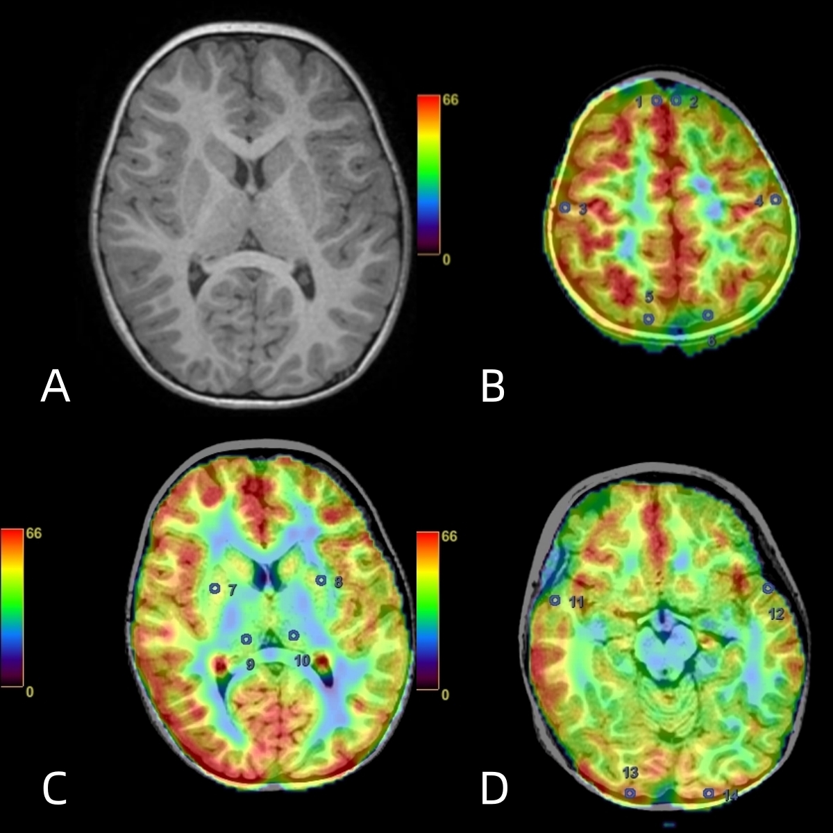
Figure 1 : A Male of 3 years old. Regions of interest(ROIS)delineation on Processed Images and 3D-T1. (A) Basis brain structure (B) Circle 1, 2: frontal lobe; circle 3, 4: postcentral gyrus; circle 5, 6: parietal lobe (C)Circle 7, 8: basal ganglia; circle 9, 10: thalamus (D)Circle 11, 12: temporal lobe; circle 13, 14: occipital lobe.
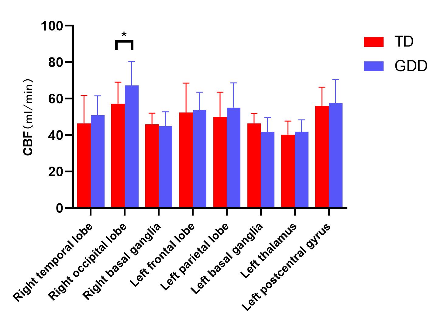
Figure 2: Comparison of the values with normal distribution of CBF from the Processed Images. *: P < 0.05.