2391
Altered association of cortical thickness with cognitive abilities in very preterm children at 6 years of age1Neuroscience Convergence Center, Institute of Green Manufacturing Technology, Korea University, Seoul, Korea, Republic of, 2Medical Device Development Center, Daegu-Gyeongbuk Medical Innovation Foundation, Daegu, Korea, Republic of, 3Division of Neonatology, Department of Pediatrics, School of Medicine, Ewha Womans University, Seoul, Korea, Republic of
Synopsis
Keywords: Normal Development, Pediatric
Motivation: Very preterm children have a risk of impaired cognitive function mediated by several risk factors. The association between structural alteration and high-order cognitive function remains unclear.
Goal(s): Very preterm children at 6 years old showed significant differences in cortical thickness associated with cognitive abilities compared with term infants.
Approach: Cortical thickness was assessed in 41 very preterm and 24 full-term children at 6 years of age. The cortical thickness analysis of structural T1-weighted images was performed using Advanced Normalization Tools.
Results: Perceptual reasoning indices, high-order cognitive function, were found to be broadly correlated with cortical thickness in both very preterm and term children
Impact: This study explores the between brain structure changes and cognitive abilities in very preterm and full-term children, providing insights for neurodevelopmental diagnosis.
INTRODUCTION
Very preterm children, born before 32 weeks of gestation, have a risk of impaired cognitive function mediated by several risk factors. [1] Cognitive impairment can be measured by various neurodevelopmental assessments and is closely associated with structural alterations of the brain morphometry such as cortical thickness. [2,3] However, the association between structural alteration and high-order cognitive function remains unclear. This study aimed to investigate the neurodevelopmental association between structural changes and cognitive abilities in very preterm and full-term children at 6 years old.METHODS
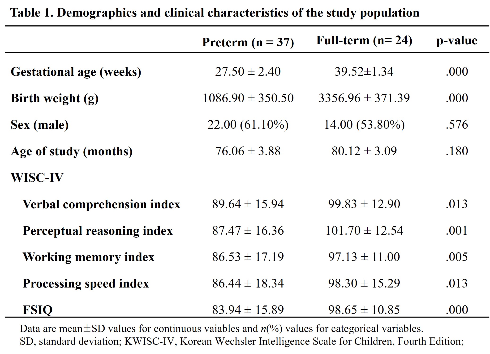
Participants: A total of 65 children (41 very preterm children and 24 full-term children) were recruited and underwent MRI brain scans. The very preterm group was recruited at 6 years of age, comprising children born preterm ≤ 32 weeks gestational age (GA) and the control group included full-term children (with ≥ 37 weeks GA at birth). The demographic and clinical characteristics of the participants are presented in Table 1.Neurodevelopmental assessment: Neurodevelopmental assessments was conducted by using the Wechsler Intelligence Scale for Children, fourth edition (WISC-IV). The WISC-IV provides not only the full-scale intelligence quotients (FSIQ), which indicates the overall cognitive abilities, but also four factor index scores based on specific cognitive profiles: the verbal comprehension index (VCI), perceptual reasoning index (PRI), working memory index (WMI), and processing speed index (PSI).
MRI acquisition: A 3.0T Siemens scanner (Verio) and a Siemens matrix coil were used. T1-magnetization-prepared rapid gradient-echo (MPRAGE) imaging parameters used were as follows: TR = 1900 ms; TE = 2.93 ms; flip angle = 8°; pixel bandwidth = 170 Hz/pixel; matrix size = 256 × 208; field-of-view = 256 mm; NEX = 1; slice thickness = 1 mm; total acquisition time = 4 min 9 s. Cortical thickness analysis: A cortical thickness analysis of structural T1-weighted (T1w) images was performed using a customized shell script, the antsCorticalThickness pipeline, based on Advanced Normalization Tools (ANTs).[4,5] Statistical analyses: Demographic and neurodevelopmental assessment scores were compared between very preterm and full-term children by a two-sample t-test using the Statistical Package for the Social Sciences, version 22.0, software. The linear regression analysis was performed to investigate the correlation between cortical thickness and the five indices of the WISC-IV. Statistical measures with p-values of < 0.05 were considered statistically significant.
RESULTS
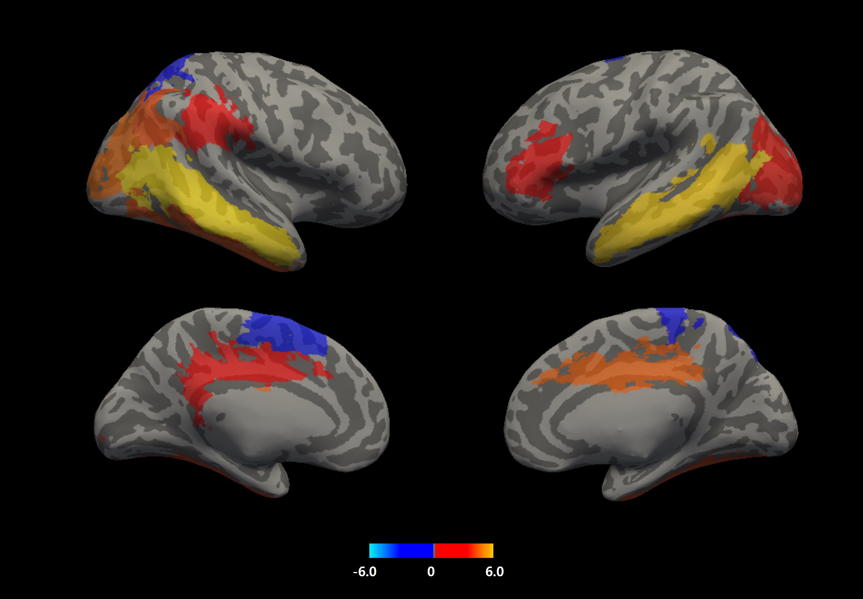
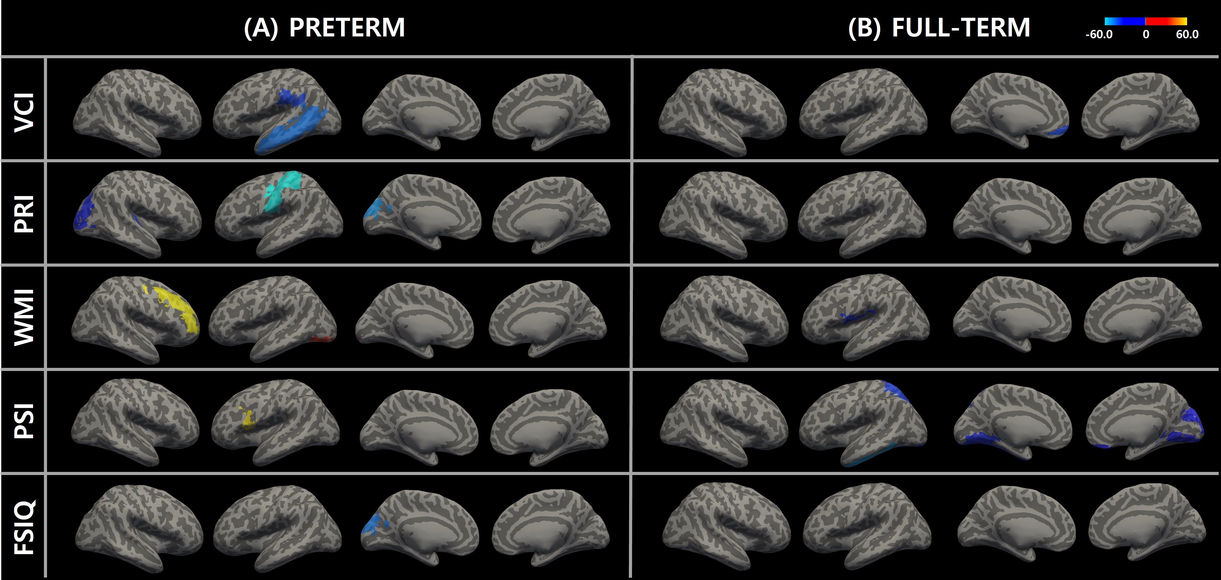
In the cortical thickness analyses, the cortex of very preterm-born children was significantly thinner than that in full-term children in the left supplementary motor area, right superior parietal gyrus, and right paracentral lobule. Conversely, very preterm-born children showed cortex that was significantly thicker than that in term-born children in the following 13 regions of interest. (Figure 1)Figure 2 describe the five WISC-IV scores and their respective correlations with regional cortical thickness. Especially, The Perceptual Reasoning Index (PRI) has been a widely used measure for high-order cognitive functions, interpreting and organizing visual information to solve problems. In both preterm and full-term children, a higher PRI score correlates with decreased cortical thickness. (Figure 2)
DISCUSSION
This study was to investigate the brain morphometry-cognitive function relationship in a sample of 41 very preterm and 24 full-term children by calculating the cortical thickness and the association between brain morphometry and cognitive abilities. We found significant differences in cortical thickness in several brain regions between the very preterm and full-term group at 6 years of age. In both groups, cortical thickness was significantly correlated with high-order cognitive functions. These findings clarify the pathophysiology of cortical thickness and its association with neurodevelopment in very preterm children, demonstrating the long-term impact of very preterm birth on structural and cognitive function.Acknowledgements
This work was supported by the National Research Foundation of Korea(NRF) grant funded by the Korea government(MSIT) (No. 2021R1C1C2003663).References
[1] Wood, N. S., et al. (2005) "The EPICure study: associations and antecedents of neurological and developmental disability at 30 months of age following extremely preterm birth." Archives of Disease in Childhood-Fetal and Neonatal Edition 90.2: F134-F140.
[2] Gogtay N, Giedd JN, Lusk L, et al. Dynamic mapping of human cortical development during childhood through early adulthood. Proc Natl Acad Sci U S A. 2004;101:8174-8179.
[3] Martinussen M, Flanders DW, Fischl B, et al. Segmental brain volumes and cognitive and perceptual correlates in 15-year-old adolescents with low birth weight. J Pediatr. 2009;155:848-853.e1.
[4] Avants BB, Tustison N, Johnson H. Advanced Normalization Tools (ANTS); 2014.
[5] Tustison NJ, Cook PA, Klein A, et al. Large-scale evaluation of ANTs and FreeSurfer cortical thickness measurements. NeuroImage. 2014;99:166-179.
Figures