2388
Longitudinal MR Elastography of Brain Mechanical Property Maturation During Adolescence1Mechanical Engineering, University of Delaware, Newark, DE, United States, 2Psychological and Brain Sciences, University of Delaware, Newark, DE, United States, 3Biomedical Engineering, Columbia University, New York, NY, United States, 4Biomedical Engineering, University of Delaware, Newark, DE, United States
Synopsis
Keywords: Adolescents, Brain
Motivation: Magnetic resonance elastography (MRE) sensitively captures structural changes in the brain. However, application of this technique has been limited to cross-sectional studies in adolescents.
Goal(s): Our aim was to examine, for the first time, longitudinal changes in MRE-derived brain tissue mechanical properties during adolescence.
Approach: Shear stiffness and damping ratio were calculated for 14 adolescents during two study visits separated by a year.
Results: Stiffness significantly decreased over one year. The largest stiffness declines were in subcortical gray matter, in agreement with cross-sectional studies. Changes in both shear stiffness and damping ratio were correlated with progression of puberty.
Impact: This longitudinal study found wide-spread softening of brain tissue during adolescence, supporting cross-sectional findings. These changes were also correlated with the progression of puberty. This confirms the sensitivity of mechanical properties to capture structural changes of brain maturation.
Introduction
Adolescence is marked by a wave of structural changes in the brain and improved cognitive function that make up brain maturation1. Notable structural changes include dendritic pruning and reorganization and axonal myelination2. Magnetic resonance elastography (MRE) has shown promise in sensitively capturing the structural changes occurring in the brain during maturation through cross-sectional studies. In a large cross-sectional study of children and adolescents, age was negatively related to tissue stiffness, particularly in cortical and subcortical gray matter3. Mechanical properties of the adolescent brain have also been associated with behavioral measures, including risk taking4. Longitudinal changes in the brain during adolescence have been tracked using volumetric measures, which show widespread cortical thinning1. No longitudinal studies have examined mechanical properties during adolescence, making it difficult to obtain a complete picture of age-related changes in brain structure5. In fact, only one longitudinal study has examined age-related changes in brain mechanical properties, which observed that stiffness decreased after one year6. However, the utility of this work was limited by a wide age range (22-61 years) that excluded adolescents6. This work presents the first longitudinal study of MRE measures in adolescence.Methods
14 adolescents (12.6 ± 0.7 years, 8M/6F) between the ages of 11-13 years participated in this study. They completed two identical study visits separated by a year (356 ± 14 days). MRE was acquired with a 1.5 mm isotropic resolution scan with 50 Hz vibrations delivered by a pneumatic actuator (Resoundant) for a total scan time of 5 minutes on a Siemens 3T Prisma scanner7. A T1-weighted MPRAGE with 0.8 mm isotropic resolution was obtained and used to segment cortical gray matter, subcortical gray matter, and white matter using Freesurfer. Complex shear modulus, $$$G = G' + iG''$$$, of the tissue was calculated using a nonlinear inversion algorithm8,9. MRE measures of shear stiffness, $$$\mu = \frac{2|G|^2}{G'+|G|}$$$, and damping ratio, $$$\xi = \frac{G''}{2G'}$$$, were calculated for each subject. All images were registered to the MNI-152 atlas using FSL to make between-subject and between-visit voxel-wise comparisons. To track pubertal status, participants completed the Pubertal Development Scale (PDS), a self-reported measure of puberty status specific to sex, which tracks changes in height, skin, hair growth, voice (male only), breasts (female only), and menarche (female only)10. Paired-samples t-tests were used to examine changes in mechanical properties over the 1-year gap. Correlations between change (over time) in mechanical properties and change in pubertal status were also examined.Results
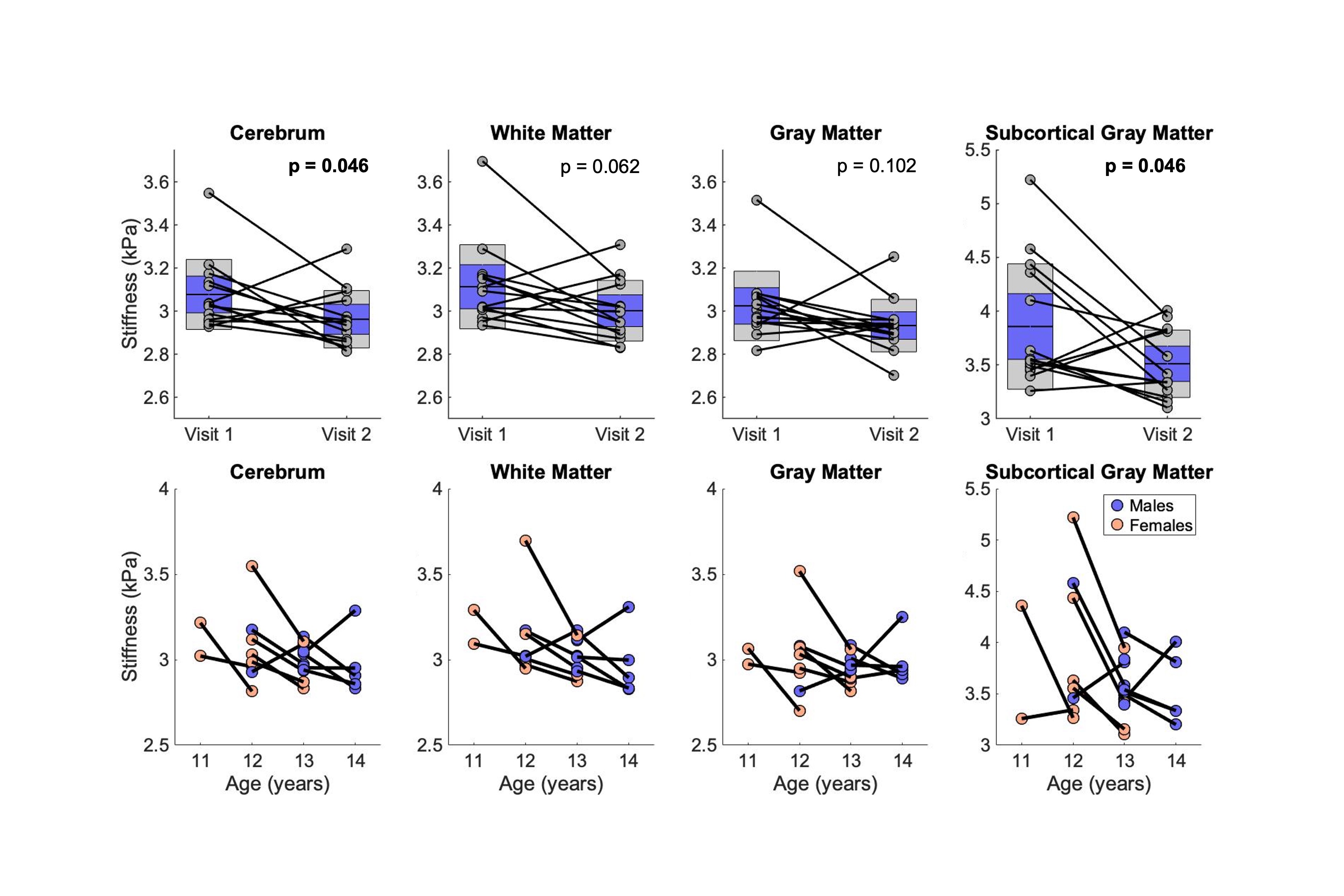
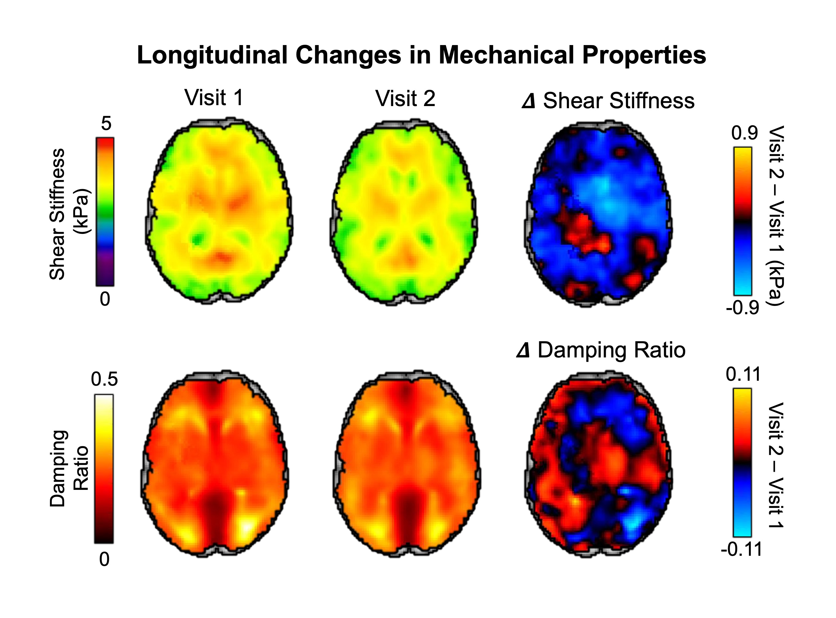
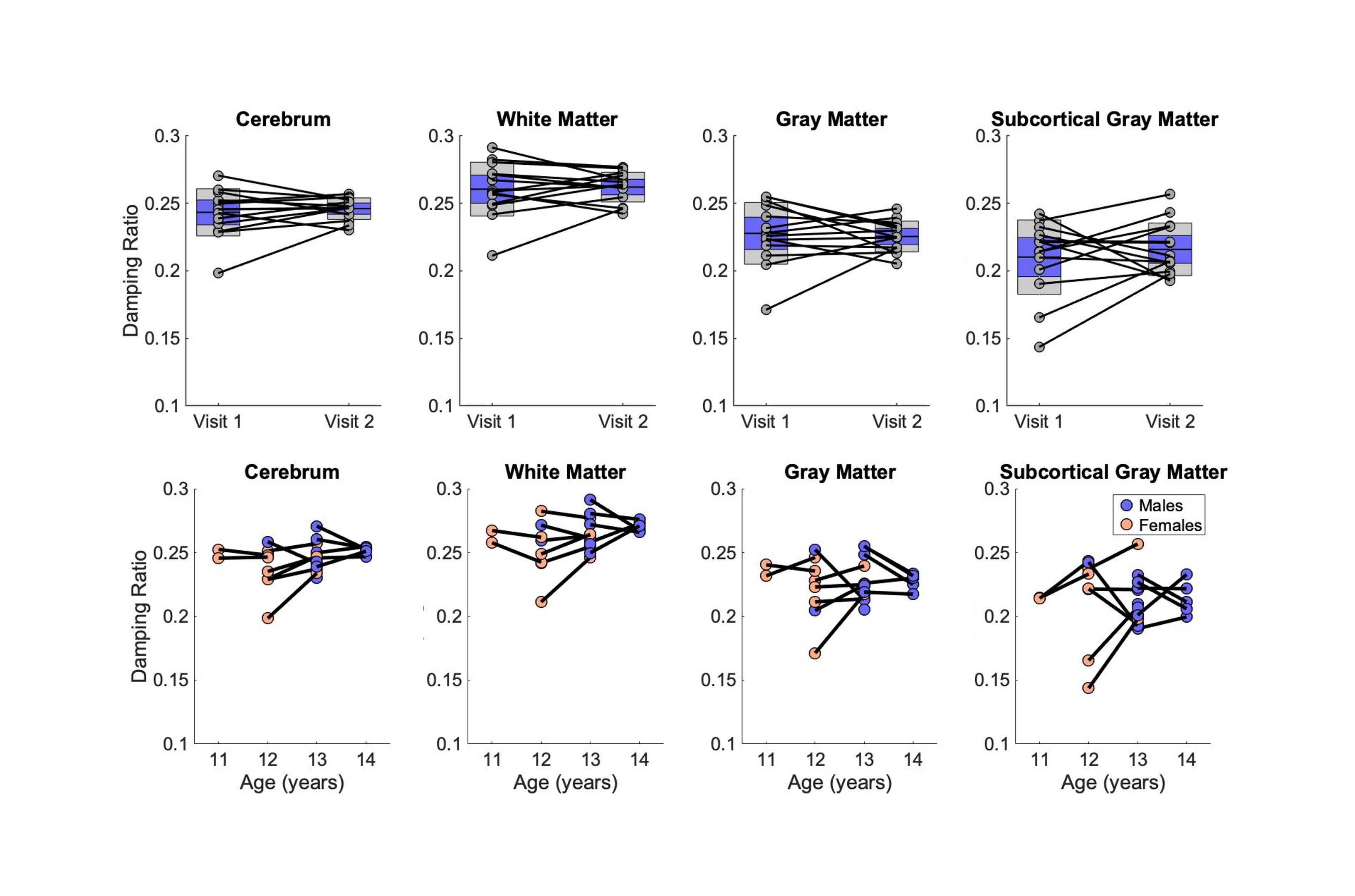
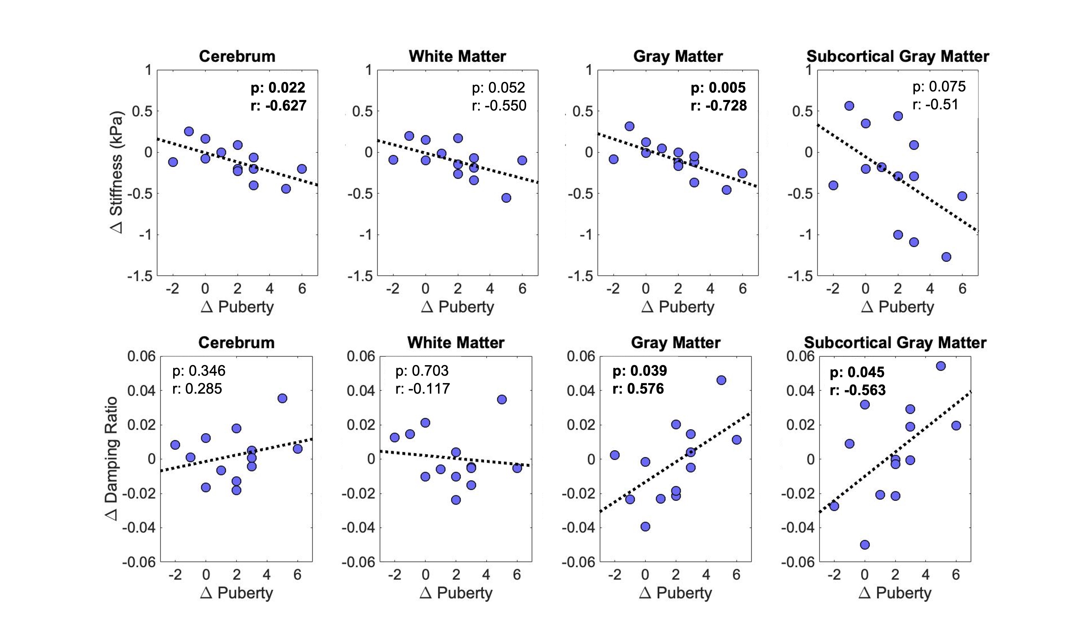
In the cerebrum, we observed a significant decrease (p=0.046) in average shear stiffness from 3.08 kPa at visit 1 (V1) to 2.96 kPa at visit 2 (V2). As illustrated in Figure 1, similar decreases were also found on the regional level in white matter (V1: 3.11 kPa, V2: 3.0 kPa, p=0.062), gray matter (V1: 3.02 kPa, V2: 2.93 kPa, p=0.102), and subcortical gray matter (V1: 3.86 kPa, V2: 3.51 kPa, p=0.046). A representative slice of the mechanical property maps averaged across all participants is shown in Figure 2 for each visit and the difference between the visits. Softening, shown in blue, occurred throughout the brain, whereas damping ratio increased along the exterior. Damping ratio did not significantly change between the visits in the cerebrum (p=0.48) as seen in Figure 3. In Figure 4, changes in both shear stiffness (p=0.005) and damping ratio (p=0.039) were correlated with pubertal progression for gray matter. Larger decreases in stiffness along with larger increases in damping ratio were seen with greater pubertal progression.Discussion & Conclusions:
This study found significant decreases in brain stiffness over one year during adolescence. This supports previous cross-sectional work that found tissue softening during childhood, adolescence, and into adulthood, particularly in gray matter3. We also observed the largest decreases in shear stiffness in gray matter at the subcortical level compared to white matter and cortical gray matter. Changes in both shear stiffness and damping ratio of the cortex were strongly correlated with changes in pubertal status, such that greater pubertal progression was associated with greater decrease in stiffness and greater increase in damping ratio. This supports previous work that identifies the cortex as the primary region of structural change during brain maturation1. Findings from this study confirm the necessity of longitudinal studies of brain tissue viscoelastic properties and could help elucidate the complex changes in brain structure occurring during brain maturation with improved sensitivity and specificity. Tracking typical development longitudinally will help provide benchmarks for studying neurodevelopmental conditions such as autism and epilepsy.Acknowledgements
This work was funded by R01MH123470 and P20GM103653.References
1. Toga AW, Thompson PM, Sowell ER. Mapping Brain Maturation. Focus (Madison). 2006;4:378–390.2. Dumontheil I. Adolescent brain development. Curr. Opin. Behav. Sci. 2016;10:39–44.
3. McIlvain G, Schneider JM, Matyi MA, et al. Mapping brain mechanical property maturation from childhood to adulthood. Neuroimage 2022;263:119590.
4. McIlvain G, Clements RG, Magoon EM, Spielberg JM, Telzer EH, Johnson CL. Viscoelasticity of reward and control systems in adolescent risk taking. Neuroimage 2020;215:116850.
5. Vijayakumar N, Mills KL, Alexander-Bloch A, Tamnes CK, Whittle S. Structural brain development: A review of methodological approaches and best practices. Dev. Cogn. Neurosci. 2018;33:129–148.
6. Herthum H, Hetzer S, Kreft B, et al. Cerebral tomoelastography based on multifrequency MR elastography in two and three dimensions. Front. Bioeng. Biotechnol. 2022;10:1056131.
7. McIlvain G, Cerjanic AM, Christodoulou AG, McGarry MDJ, Johnson CL. OSCILLATE: A low-rank approach for accelerated magnetic resonance elastography. Magn. Reson. Med. 2022;88:1659–1672.
8. Houten EEW Van, Miga MI, Weaver JB, Kennedy FE, Paulsen KD. Three-dimensional subzone-based reconstruction algorithm for MR elastography. Magn. Reson. Med. 2001;45:827–837.
9. McGarry MDJ, Houten EEW Van, Johnson CL, et al. Multiresolution MR elastography using nonlinear inversion. Med. Phys. 2012;39:6388–6396.
10. Petersen AC, Crockett L, Richards M, Boxer A. A self-report measure of pubertal status: Reliability, validity, and initial norms. J. Youth Adolesc. 1988;17:117–133.
Figures