2387
Assessment of individualised cortical gyrification in infants with congenital heart disease1Centre for the Developing Brain, King's College London, London, United Kingdom, 2GKT Medical School, King's College London, London, United Kingdom, 3Department of Cardiovascular Imaging, School of Biomedical Engineering & Imaging Science, King's College London, London, United Kingdom
Synopsis
Keywords: Neuro, Pediatric, Cortex
Motivation: Congenital heart disease (CHD) is common and associated with impaired early brain development.
Goal(s): To assess whether preoperative cortical Gyrification Index (GI) in infants with CHD deviates from the normal trajectory.
Approach: GI trajectories were normatively-modelled using reference control MRI data from 320 healthy infants, enabling calculation of GI Z-scores for the whole brain and frontal, occipital, parietal, temporal, cingulate and insular cortices for 130 infants with CHD, after accounting for sex, postmenstrual age at scan and days since birth at scan.
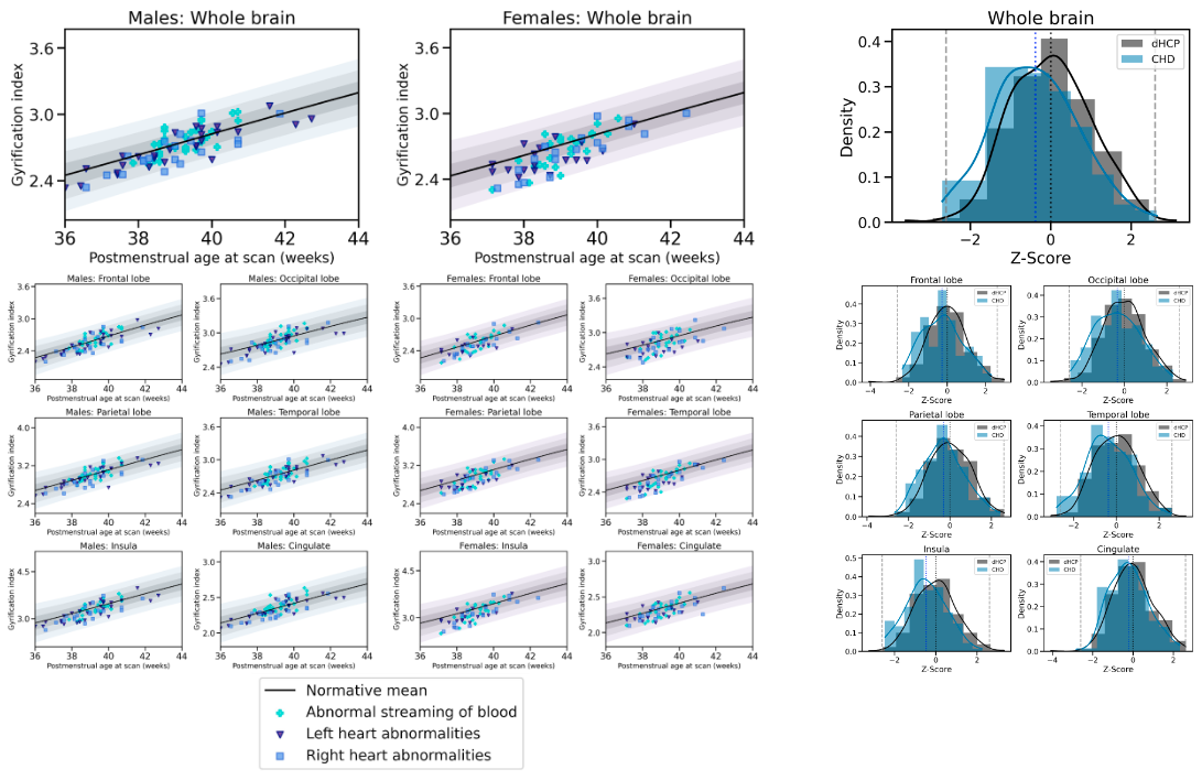
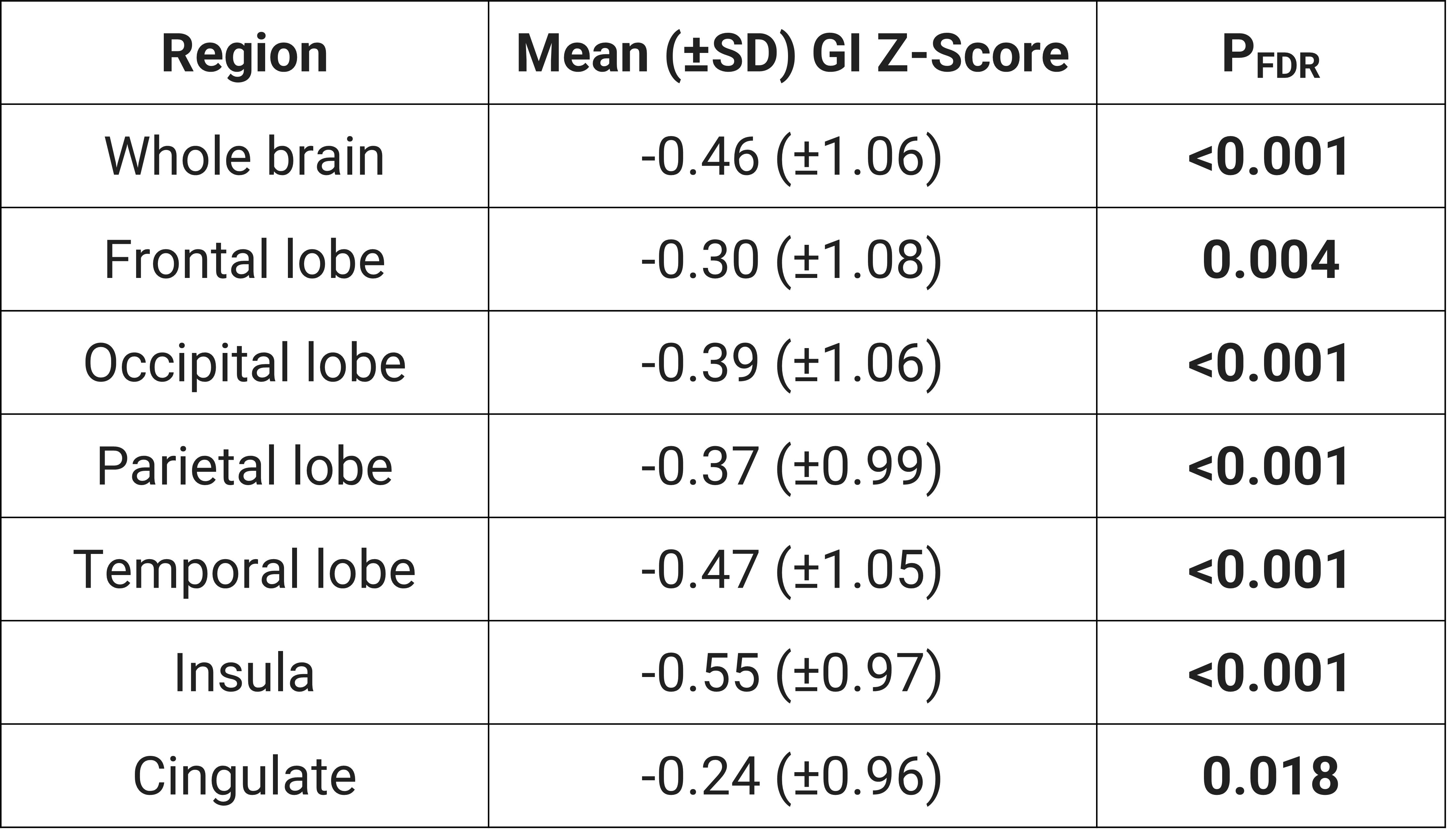
Results: Mean GI Z-scores were significantly lower for the whole brain and all cortical regions in infants with CHD (All PFDR<0.018)
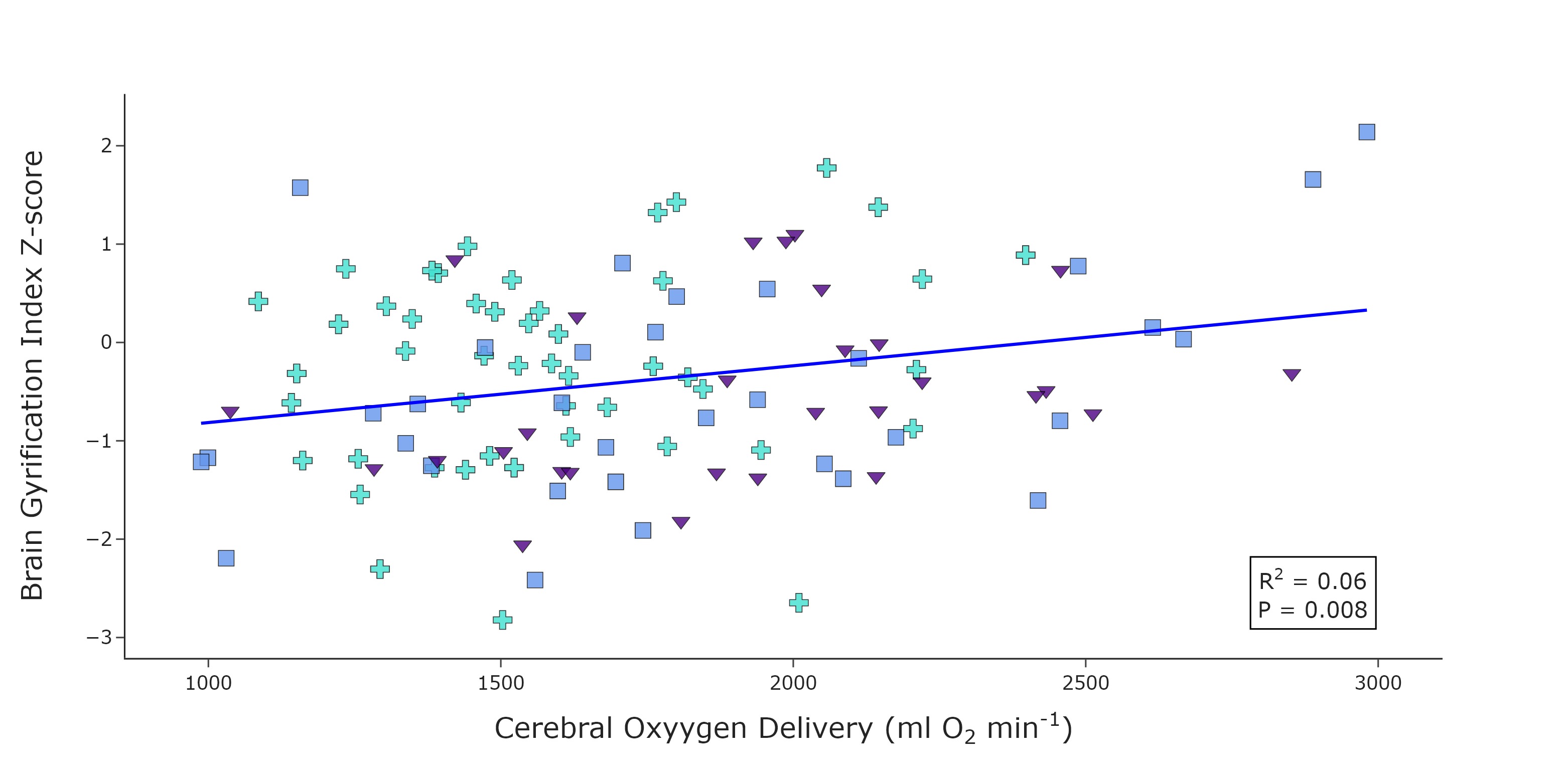
Impact: Global and regional brain gyrification is reduced in infants with critical or severe CHD in the neonatal period, prior to cardiac surgery, and these individualised measures of cortical folding are significantly associated with cerebral oxygen delivery in the neonatal period.
Introduction
Congenital heart disease (CHD) is common and is associated with impaired early brain development and adverse neurodevelopmental outcomes. Previous neuroimaging studies have identified altered cortical development and reduced cerebral oxygen delivery (CDO2) in infants with CHD1,2. However, these studies were limited by small cohorts and used traditional case-control comparison approaches. Normative modelling involves mapping individual datapoints from a reference distribution, derived from a control cohort, allowing the characterization and assessment of deviations from this typically developing population in individual subjects.This study aimed to test the hypothesis that gyrification index (GI) in infants with CHD deviates from the normal trajectory, by calculating cortical GI Z-scores in individuals with CHD and quantifying how much each individual GI measurement deviates from the control population mean; and explore how GI Z-scores related to CDO2 in infants with CHD.
Methods
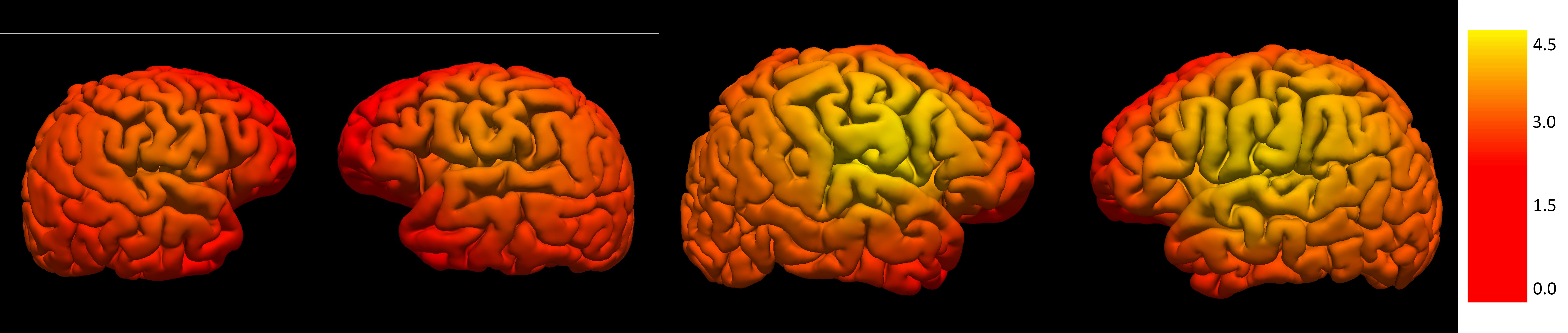
Infants with CHD were prospectively recruited between 2015 and 2022 as part of the Congenital Heart Disease Imaging Programme at St. Thomas' Hospital, London. Each infant was allocated to one of three diagnostic categories based on the hemodynamic impact of the underlying cardiac diagnosis, using the sequential segmental approach: (1) abnormal streaming of blood; (2) left-sided heart lesions; or (3) right-sided heart lesions. Control infants were scanned as part of the developing human connectome project (dHCP). Informed, written consent was acquired prior to MRI (Ethical approval CHD:07/H0707/105&21/WA/0075; dHCP:14/LO/1169). Imaging was performed on a Philips 3T MRI scanner.Structural (T2-weighted) multi-slice turbo spin-echo scans were acquired in two stacks (axial and sagittal planes) (TR/TE=12,000/156ms; flip-angle=90°; slice thickness=1.6mm; slice overlap=0.8mm; in-plane resolution=0.8×0.8mm; SENSE factor=2.11/2.58 [axial/sagittal]). These stacks were reconstructed using a dedicated algorithm to correct motion and integrate data from both acquired stacks (reconstructed voxel size=0.5mm3)3,4. Structural data were processed with the dHCP pipeline5 and cortical surfaces were used to derive mean GI values for the whole brain and frontal, occipital, parietal, temporal, cingulate and insular cortices6,7 (Figure 1). Typical brain GI trajectories were normatively-modelled, after accounting for sex, postmenstrual age at scan and days since birth at scan, using reference control MRI data acquired for the dHCP8. This enabled calculation of GI Z-scores for the whole brain and for all cortical regions for each infant with CHD, quantifying the degree of atypicality for cortical gyrification.
Quantitative flow imaging was performed using velocity-sensitised phase contrast imaging, with a single-slice T1-weighted fast-field-echo sequence (Field of view=100×100mm2; resolution=0.6×0.6×4.0mm2; TR/TE=6.4/4.3ms; flip angle=10°;20 repetitions; maximal encoding velocity=140cm/s; scan time=71s. Cerebral blood flow (CBF) was calculated by summing phase-contrast derived flows through the left and right internal carotid and basilar arteries. Haemoglobin (Hb) levels were measured as part of routine clinical care in CHD participants. Pre-ductal arterial oxygen saturation (SaO2) was measured during the scan. CDO2 was calculated using: CDO2 = SaO2 x [Hb] x 1.36 x [CBF]
A T-test was used to compare GI Z-scores between infants with CHD and control infants. Pearson's correlation coefficient was calculated to determine the relationship between brain GI z-scores and CDO2. Benjamini & Hochberg False Discovery Rate (PFDR) was applied to correct for multiple comparisons. PFDR values<0.05 were considered significant.
Results
Cortical GI measurements were obtained for 130 infants with CHD [59 male; median age at birth=38.43 (37.57-38.86) weeks, median age at scan= 39.00 (38.43-39.71) weeks] and 320 healthy infants [158 male; median age at birth=40.29 (39.29-41.00) weeks; median age at scan=41.50 (40.43-43.00) weeks]. Mean GI Z-scores were significantly lower for the whole brain and all cortical regions in infants with CHD (All PFDR<0.018) (Figure 2, Figure 3). Cerebral blood flow data were obtained in 111 infants with CHD. CDO2 [mean (±SD)=1640 (±1436-2043) ml O2/min)] was significantly positively correlated with brain GI Z-score (R2=0.06, p=0.008), after accounting for infant sex, postmenstrual age at scan and days since birth at scan (Figure 4).Conclusion
Global and regional brain gyrification is reduced in infants with CHD in the neonatal period before cardiac surgery, with the temporal lobe and the insular showing the greatest reduction compared to healthy infants. On average, whole brain GIs are half a standard deviation lower than would be expected when compared to typical development.Individualised measures of cortical folding are significantly associated with cerebral oxygen delivery in the early neonatal period. To our knowledge, this is the first use of a normative modelling approach to assess cortical gyrification indices in individual infants with CHD. Strategies to improve cerebral oxygen delivery may help correct the altered trajectory of impaired brain development found in this population. Further studies with longitudinal analysis are required to determine whether preoperative neuroimaging findings relate to neurodevelopmental outcomes in later childhood and beyond.
Acknowledgements
We thank the families who participated in this study. We also thank our research radiologists, our research radiographers, and our neonatal scanning team at the Centre for the Developing Brain at King's College London. In addition, we thank the staff from the St Thomas’ Hospital Neonatal Intensive Care Unit; the Evelina London Children’s Hospital Fetal and Paediatric Cardiology Departments and the Evelina London Paediatric Intensive Care Unit. This research was funded by the Medical Research Council UK (MR/ L011530/1; MR/V002465/1), the British Heart Foundation (FS/15/ 55/31649) and Action Medical Research (GN2630). The normative sample was collected as part of the Developing Human Connectome Project, funded by the European Research Council under the European Union’s Seventh Framework Program (FP7/20072013)/European Research Council grant agreement no. 319456. This research was also supported by the Wellcome Engineering and Physical Sciences Research Council Centre for Medical Engineering at King’s College London (WT 203148/Z/16/Z), Medical Research Council UK strategic grant (MR/K006355/1), Medical Research Council UK Centre grant (MR/N026063/1) and by the National Institute for Health Research (NIHR) Biomedical Research Centre based at Guy’s and St Thomas’ NHS Foundation Trust and King’s College London.References
1. Kelly CJ, Makropoulos A, Cordero-Grande L, et al. Impaired development of the cerebral cortex in infants with congenital heart disease is correlated to reduced cerebral oxygen delivery. Sci Rep. 2017;7(1):15088. doi:10.1038/s41598-017-14939-z
2. Kelly CJ, Christiaens D, Batalle D, et al. Abnormal Microstructural Development of the Cerebral Cortex in Neonates With Congenital Heart Disease Is Associated With Impaired Cerebral Oxygen Delivery. J Am Heart Assoc Cardiovasc Cerebrovasc Dis. 2019;8(5). doi:10.1161/JAHA.118.009893 3. Cordero-Grande L, Teixeira RPAG, Hughes EJ, Hutter J, Price AN, Hajnal JV. Sensitivity Encoding for Aligned Multishot Magnetic Resonance Reconstruction. IEEE Trans Comput Imaging. 2016;2(3):266-280. doi:10.1109/TCI.2016.2557069
4. Cordero‐Grande L, Hughes EJ, Hutter J, Price AN, Hajnal JV. Three‐dimensional motion corrected sensitivity encoding reconstruction for multi‐shot multi‐slice MRI: Application to neonatal brain imaging. Magn Reson Med. 2018;79(3):1365-1376. doi:10.1002/mrm.26796
5. Makropoulos A, Robinson EC, Schuh A, et al. The developing human connectome project: A minimal processing pipeline for neonatal cortical surface reconstruction. NeuroImage. 2018;173:88-112. doi:10.1016/j.neuroimage.2018.01.054
6. Armstrong E, Schleicher A, Omran H, Curtis M, Zilles K. The ontogeny of human gyrification. Cereb Cortex N Y N 1991. 1995;5(1):56-63. doi:10.1093/cercor/5.1.56
7. Schaer M, Cuadra MB, Tamarit L, Lazeyras F, Eliez S, Thiran JP. A surface-based approach to quantify local cortical gyrification. IEEE Trans Med Imaging. 2008;27(2):161-170. doi:10.1109/TMI.2007.903576
8. Dimitrova R, Arulkumaran S, Carney O, et al. Phenotyping the Preterm Brain: Characterizing Individual Deviations From Normative Volumetric Development in Two Large Infant Cohorts. Cereb Cortex N Y N 1991. 2021;31(8):3665-3677. doi:10.1093/cercor/bhab039
Figures