2385
Prenatal Associations of Maternal Psychological State with Infant Gray Matter Microstructure1Waisman Center, University of Wisconsin-Madison, Madison, WI, United States, 2Neuroscience Training Program, University of Wisconsin-Madison, Madison, WI, United States, 3Department of Pediatrics, University of Wisconsin-Madison, Madison, WI, United States, 4Department of Medical Physics, University of Wisconsin-Madison, Madison, WI, United States
Synopsis
Keywords: Neuro, Diffusion/other diffusion imaging techniques
Motivation: Given known associations between maternal distress during pregnancy and increased risk of offspring to develop psychopathology, it is critical to assess the influences of prenatal maternal depression & anxiety (pMDA) on infant brain organization.
Goal(s): To investigate the relationship between pMDA and GM organization and assess differences in this relationship between male and female infants.
Approach: In this study, we apply the NODDI GM- Based Spatial Statistics framework adapted for the infant brain to assess the relationship with pMDA and infant GM organization and investigate sex-related differences within this relationship.
Results: Our findings suggest a sex-dependent association between pMDA and infant GM microstructure.
Impact: Results may inform the development of interventions for maternal support during pregnancy.
Introduction
Neurodevelopment during the prenatal and neonatal periods establishes the brain’s structural framework supporting behavior and cognitive abilities1, but is also a period of vulnerability in which various influences can shape and contribute to differences in brain and behavioral outcomes2. Evidence links prenatal exposure to maternal depression and anxiety (pMDA) to negative behavioral reactivity in infants3 and differential childhood behavioral outcomes4. Given known associations between maternal distress during pregnancy and increased risk of offspring to develop psychopathology5, it is critical to assess the influences of pMDA on infant brain organization. Previous work has linked maternal depression and anxiety to infant white matter (WM)6-10 and gray matter (GM) microstructure11, 12, particularly highlighting sex-dependent effects6, 12. While work linking pMDA to WM microstructure have employed biophysical modeling techniques of diffusion MRI, such as neurite orientation and dispersion density imaging (NODDI)13, NODDI has not been used to investigate this relationship in GM despite the importance of the early organization of the cortex. In this study, we apply the NODDI GM- Based Spatial Statistics (NODDI-GBSS)14, 15 framework adapted for the infant brain to assess the relationship with pMDA and infant GM organization and investigate sex-related differences within this relationship.Methods
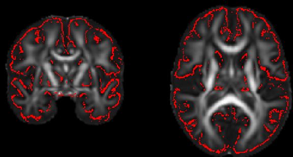
Mothers completed Edinburgh Postnatal Depression Scale (EPDS)16 and State-Trait Anxiety Inventory (STAI)17 at 28 and 35 weeks’, with higher scores indicative of increased depression and anxiety symptoms, respectively. A composite score of prenatal maternal depression and anxiety (pMDA) was calculated by PCA utilizing EPDS and STAI scores across the second and third trimesters. Diffusion MRI (dMRI) data was collected from 89 infants (47 female) whose mothers completed prenatal EPDS and STAI questionnaires. Infants were scanned within the first month of life (average 32 days ((gestation corrected)) on a 3T GE MR750 scanner equipped with a 32-channel head RF array coil (Nova Medical). A three-shell protocol was acquired in 9/18/36 diffusion-encoding gradient directions at b = 350/800/1500 s/mm2, respectively, and 6 with no (b = 0 s/mm2) diffusion weighting. Images were corrected for eddy-current distortions and head motion18, Gibbs ringing, and EPI distortions using an in house-processing pipeline and software developed through FSL19 and MRTrix320 and were fit to DTI and NODDI13 models.We previously adapted the NODDI-GBSS framework14 for infants21. Briefly, as the ODI map from NODDI provides improved contrast between tissue types than the FA map at this age range, ODI was used for GM segmentation. Further, based on the reduced average GM fraction values in the 1-month brain compared to adults, an adjusted GM fraction threshold of 0.45 was used to construct the GM skeleton (Fig1).
General linear models were used to investigate pMDA relationships to the cortical GM microstructure across DTI and NODDI parameters controlling for the effects of age and sex. Given sex-dependencies reported in previous literature, sex by pMDA interactions were also investigated. Non-parametric permutation testing was carried out using Permutation Analysis of Linear Models (PALM)14-15 and threshold free cluster enhancement16 were used to identify significant regions at p < 0.05, FWER-corrected within modality and contrast.
Results
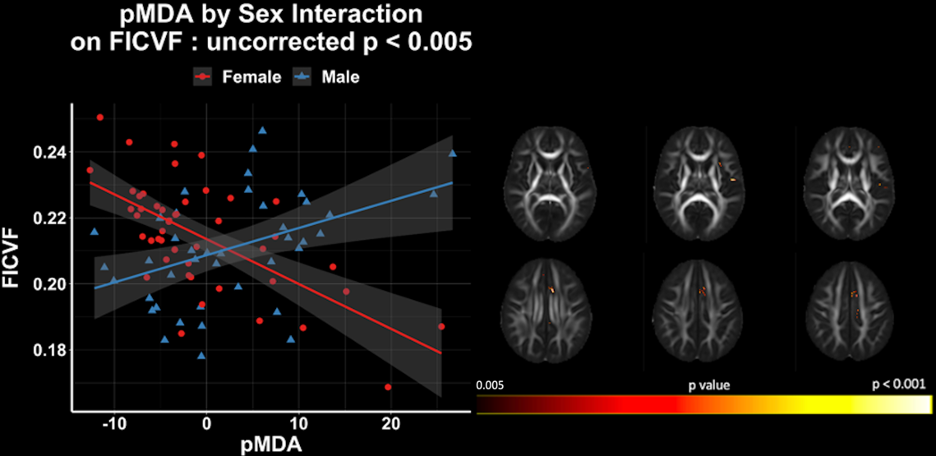
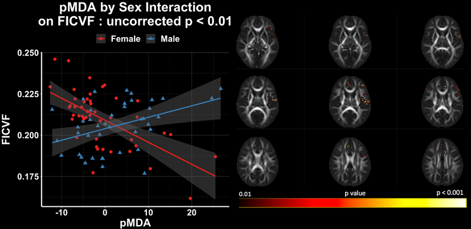
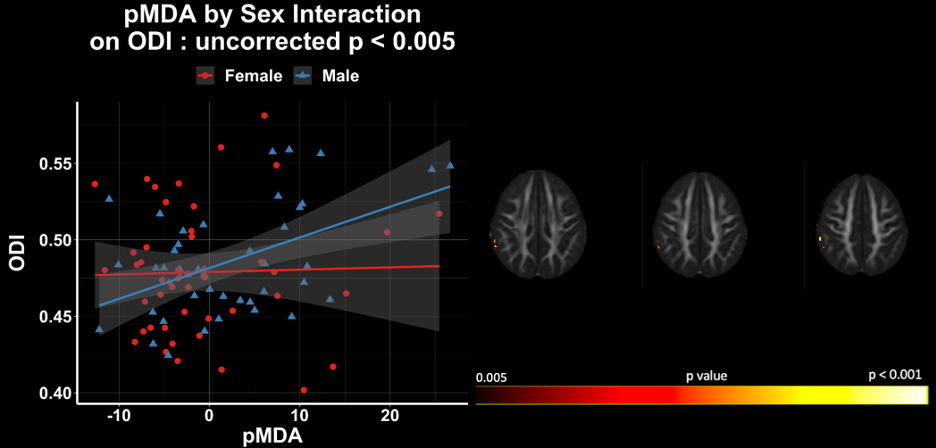
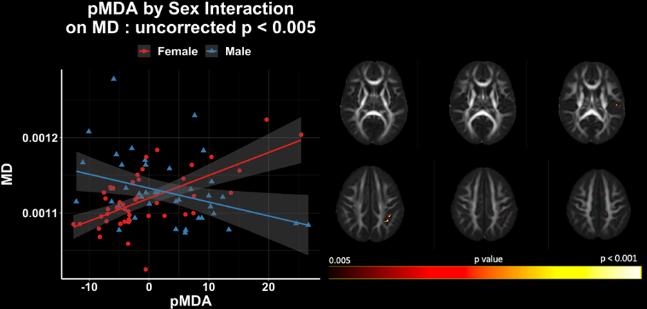
Across the GM skeleton, pMDA was not associated with DTI and NODDI GM microstructure after accounting for age and sex. A pMDA by sex interaction (p < 0.005; uncorrected) was observed with FICVF in frontal left hemisphere GM, with a small number of voxels surviving multiple comparison correction (Fig2). To examine the extent of this sex-interaction, we relaxed our statistical threshold (p<0.01 uncorrected) and observed more widespread regions of the left hemisphere to be associated (Fig3). Exploratory sex-dependent relationships were also detected between pMDA and ODI and MD (uncorrected; p < 0.005) (Figs4&5).Discussion
Our findings suggest a sex-dependent association between pMDA and infant GM microstructure. This differential relationship between brain microstructure and maternal psychological state between male and female infants has been reported in studies of WM microstructure6 and GM microstructure of the amygdala12 where compared to females, male infants exposed to higher levels of maternal symptoms of depression and/or anxiety were associated with higher WM neurite density and dispersion, and higher amygdala mean diffusivity. Our findings suggest that compared to females, male infants exposed to higher pMDA have higher neurite density (FICVF) in frontal and parietal GM of the left hemisphere and higher right hemisphere GM neurite dispersion (ODI). These findings are in conjunction with WM findings from Dean et al., 20186, however, our GM MD findings are in opposing directions compared to those reported in Hashempour et al., 2023, potentially highlighting sex-related differences in this relationship between cortical and subcortical structures which should be explored in future work.Acknowledgements
We sincerely thank our research participants and their families who participated in this research as well as the dedicated research staff who made this work possible. This work was supported by grants by the National Institutes of Mental Health (P50 MH100031; Dr. Richard Davidson) and R00 MH11056 (Dr. Douglas Dean) from the National Institute of Mental Health, National Institutes of Health. Infrastructure support was also provided, in part, by grant U54 HD090256 from the Eunice Kennedy Shriver NICHD, National Institutes of Health (Waisman Center) First author, Marissa DiPiero was also supported in part by NIH/NINDS T32 NS105602 and The Morse Society Graduate Student Fellowship for training in childhood mental health and developmental disabilities at the Waisman Center.
References
1. Knudsen, E.I., Sensitive Periods in the Development of the Brain and Behavior. Journal of Cognitive Neuroscience, 2004. 16(8): p. 1412-1425. DOI: 10.1162/0898929042304796.
2. Davidson, R.J. and B.S. McEwen, Social influences on neuroplasticity: stress and interventions to promote well-being. Nat Neurosci, 2012. 15(5): p. 689-95. DOI: 10.1038/nn.3093.
3. Davis, E.P., et al., Prenatal Maternal Anxiety and Depression Predict Negative Behavioral Reactivity in Infancy. Infancy, 2004. 6(3): p. 319-331. DOI: https://doi.org/10.1207/s15327078in0603_1.
4. Rogers, A., et al., Association Between Maternal Perinatal Depression and Anxiety and Child and Adolescent Development: A Meta-analysis. JAMA Pediatr, 2020. 174(11): p. 1082-1092. DOI: 10.1001/jamapediatrics.2020.2910.
5. Chan, J.C., B.M. Nugent, and T.L. Bale, Parental Advisory: Maternal and Paternal Stress Can Impact Offspring Neurodevelopment. Biol Psychiatry, 2018. 83(10): p. 886-894. DOI: 10.1016/j.biopsych.2017.10.005.
6. Dean, D.C., 3rd, et al., Association of Prenatal Maternal Depression and Anxiety Symptoms With Infant White Matter Microstructure. JAMA Pediatr, 2018. 172(10): p. 973-981. DOI: 10.1001/jamapediatrics.2018.2132.
7. Borchers, L.R., et al., Prenatal and postnatal depressive symptoms, infant white matter, and toddler behavioral problems. J Affect Disord, 2021. 282: p. 465-471. DOI: 10.1016/j.jad.2020.12.075.
8. Graham, R.M., et al., Maternal Anxiety and Depression during Late Pregnancy and Newborn Brain White Matter Development. AJNR Am J Neuroradiol, 2020. 41(10): p. 1908-1915. DOI: 10.3174/ajnr.A6759.
9. Lautarescu, A., et al., Maternal depressive symptoms, neonatal white matter, and toddler social-emotional development. Transl Psychiatry, 2022. 12(1): p. 323. DOI: 10.1038/s41398-022-02073-y.
10. Demers, C.H., et al., Exposure to prenatal maternal distress and infant white matter neurodevelopment. Dev Psychopathol, 2021. 33(5): p. 1526-1538. DOI: 10.1017/s0954579421000742.
11. Wen, D.J., et al., Influences of prenatal and postnatal maternal depression on amygdala volume and microstructure in young children. Transl Psychiatry, 2017. 7(4): p. e1103. DOI: 10.1038/tp.2017.74.
12. Hashempour, N., et al., Prenatal maternal depressive symptoms are associated with neonatal left amygdala microstructure in a sex-dependent way. European Journal of Neuroscience, 2023. 57(10): p. 1671-1688. DOI: https://doi.org/10.1111/ejn.15989.
13. Zhang, H., et al., NODDI: practical in vivo neurite orientation dispersion and density imaging of the human brain. Neuroimage, 2012. 61(4): p. 1000-16. DOI: 10.1016/j.neuroimage.2012.03.072.
14. Nazeri, A., et al., Gray Matter Neuritic Microstructure Deficits in Schizophrenia and Bipolar Disorder. Biol Psychiatry, 2017. 82(10): p. 726-736. DOI: 10.1016/j.biopsych.2016.12.005.
15. Nazeri, A., et al., Functional consequences of neurite orientation dispersion and density in humans across the adult lifespan. J Neurosci, 2015. 35(4): p. 1753-62. DOI: 10.1523/JNEUROSCI.3979-14.2015.
16. Cox, J.L., J.M. Holden, and R. Sagovsky, Detection of Postnatal Depression: Development of the 10-item Edinburgh Postnatal Depression Scale. The British Journal of Psychiatry, 1987. 150(6): p. 782-786. DOI: 10.1192/bjp.150.6.782.
17. Spielberger, C.D., State-Trait Anxiety Inventory, in The Corsini Encyclopedia of Psychology. 2010. p. 1-1.
18. Andersson, J.L.R. and S.N. Sotiropoulos, An integrated approach to correction for off-resonance effects and subject movement in diffusion MR imaging. Neuroimage, 2016. 125: p. 1063-1078. DOI: 10.1016/j.neuroimage.2015.10.019.
19. Jenkinson, M., et al., Fsl. Neuroimage, 2012. 62(2): p. 782-90. DOI: 10.1016/j.neuroimage.2011.09.015.
20. Tournier, J.D., et al., MRtrix3: A fast, flexible and open software framework for medical image processing and visualisation. Neuroimage, 2019. 202: p. 116137. DOI: 10.1016/j.neuroimage.2019.116137.
21. Marissa DiPiero, P.G.R., Hassan Cordash, Jose Guerrero Gonzalez, Richard J. Davidson, Andrew Alexander, and Douglas C. Dean III, Adapting the NODDI-GBSS Framework for the 1-Month Infant Brain. Proceedings of the Annual Meeting of the International Society for Magnetic Resonance in Medicine., 2023.
Figures