2381
MRI-Guided Transcranial Magnetic Stimulation in the Treatment of Pediatric Cases with Spastic Cerebral Palsy1The First Affiliated Hospital of Xi'an Jiaotong University, Xi'an, China
Synopsis
Keywords: Neuro, MR-Guided Interventions
Motivation: Transcranial magnetic stimulation (TMS)-assisted rehabilitation therapy has demonstrated the potential to enhance prognoses in spastic cerebral palsy (SCP). However, there are still great differences in the efficacy among individuals.
Goal(s): We employed personalized MRI-guided TMS to precisely target symptomatic neural circuits in children with SCP.
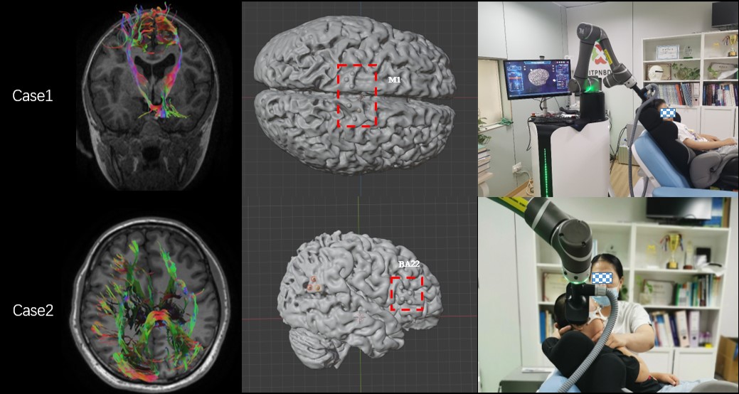
Approach: We used DSI Studio software to track symptom-related fibre bundles within the cortex for targeting and analysed MRI structural and functional differences before and after treatment.
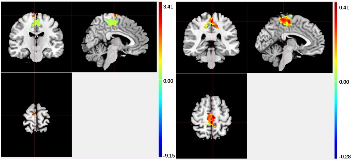
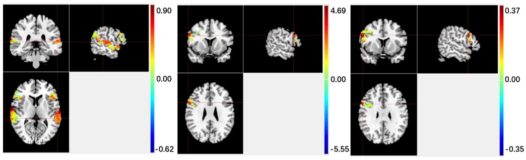
Results: Our findings revealed post-treatment improvements in the quantity, maximum length, and mean length of fiber bundles. Additionally, enhanced neural activity and improved consistency of neural signals were observed after treatment.
Impact: Individualized MRI-guided TMS interventions have shown promise in ameliorating neurological symptoms in individuals with SCP.
Introduction
Spastic cerebral palsy (SCP), often resulting from developmental brain damage, underscores the significance of neural circuit remodeling in cerebral palsy rehabilitation[1]. Transcranial magnetic stimulation (TMS) offers the capacity to modulate neural electrical activity, thereby augmenting brain plasticity[2]. In our investigation, we employed personalized MRI-guided transcranial magnetic stimulation (TMS) to precisely target symptomatic sites[3]. Our objective was to enhance the rate of symptomatic improvement in TMS-treated SCP individuals while examining structural and functional alterations before and after treatment.Methods
We enrolled two 6-year-old male children diagnosed with SCP in our study. The first child presented with lower limb motor deficits, while the second child exhibited speech dysfunction. To localize specific neural circuits, we employed DSI Studio software for tracking motor-related fiber tracts (i.e., corticospinal tracts) and speechrelated fiber tracts, including branches of the arcuate fasciculus and superior longitudinal fasciculus (branches II and III), within the cortex. Utilizing TMS expert recommendations and considering the individual lesion characteristics of each child, we applied high-frequency bilateral stimulation to the lower limb distribution area of the primary motor cortex for Child 1[4]. For Child 2, we administered high-frequency stimulation to the right inferior frontal gyrus within neural pathways associated with the language function[5]. Additionally, Child 1's evaluation involved the Gross Motor Scale, while Child 2 was assessed using the Speech Delay Checklist.Results
Our study involved two 6-year-old boys with SCP, one presenting with lower limb motor deficits and the other with speech dysfunction. Following individualized MRI-guided transcranial magnetic stimulation (TMS) treatments. Scale assessment indicated a slight increase in gross motor scores in Child 1 (dimension E: 55 to 57) and no change in language scores in Child 2. MRI evaluations indicated positive outcomes for both children. In Child 1, there was an increase in the number, maximum length, and mean length of corticospinal tracts, accompanied by enhanced neural activity in the M1 region. Child 2 demonstrated post-treatment enhancements in fiber bundle characteristics in the language-related neural circuits, including increased functional connectivity and enhanced neural signals. These findings suggest that individualized MRI-guided TMS interventions hold promise in ameliorating symptoms in children with SCP, addressing both motor and language-related deficits.Discussion
Our study demonstrated a modest improvement in gross motor scores for Child 1, indicating the effectiveness of the treatment in enhancing lower limb movement. These results suggest that the treatment protocol is not only beneficial but also suitable for continued treatment and longitudinal comparisons. In the case of Child 2, while there was no observed increase in language scores, noteworthy improvements in fiber bundles within the language loop and enhanced functional connectivity were evident. This implies that TMS stimulation yields positive feedback within the brain, warranting consideration for the addition of temporal lobe stimulation (BA39) to complement the existing dorsal inferior frontal gyrus regions (BA44 and BA45). Such an extended intervention may address both sensory and motor aphasia bidirectionally, further improving language deficits.Conclusion
Our study demonstrates the feasibility and validity of individualized MRI-guided transcranial magnetic stimulation (TMS) in accurately targeting stimulation sites, enhancing its efficacy, activating specific neural circuits, and ultimately ameliorating symptoms associated with SCPAcknowledgements
Yitong Bian, Liang Wu, and Zhen Jia contribute equally to this work. This work was supported by the National Natural Science Foundation of China (82272618, 81971581) . Please address correspondence to Jian Yang, e-mail: yj1118@mail.xjtu.edu.cn and Xianjun Li, e-mail: xianj.li@mail.xjtu.edu.cn.References
[1] C. Morgan, L. Fetters, L. Adde, et al. Early intervention for children aged 0 to 2 years with or at high risk of cerebral palsy: international clinical practice guideline based on systematic reviews [J]. JAMA Pediatr, 2021, 175: 846-858.
[2] V. Kozyrev, R. Staadt, U. T. Eysel, et al. TMS-induced neuronal plasticity enables targeted remodeling of visual cortical maps [J]. Proc Natl Acad Sci U S A, 2018, 115: 6476-6481.
[3] Yingying S, Lei W, Jinlin P, et al. Effects of repetitive transcranial magnetic stimulation on motor function and language ability in cerebral palsy: A systematic review and meta-analysis. Front Pediatr, 2023 Feb 16:11:835472.
[4] Sasegbon A, Watanabe M, Simons A, et al. Cerebellar repetitive transcranial magnetic stimulation restores pharyngeal brain activity and swallowing behaviour after disruption by a cortical virtual lesion. J Physiol. 2019. 597(9):2533-2546.
[5] Zewdie E, Ciechanski P, Kuo HC, et al. Safety and tolerability of transcranial magnetic and direct current stimulation in children: Prospective single center evidence from 3.5 million stimulations. Brain Stimul, 2020,13(3):565-575.
Figures