2379
Comparative Analysis of Three Rapid T1WI Imaging Techniques in Fetal Gastrointestinal Magnetic Resonance Imaging1Department of Radiology, Qilu Hospital of Shandong University, Jinan, China, 2MR Research Collaboration, Siemens Healthineer, Shanghai, China, 3MRI clinical application,Customer Service Department,Siemens Healthineers Digital Technology (Shanghai) Co., Ltd., Shanghai, China
Synopsis
Keywords: Fetal, Fetus, gastrointestinal disorders
Motivation: In the prenatal diagnosis of gastrointestinal abnormality,fetal magnetic resonance imaging (MRI) is becoming increasingly relevant.
Goal(s): T1-weighted sequences enable assessment of the presence of meconium, allowing diagnosis of gastrointestinal abnormality, a method with a higher success rate and better image quality is necessary.
Approach: In this work, we compared the performance of three rapid 3D T1-weighted imaging sequences for fetal gastrointestinal MRI at 1.5 T MRI scanner, including free-breathing radial and breath-hold, as well as the free-breathing multi-average 3D VIBE sequences.
Results: Our results demonstrated that free-breathing multi-average VIBE sequence not only demonstrates a higher success rate but also delivers superior image quality.
Impact: The T1WI images of fetal gastrointestinal were barely satisfactory , which accompanied by motion and other artifacts. It is necessary to find a scanning method that is tolerable to the mother and has good image quality.
Introduction
Fetal gastrointestinal magnetic resonance imaging has improved with the use of motion correction technologies and data collection techniques, and is now a useful supplement to prenatal ultrasound diagnoses[1-5]. Recently, VIBE (Volumetric Interpolated Breath-hold Examination) has been effectively used in abdominal, breast and Fetus imaging, where short ”breath-hold” length acquisitions are required to obtain dynamic, high-resolution T1 weighted tissue imaging minimally affected by respiratory movement[6-9]. In the present work, we aimed to compare the physicians’ score, SNR and CNR values of the free breathing radial, breath-hold and multi-average VIBE sequences at 1.5 T MRI scanner.Methods
All experimental protocols received approval from the Ethics Committee of Shandong University Qilu hospital. The study comprised 65 fetal MRI evaluations conducted between the 28th and 36th weeks of gestation, alongside prenatal MRI and ultrasound assessments. The study group consisted of 43 healthy control fetuses and 22 fetuses diagnosed with various aberrations in gastrointestinal development, including intestinal blockage, intestinal atresia, and intestinal cysts, as detected during ultrasound examinations. Data Acquisition: All fetal MRI examinations were performed on a 1.5 T scanner (MAGNETOM Area, Siemens Healthcare, Erlangen, Germany) with a body coil in concordance with the ISUOG fetal MRI guidelines. T2-weighted images of fetus were acquired a Fast Imaging with Steady-state Precession (True-FISP) sequence with the following parameters: TR/TE, 3.8/1.9 ms; flip, angle 50°; readout bandwidth, 781 Hz; FOV, 195 × 280 mm2. The free breathing radial VIBE images were obtained with parameters: TR/TE, 4.48/2.15ms; Flip angle, 10°; spatial resolution, 1 × 1 × 3 mm3; average 1; TA, 90s. For the free breathing multi-average VIBE sequence, the following acquisition parameters were used: TR/TE, 7.23/2.39ms; Flip angle, 10°; spatial resolution, 1 × 1 × 3 mm3; average 5; TA, 92s. For the breath-hold VIBE sequence, the following acquisition parameters were used: TR/TE, 7.23/2.39ms; Flip angle, 10°; spatial resolution, 1 × 1 × 3 mm3; average 1; TA, 16s. Data analysis: For image comparison and evaluation, MPR and maximum intensity projection (MIP) were performed using the standard software in the Siemens imaging system. For quality assessment, two readers (with up 5 year of experience in fetus imaging) evaluated images acquired by the three different methods according to previously published methods. Quantitative assessment of apparent contrast-to-noise ratio (CNR), signal-to-noise ratio (SNR), and noise was performed. MRI data sets between the independent samples t-test, set a significant level of p < 0.05.Results
The results show that MA-vibe images have around 33% less average noise intensity compared to BH-vibe images and 70% less than Star-vibe images. Additionally, the average SNR and CNR in MA-vibe images for various intestinal segments (bladder, rectum, sigmoid colon, ascending colon, descending colon, and transverse colon) are significantly higher than those in BH-vibe and Star-vibe images (P<0.05).Figure 1 displays images of a healthy fetal digestive system captured using various imaging techniques: True-FISP, Multi-average VIBE (MA-vibe), Breath-Hold VIBE (BH-vibe), and free-breathing radial VIBE (Star-vibe). In the MA-vibe image (Figure 1b), the fetal colon is distinctly visible, contrasting with the less defined and grainy appearance in the BH-vibe (Figure 1c) and Star-vibe (Figure 1d) images. Figure 2 showcases four images of a fetal digestive system with intestinal malrotation obtained using different imaging sequences. In the MA-vibe image (Figure 2b), the fetal colon exhibits enhanced contrast compared to the BH-vibe (Figure 2c) and Star-vibe (Figure 2d) images. Figure 3 presents data on average noise intensities (Figure 3a), quality scores from experienced readers (Figure 3b), SNR (Figure 3c), and CNR (Figure 3d) of three methods.Discussion
Star-vibe employs spiral K-space sampling, which reduces motion artifacts to some extent. However, due to the fetal intestine's irregular peristalsis and under-sampling in K-space typical of VIBE technology, granular artifacts may appear in fetal Star-vibe images[10].Maternal breath-holding issues and unpredictable fetal movements frequently cause motion artifacts in BH-vibe images. Moreover, when the mother is overweight, ASSET artifacts can also manifest in the image center due to parallel acceleration. The use of a multi-averaging approach in free-breathing MA-vibe effectively reduces motion artifacts and image noise, thereby improving the examination's success rate.Conclusion
The free-breathing MA-VIBE sequence reduces noise and enhances image contrast by addressing maternal and fetal motion. When it's challenging to obtain optimal fetal T1WI images, the MA-VIBE method is a viable option.Acknowledgements
This study was supported by the Shandong Provincial Natural Science Foundation (ZR2021MH237 and ZR2021QH125).References
1. Farhataziz N, Engels JE, Ramus RM, Zaretsky M, Twickler DM. Fetal MRI of urine and meconium by gestational age for the diagnosis of genitourinary and gastrointestinal abnormalities. American Journal of Roentgenology 2005;184(6):1891-1897.
2. Furey EA, Bailey AA, Twickler DM. Fetal MR imaging of gastrointestinal abnormalities. Radiographics 2016;36(3):904-917.
3. Aertsen M, Diogo MC, Dymarkowski S, Deprest J, Prayer D. Fetal MRI for dummies: what the fetal medicine specialist should know about acquisitions and sequences. Prenatal diagnosis 2020;40(1):6-17.
4. Prayer D, Malinger G, Brugger P, Cassady C, De Catte L, De Keersmaecker B, Fernandes G, Glanc P, Gonçalves L, Gruber G. ISUOG Practice Guidelines: performance of fetal magnetic resonance imaging. Ultrasound in Obstetrics & Gynecology 2017;49(5):671-680.
5. Manganaro L, Saldari M, Bernardo S, Vinci V, Aliberti C, Sollazzo P, Giancotti A, Capozza F, Porpora MG, Cozzi DA. Role of magnetic resonance imaging in the prenatal diagnosis of gastrointestinal fetal anomalies. La radiologia medica 2015;120:393-403.
6. Azevedo RM, de Campos RO, Ramalho M, Herédia V, Dale BM, Semelka RC. Free-breathing 3D T1-weighted gradient-echo sequence with radial data sampling in abdominal MRI: preliminary observations. American journal of roentgenology 2011;197(3):650-657.
7. Chandarana H, Block TK, Rosenkrantz AB, Lim RP, Kim D, Mossa DJ, Babb JS, Kiefer B, Lee VS. Free-breathing radial 3D fat-suppressed T1-weighted gradient echo sequence: a viable alternative for contrast-enhanced liver imaging in patients unable to suspend respiration. Investigative radiology 2011;46(10):648-653.
8. Inaoka T, Sugimori H, Sasaki Y, Takahashi K, Sengoku K, Takada N, Aburano T. VIBE MRI for evaluating the normal and abnormal gastrointestinal tract in fetuses. American Journal of Roentgenology 2007;189(6):W303-W308.
9. Sun T, Jiang L, Zhang Z, Zhang C, Zhang H, Wang G, Qian Z. Feasibility of free-breathing T1-weighted 3D radial VIBE for fetal MRI in various anomalies. Magnetic Resonance Imaging 2020;69:57-64.
10. Block KT, Chandarana H, Milla S, Bruno M, Mulholland T, Fatterpekar G, Hagiwara M, Grimm R, Geppert C, Kiefer B. Towards routine clinical use of radial stack-of-stars 3D gradient-echo sequences for reducing motion sensitivity. Journal of the Korean Society of Magnetic Resonance in Medicine 2014;18(2):87-106.
Figures
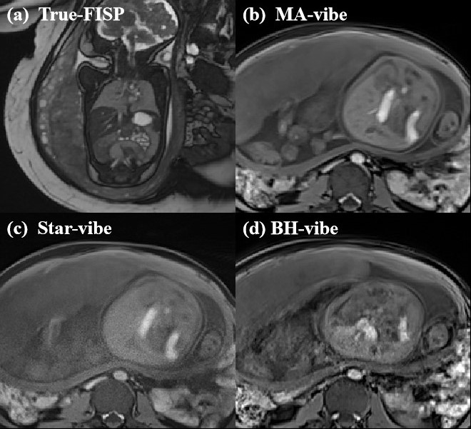
Figure 1. (a) - (d) The images of the healthy fetal (28 weeks) intestinal tract acquired using the True-FISP, MA-vibe, Star-vibe, and BH-vibe sequences, respectively.
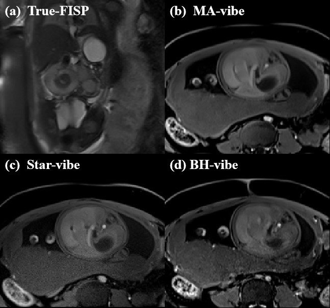
Figure 2. (a) - (d) The images of the fetal intestinal tract with malrotation (at 29 weeks) acquired by the True-FISP, MA-vibe, Star-vibe, and BH-vibe sequences, respectively.
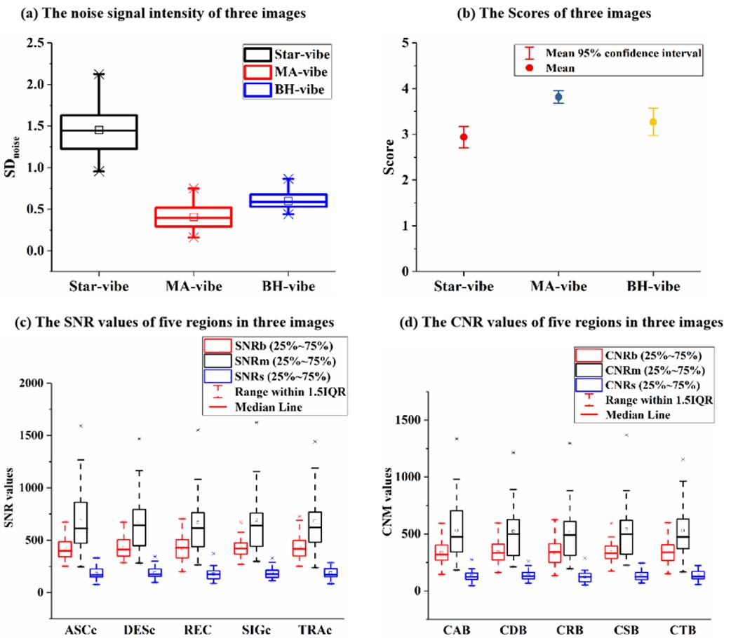
Figure 3. (a) The noise signal intensities of fetal intestinal tract images acquired by Star-vibe, MA-vibe and BH-vibe sequences. (b) quality scores from experienced readers of fetal images acquired by three sequences. (c) the SNR values of five regions in three images; (d) the CNR values of five regions in three images. SDnoise: the noise signal intensity; SNRb: the signal-to-noise ratio of BH-vibe image; SNRm: the signal-to-noise ratio of MA-vibe image; SNRs: the signal-to-noise ratio of star-vibe image;