2377
Developmental Changes of N-Acetyl-Aspartyl-Glutamate (NAAG) in the Human Brain1Radiology, Children's Hospital Los Angeles/USC, Los Angeles, CA, United States, 2Rudi Schulte Research Institute, Santa Barbara, CA, United States
Synopsis
Keywords: Neuro, Brain, metabolism, development
Motivation: N-acetyl-aspartyl-glutamate (NAAG) is a dipeptide and glutamate neuromodulator with possible significance as therapeutical target for injuries and diseases associated with glutamate neurotoxicity.
Goal(s): Determine age-dependent differences in its accumulation in white and grey matter in the human brain.
Approach: Existing MR spectra from 410 “closest-to-normal” pediatric patients and controls were evaluated.
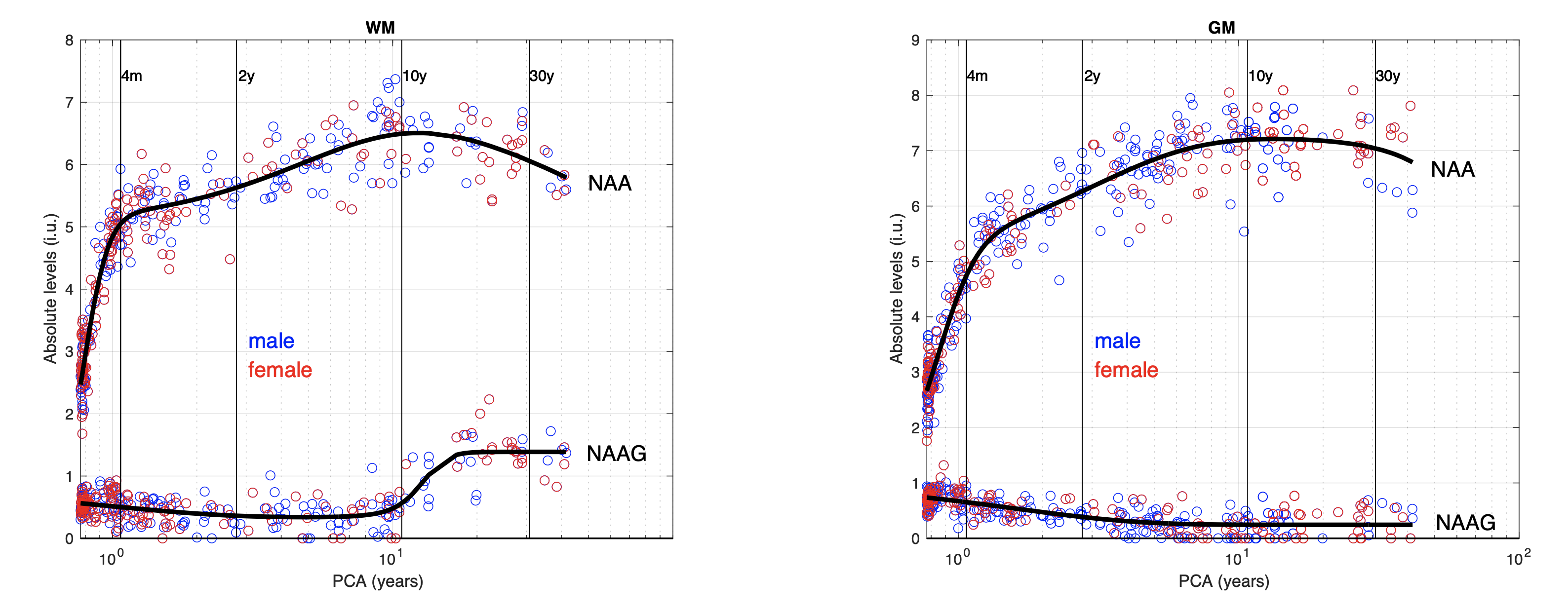
Results: NAAG concentrations in the human brain were low and borderline detectable from birth to early childhood but then increased in late childhood in the parietal white matter whereas NAAG remained low in parietal grey matter.
Impact: This study demonstrates that NAAG accumulates at late childhood predominantly in WM. Furthermore, it suggests that in vivo MRS can be used to assess NAAG levels in disorders associated with excessive glutamatergic neurotransmission, such as childhood seizures.
Introduction
N-acetyl-aspartyl-glutamate (NAAG) is a dipeptide that is present in the human brain in relatively high concentrations. NAAG is synthesized in the neurons from NAA and glutamate. It has been suggested that it may function as a regulator of glutamate signaling and be a possible therapeutic target for brain disorders and injuries that are associated with glutamate excitotoxicity1,2. For possible applications aiming to regulate glutamate, monitoring of in vivo NAAG levels may be crucial. The goal of this study was to demonstrate the feasibility of observing age-dependent changes of NAAG in the human brain by in vivo MRS.Methods
The analysis included 725 spectra obtained from 410 controls and term-born patients with minor indications for MRI (MRI reported as unremarkable) extracted from a database of ≈ 6,500 patients studied at our institution. Ages ranged from 0-41 years and 212 subjects were male whereas 198 subjects were female. All spectra were acquired from parietal white matter (WM, 333 spectra) and parietal grey matter (GM, 392 spectra) on clinical 3T MR scanners using single-voxel PRESS, TE = 35ms, TR = 2000ms, and NAV=128. All spectra were processed with fully automated LCModel software3 and absolute institutional levels of NAA and NAAG were tabulated with no further corrections. To yield developmental curves, NAAG and NAA were plotted versus post-conceptional age and fitted with model functions (not explained in detail).Results
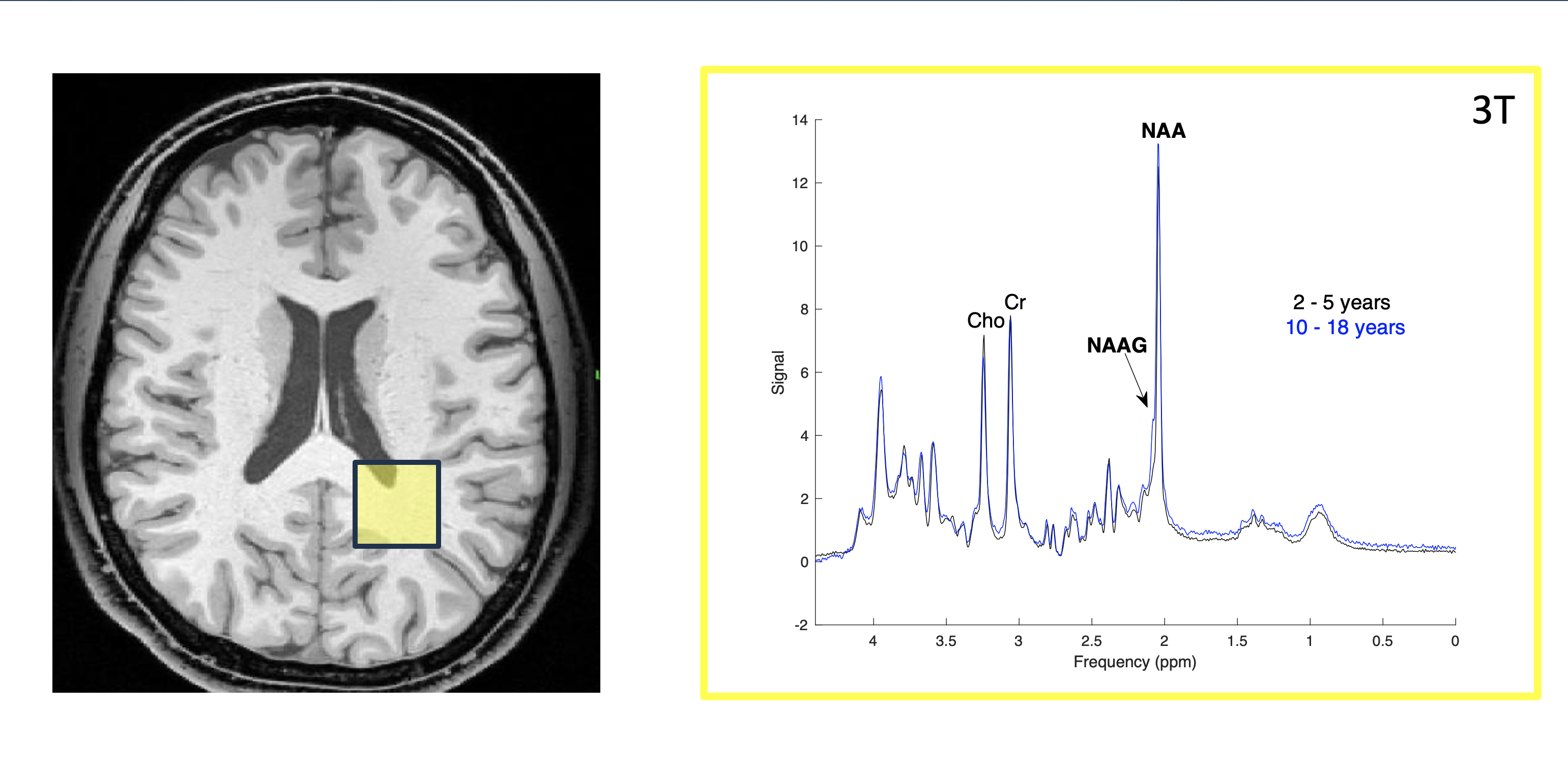
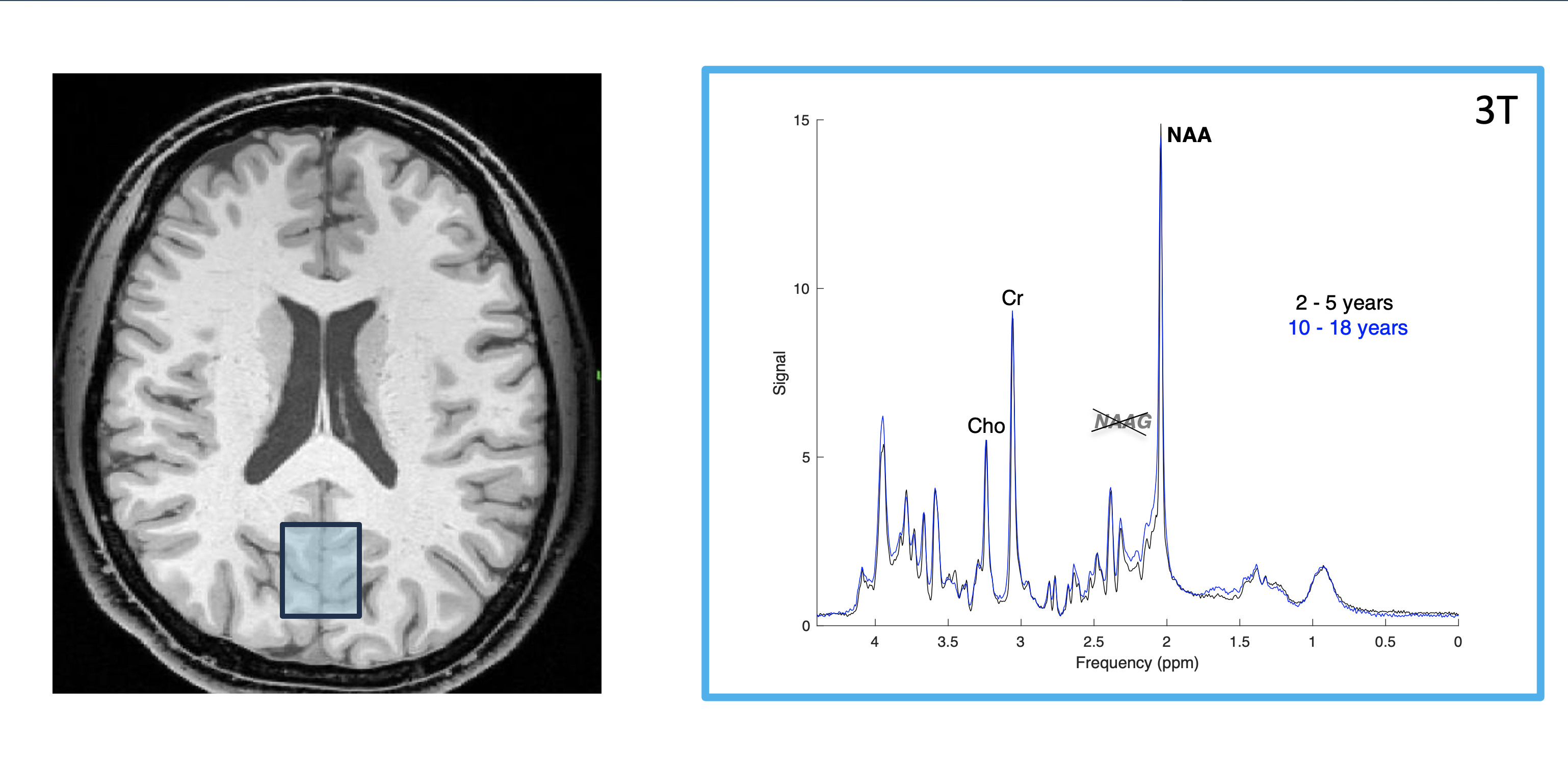
The observed time-courses of NAA in WM and GM were consistent with earlier reports4-6. After birth until early childhood, NAAG levels were low and borderline detectable in both WM and GM. At late childhood (≈ 10 years), NAAG increased in WM but remained low in GM (Figs. 1-3). No gender differences were noted.Discussion
In this study we analyzed the time course of NAAG in the human brain. We found that NAAG levels were low and not different in parietal WM and GM in early brain development well into childhood. NAAG then increased in WM consistent with previous studies that reported higher NAAG in WM of young adults7. Yet, the switch, during late childhood, is remarkably delayed when compared with the much earlier maturational changes of other metabolites reported in previous studies4-6.NAAG is believed to be neuroprotective via the activation of the metabotropic glutamate receptor 3 and subsequent reduced glutamate release2. Thus, the increased availability of NAAG at early adolescence could explain the alleviation of childhood seizures caused by excessive glutamatergic neurotransmission. Reliable separation and quantitation of NAAG (and NAA), for possible clinical applications, is compromised in low-quality spectra by the proximity of their main signals on the chemical shift axis. However, the routine use of 3T scanners appear to make the monitoring of NAAG levels in individual patients increasingly feasible.Conclusions
Developmental changes of NAAG occur in late childhood and appear to be predominantly in white matter.Acknowledgements
Rudi Schulte Research InstituteReferences
1. Baslow MH. Functions of N-acetyl-L-aspartate and N-acetyl-L-aspartylglutamate in the vertebrate brain: role in glial cell-specific signaling. Journal of Neurochemistry. 2000;75(2):453-9.
2. Morland C, Nordengen K. -Acetyl-Aspartyl-Glutamate in Brain Health and Disease. Int J Mol Sci. Jan 23 2022;23(3)doi:10.3390/ijms23031268
3. Provencher SW. Estimation of metabolite concentrations from localized in vivo proton NMR spectra. Magn Reson Med. 1993;30(6):672-9.
4. Blüml S, Wisnowski JL, Nelson MD, et al. Metabolic Maturation of the Human Brain From Birth Through Adolescence: Insights From In Vivo Magnetic Resonance Spectroscopy. Cereb Cortex. Sep 2012;doi:10.1093/cercor/bhs283
5. Kreis R, Ernst T, Ross BD. Development of the human brain: in vivo quantification of metabolite and water content with proton magnetic resonance spectroscopy. Magn Reson Med. Oct 1993;30(4):424-37.
6. Pouwels PJ, Brockmann K, Kruse B, et al. Regional age dependence of human brain metabolites from infancy to adulthood as detected by quantitative localized proton MRS. Pediatr Res. Oct 1999;46(4):474-85.
7. Pouwels PJ, Frahm J. Differential distribution of NAA and NAAG in human brain as determined by quantitative localized proton MRS. NMR Biomed. Apr 1997;10(2):73-8. doi:10.1002/(sici)1099-1492(199704)10:2<73::aid-nbm448>3.0.co;2-4
Figures