2374
Fetal GAs prediction via geometric descriptors of cortical development1NeuroImaging Lab., IRCCS Eugenio Medea, Bosisio Parini (LC), Italy, 2University of Padua, Padova, Italy, 3University of Milan, Milano, Italy, 4IRCCS Ca’ Granda Ospedale Maggiore Policlinico, Milano, Italy, 5University of Milano-Bicocca, Milano, Italy
Synopsis
Keywords: Fetal, Fetus, Cortical development, gestational age prediction, shape signatures
Motivation: Capture more nuanced aspects of fetal brain cortex development.
Goal(s): Investigate the cortical surface of 65 fetal brain reconstructions from MRI examinations with global descriptors derived from scalar point-wise curvature-based metrics (H, K, SI, C, FI) and multidimensional point-wise shape signatures (HKS, WKS, SHOT).
Approach: The morphometric properties extracted by these descriptors were provided as input to SVR models to predict the gestational age. Two public atlases and one dataset were adopted to train and test the models, respectively.
Results: SHOT better encode the cerebral cortex development during pregnancy, achieving a prediction R2 of 0.89 and MAE of 6.3 days.
Impact: SHOT provides researchers with sophisticated tool to capture more nuanced aspects of the fetal brain cortex development across gestational ages.
Introduction
Fetal Magnetic Resonance Imaging (MRI) is a pivotal tool in studying brain morphology, characterizing its development and identifying pathological conditions1,2. Geometric shape descriptors can be exploited to compactly represent the morphometric information, providing valuable insights for diagnosis.Here, we exploited these descriptors to extract the morphometric properties of the fetal brain cortical surface and estimate with them the gestational age (GA), in terms of weeks. Experiments have led to promising prediction results, providing an advanced tool to monitor the gyrification process.
Materials and Methods
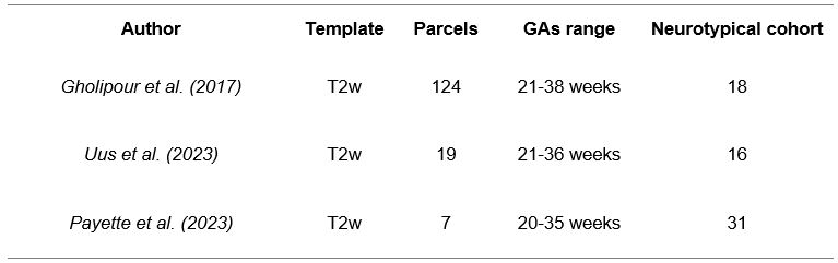
Dataset:65 brain MRI reconstructions of healthy fetuses were identified from two publicly available atlases3,4 and one public dataset5 (Table 1).
Fetal brain cortical surface:
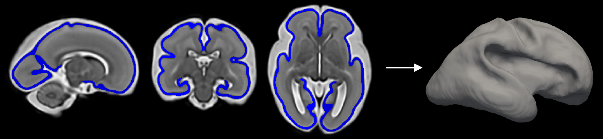
The focus lies on the inner cortical surface, which is the innermost part of the brain's Gray Matter (GM) (Figure 1). The surface was reconstructed from the brain-segmented images using an in-house pipeline, followed by manual refinement.
Global descriptors:
To capture the most informative properties of the inner cortical surface shape, we computed different global descriptors: scalar point-wise curvature-based metrics (curvedness (C), mean curvature (H), Gaussian curvature (K), shape index (SI), folding index (FI)) and multidimensional point-wise shape signatures (Heat Kernel Signature (HKS)6, Wave Kernel Signature (WKS)7, Signature of Histograms of OrienTations (SHOT)8).
The scalar point-wise curvature-based metrics are computed using the FreeSurfer function mris_curvature_stats9. Consequently, we partitioned the values of the vertices into 100 bins to characterize their distribution, and we normalized the derived distribution for the number of associated vertices10.
HKS and WKS descriptors are implemented in MATLAB. Notably, we used k=100 eigenvalues and scaled the temporal domain logarithmically in n=10 time values6. SHOT descriptor is implemented in Python (https://github.com/uhlmanngroup/pyshot) and applied with the default options.
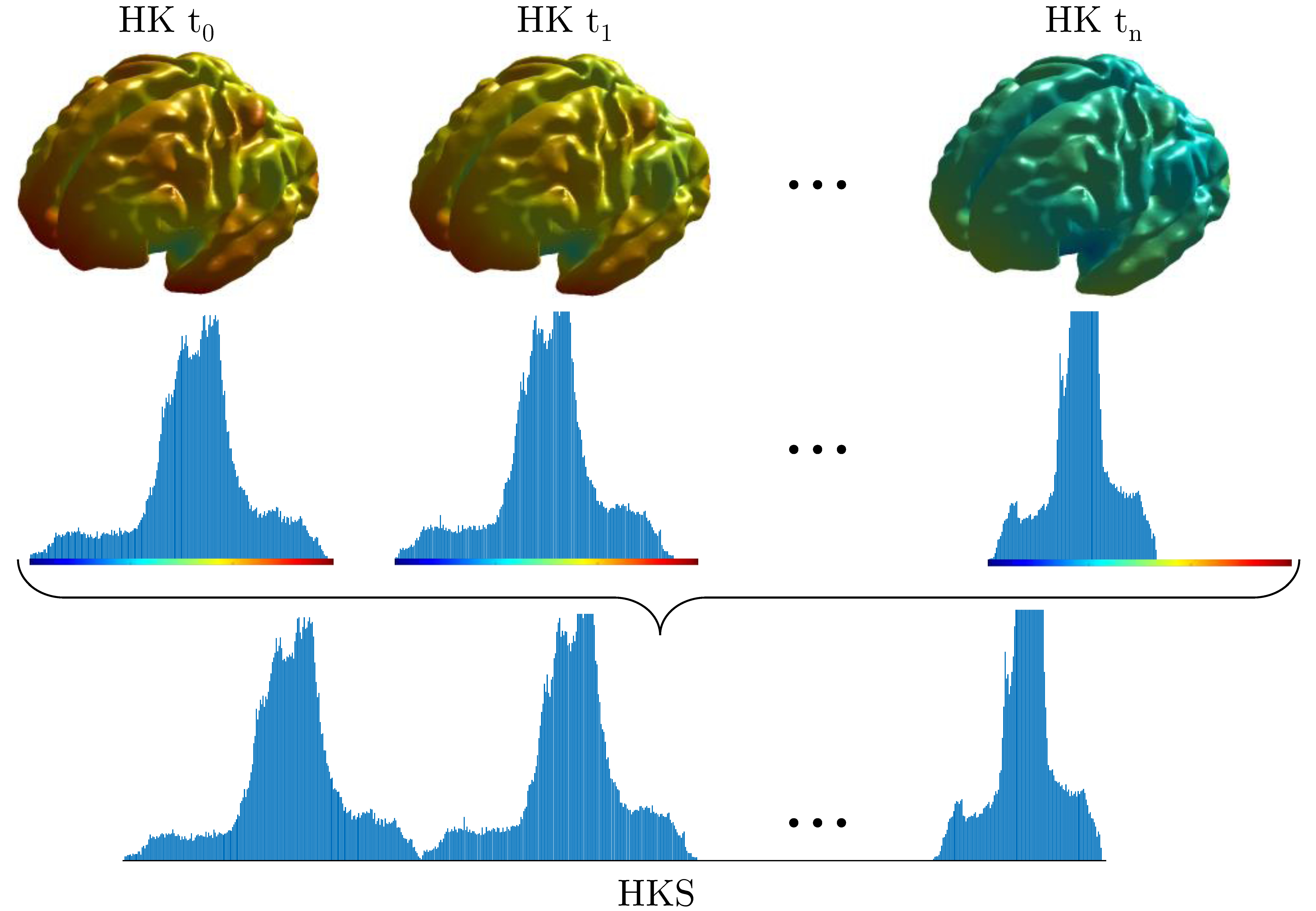
Consequently, we partitioned into 100 bins the values of the vertices obtained for each signature, and we normalized the derived distribution for the number of associated vertices. Finally, we concatenated the obtained distributions for each time point to derive its global signature11 (Figure 2).
Support Vector Regression:
The shape metrics extracted by each descriptor are normalized via z-score and evaluated using a linear kernel SVR algorithm to predict the GA. Notably, we trained the models on the metrics extracted by descriptors from the atlas’ fetuses3,4 and tested them on the metrics extracted from the healthy fetuses included in the FeTA dataset5. We evaluated the goodness-of-fit of the SVR models by measuring the mean absolute error (MAE), the root mean square error (RMSE), and the coefficient of determination (R2). The concordance between predicted and true GAs was determined using Lin’s concordance correlation coefficient, with strength of agreement assessed by McBride’s criteria (poor <0.90; moderate 0.90–0.95; substantial 0.95–0.99; almost perfect >0.99)12,13.
Results
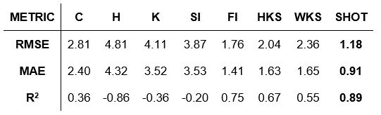
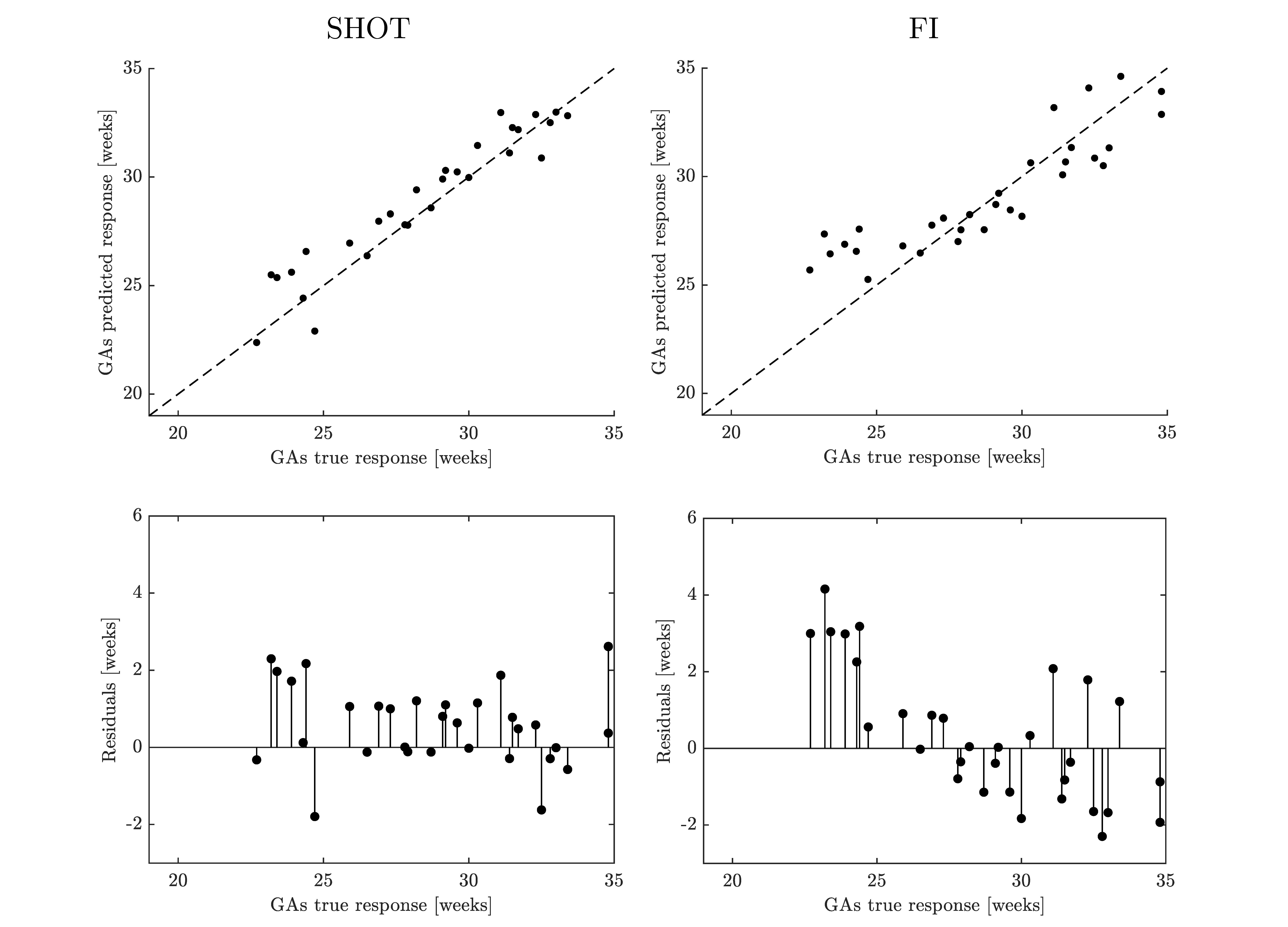
Table 2 shows the performance of the considered individual SVR models in the prediction of GA for neurotypical fetuses. SHOT outperforms the other descriptors, achieving a prediction R2 of 0.89 and a corresponding MAE of 6.3 days (substantial concordance agreement, ρc=0.95, 95% CI 0.89–0.97). FI, which represents the best curvature-based descriptor, achieved a prediction R2 of 0.75 and MAE of 9.9 days (poor concordance agreement, ρc=0.84, 95% CI 0.72–0.91).The GA prediction obtained with the best global multidimensional point-wise shape signature (SHOT) and scalar point-wise curvature-based metric (FI) are visualized in the true versus predicted response plot, and their prediction model is evaluated using the residual plot (Figure 3).
Discussion and Conclusion
Several studies have been proposed to analyze the fetal brain gyrification process through the extraction of cortical surface morphometric properties14-17. In this context, curvature-based measures represent the gold standard for assessing neurodevelopment18-22. Here, we introduced to the fetal context novel multidimensional point-wise shape signatures (HKS, WKS, SHOT) to analyze the inner cortical surface development in the fetal brain. These descriptors have rarely been applied in the medical field due to the intricate nature of medical data, the heavy workload and expertise required from operators, and the need for thorough validation and approval processes to comply with regulatory standards11,23. The results obtained indicate that SHOT outperforms all other descriptors investigated, better capturing the morphologic changes that occur during fetal neurodevelopment with an estimated error of less than a week. Figure 3 shows that this error remains consistent along the GAs.SHOT provides researchers with more sophisticated tool to capture more nuanced aspects of shapes. Thus, a novel exploration of fetal brain morphology based on multidimensional point-wise shape signatures can potentially uncover new insight into the structure and shape of the brain.
Acknowledgements
This work was supported by Italian Ministry of Health, “Ricerca Corrente 2023” funds, grant #RF-2019-12371349. MUR also supported this work by grant “Dipartimenti di Eccellenza 2023-2027” to the Department of Informatics, Systems and Communication of the University of Milano-Bicocca, Italy. We gratefully acknowledge the support of NVIDIA Corporation with the RTX A5000 GPUs granted through the Academic Hardware Grant Program to the University of Milano-Bicocca for the project "Learned representations for implicit binary operations on real-world 2D-3D data".
References
Habas, P. A., Scott, J. A., Roosta, A., Rajagopalan, V., Kim, K., Rousseau, F., Barkovich, A. J., Glenn, O. A., & Studholme, C. (2012). Early folding patterns and asymmetries of the normal human brain detected from in utero MRI. Cerebral cortex (New York, N.Y. : 1991), 22(1), 13–25. https://doi.org/10.1093/cercor/bhr053
Tarui, T., Im, K., Madan, N., Madankumar, R., Skotko, B. G., Schwartz, A., Sharr, C., Ralston, S. J., Kitano, R., Akiyama, S., Yun, H. J., Grant, E., & Bianchi, D. W. (2020). Quantitative MRI Analyses of Regional Brain Growth in Living Fetuses with Down Syndrome. Cerebral cortex (New York, N.Y. : 1991), 30(1), 382–390. https://doi.org/10.1093/cercor/bhz094
Gholipour, A., Rollins, C. K., Velasco-Annis, C., Ouaalam, A., Akhondi-Asl, A., Afacan, O., Ortinau, C. M., Clancy, S., Limperopoulos, C., Yang, E., Estroff, J. A., & Warfield, S. K. (2017). A normative spatiotemporal MRI atlas of the fetal brain for automatic segmentation and analysis of early brain growth. Scientific reports, 7(1), 476. https://doi.org/10.1038/s41598-017-00525-w
Uus, A., Kyriakopoulou, V., Cordero Grande, L., Christiaens, D., Pietsch, M., Price, A., Wilson, S., Patkee, P., Karolis, S., Schuh, A., Gartner, A., Williams, L., Hughes, E., Arichi, T., O'Muircheartaigh, J., Hutter, J., Robinson, E., Tournier, JD., Rueckert, D., Counsell, S., Rutherford, M., Deprez, M., Hajnal, JV., Edwards, AD. (2023) Multi-channel spatio-temporal MRI atlas of the normal fetal brain development from the developing Human Connectome Project. G-Node. https://doi.org/10.12751/g-node.ysgsy1
Payette, K., Li, H. B., de Dumast, P., Licandro, R., Ji, H., Siddiquee, M. M. R., Xu, D., Myronenko, A., Liu, H., Pei, Y., Wang, L., Peng, Y., Xie, J., Zhang, H., Dong, G., Fu, H., Wang, G., Rieu, Z., Kim, D., Kim, H. G., … Jakab, A. (2023). Fetal brain tissue annotation and segmentation challenge results. Medical image analysis, 88, 102833. Advance online publication. https://doi.org/10.1016/j.media.2023.102833
Sun, J., Ovsjanikov, M., & Guibas, L. (2009). A concise and provably informative multi‐scale signature based on heat diffusion. In Computer graphics forum (Vol. 28, No. 5, pp. 1383-1392). Oxford, UK: Blackwell Publishing Ltd.
Aubry, M., Schlickewei, U., & Cremers, D. (2011). The wave kernel signature: A quantum mechanical approach to shape analysis. In 2011 IEEE International Conference on Computer Vision Workshops (ICCV workshops) (pp. 1626-1633). IEEE.
Salti, S., Tombari, F., & Di Stefano, L. (2014). SHOT: Unique signatures of histograms for surface and texture description. Computer Vision and Image Understanding, 125, 251-264.
Pienaar, R., Fischl, B., Caviness, V., Makris, N., & Grant, P. E. (2008). A METHODOLOGY FOR ANALYZING CURVATURE IN THE DEVELOPING BRAIN FROM PRETERM TO ADULT. International journal of imaging systems and technology, 18(1), 42–68. https://doi.org/10.1002/ima.v18:1
Rodriguez-Carranza, C. E., Mukherjee, P., Vigneron, D., Barkovich, J., & Studholme, C. (2008). A framework for in vivo quantification of regional brain folding in premature neonates. NeuroImage, 41(2), 462–478. https://doi.org/10.1016/j.neuroimage.2008.01.008
Castellani, U., Mirtuono, P., Murino, V., Bellani, M., Rambaldelli, G., Tansella, M., & Brambilla, P. (2011). A new shape diffusion descriptor for brain classification. Medical image computing and computer-assisted intervention : MICCAI ... International Conference on Medical Image Computing and Computer-Assisted Intervention, 14(Pt 2), 426–433. https://doi.org/10.1007/978-3-642-23629-7_52
Lawrence, I., & Lin, K. (1989). A concordance correlation coefficient to evaluate reproducibility. Biometrics, 255-268. https://doi.org/10.2307/2532051
McBride, G. B. (2005). A proposal for strength-of-agreement criteria for Lin’s concordance correlation coefficient. NIWA client report: HAM2005-062, 45, 307-310.
Benkarim, O. M., Hahner, N., Piella, G., Gratacos, E., González Ballester, M. A., Eixarch, E., & Sanroma, G. (2018). Cortical folding alterations in fetuses with isolated non-severe ventriculomegaly. NeuroImage. Clinical, 18, 103–114. https://doi.org/10.1016/j.nicl.2018.01.006
Tarui, T., Madan, N., Farhat, N., Kitano, R., Ceren Tanritanir, A., Graham, G., Gagoski, B., Craig, A., Rollins, C. K., Ortinau, C., Iyer, V., Pienaar, R., Bianchi, D. W., Grant, P. E., & Im, K. (2018). Disorganized Patterns of Sulcal Position in Fetal Brains with Agenesis of Corpus Callosum. Cerebral cortex (New York, N.Y. : 1991), 28(9), 3192–3203. https://doi.org/10.1093/cercor/bhx191
Tarui, T., Madan, N., Graham, G., Kitano, R., Akiyama, S., Takeoka, E., Reid, S., Yun, H. J., Craig, A., Samura, O., Grant, E., & Im, K. (2023). Comprehensive quantitative analyses of fetal magnetic resonance imaging in isolated cerebral ventriculomegaly. NeuroImage. Clinical, 37, 103357. https://doi.org/10.1016/j.nicl.2023.103357
Demirci, N., & Holland, M. A. (2022). Cortical thickness systematically varies with curvature and depth in healthy human brains. Human brain mapping, 43(6), 2064–2084. https://doi.org/10.1002/hbm.25776
Batchelor, P. G., Castellano Smith, A. D., Hill, D. L., Hawkes, D. J., Cox, T. C., & Dean, A. F. (2002). Measures of folding applied to the development of the human fetal brain. IEEE transactions on medical imaging, 21(8), 953–965. https://doi.org/10.1109/TMI.2002.803108
Clouchoux, C., Kudelski, D., Gholipour, A., Warfield, S. K., Viseur, S., Bouyssi-Kobar, M., Mari, J. L., Evans, A. C., du Plessis, A. J., & Limperopoulos, C. (2012). Quantitative in vivo MRI measurement of cortical development in the fetus. Brain structure & function, 217(1), 127–139. https://doi.org/10.1007/s00429-011-0325-x
Hu, H. H., Chen, H. Y., Hung, C. I., Guo, W. Y., & Wu, Y. T. (2013). Shape and curvedness analysis of brain morphology using human fetal magnetic resonance images in utero. Brain structure & function, 218(6), 1451–1462. https://doi.org/10.1007/s00429-012-0469-3
Shimony, J. S., Smyser, C. D., Wideman, G., Alexopoulos, D., Hill, J., Harwell, J., Dierker, D., Van Essen, D. C., Inder, T. E., & Neil, J. J. (2016). Comparison of cortical folding measures for evaluation of developing human brain. NeuroImage, 125, 780–790. https://doi.org/10.1016/j.neuroimage.2015.11.001
Wu, J., Awate, S. P., Licht, D. J., Clouchoux, C., du Plessis, A. J., Avants, B. B., Vossough, A., Gee, J. C., & Limperopoulos, C. (2015). Assessment of MRI-Based Automated Fetal Cerebral Cortical Folding Measures in Prediction of Gestational Age in the Third Trimester. AJNR. American journal of neuroradiology, 36(7), 1369–1374. https://doi.org/10.3174/ajnr.A4357
Dahdouh, S., & Limperopoulos, C. (2016). Unsupervised fetal cortical surface parcellation. Proceedings of SPIE--the International Society for Optical Engineering, 9784, 97840J. https://doi.org/10.1117/12.2212805
Figures