2373
Sex difference in brain white matter and relevant neurobehavioral development: preterm vs. term neonates1Department of Radiology, The First Affiliated Hospital of Xi'an Jiaotong University, Xi'an, China, xi'an, China, 2Department of Radiology, The First Affiliated Hospital of Xi'an Jiaotong University, Xi'an, China, Xi'an, China, 3Department of Radiology, the First Affiliated Hospital of Xi’an Jiaotong University, Xi’an, China
Synopsis
Keywords: Neuro, Brain
Motivation: Preterm male present high risk for neurodevelopmental disability. However, sex differences in preterm neurodevelopmental characterization is not yet known.
Goal(s): Investigate sex differences in neurodevelopmental evolution of preterm by exploring the postnatal age-related changes of brain white matter (WM) and neurobehavioral abilities.
Approach: The diffusion tensor imaging (DTI) parameter FA was applied to indirectly reflect the changes of brain white matter microstructure.
Results: The FA in preterm neonates is lower than term neonates which increased with postnatal age in both. Male presented significantly higher growth rate in certain brain regions compared with female especially for preterm male.
Impact: Preterm male showed higher growth rate in certain brain regions which indicate a “catch-up” pattern toward the term neonates and this finding may suggest that certain intervention would be expected to work well in improving the preterm male outcome.
abstract
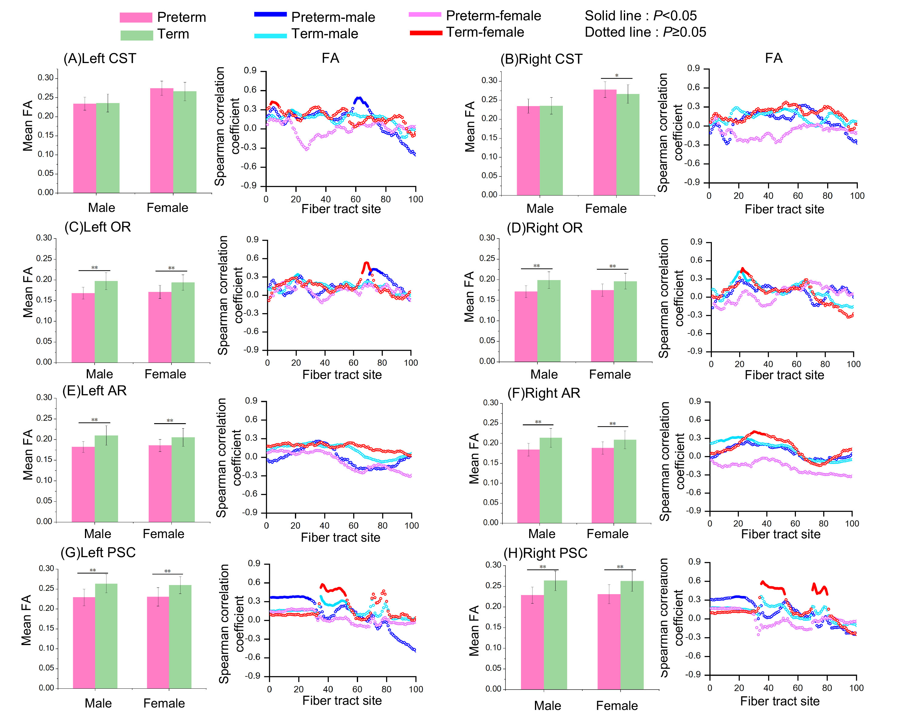
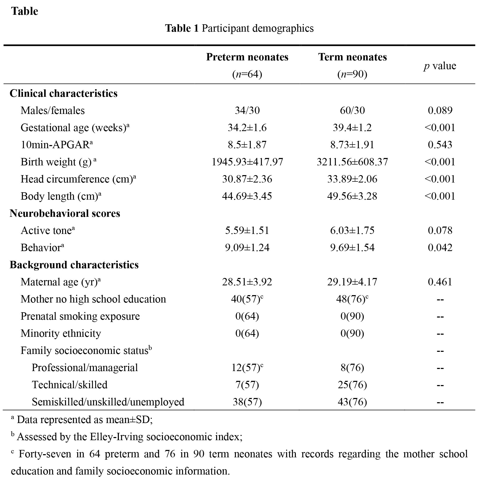
Introduction:Preterm male present high risk for neonatal mortality and morbidity, subsequent neurodevelopmental disorders is well known[1,2]. Many neuroimaging studies have revealed sex differences in the brain structure of premature infants such as brain tissue volume differences and white matter structure alterations[3-5]. Human brain initiates a rapid growth trajectory following birth which is crucial for the brain anatomical remodeling and improving neurobehavioral outcomes[6,7]. However, sex differences in preterm neurodevelopmental characterization is not yet known. Diffusion tensor imaging (DTI) allows quantitative characterization of in-vivo water diffusion pattern to reveal the brain development, particularly, DTI-derived fractal anisotropy (FA) could reflect the myelination and myelination processes[8]. This study aims to elaborate the sex difference in early evolution of preterm sensorimotor function by exploring the postnatal age-related changes of brain white matter (WM) microstructure and neurobehavioral abilities during the neonatal stage.Methods: 284 neonates (GA range, 30-42 weeks) who underwent MRI examination between October 2014 and June 2015 were included in this study. DTI were acquired with 35 directions using the following parameters: b value =1000 s/mm2; TR, 5500ms; TE, 95ms; section thickness, 4mm; field of view, 180mm. Neonatal behavioral neurological assessment (Chinese)[9] and Dubowitz neurological assessment[10] was applied to assessment the neonatal neurobehavioral development within 5 days before or after MRI exam. All DTI images were processed using Oxford Centre for Functional Magnetic Resonance Imaging of the Brain Software Library (FSL) version 5.0. Four WMs were selected as regions of interest, i.e. OR (optical radiation), AR (auditory radiation), CST (corticospinal tract), and thal-PSC (thalamus-primary somatosensory cortex). Scatterplots with linear fitting was respectively used to detail the between FA, neurobehavioral scores and postnatal age.Results: Of 284 neonates recruited, 154 neonates met the inclusion criteria and were included. There were 64 preterm (male/female, 34/30) and 90 term (male/female, 60/30) (Fig.1 and Table 1).Scatterplots between postnatal age and brain WM FA (Fig.2). For bilateral OR, AR, and thal-PSC, term neonates showed higher Pearson correlation between adjusted FA values and postnatal age than preterm neonates both in male and female. While for left CST, preterm female showed higher Pearson correlation between adjusted FA values and postnatal age than term. However, Premature male showed higher Pearson correlation in the posterior segment of left CST, the posterior segment of left OR, and the proximal segment of bilateral PSC, respectively. Term male showed higher Pearson correlation in the proximal segment of right AR. Scatterplots between postnatal age and neurobehavioral scores For active tone, and behavior female showed higher linear relation between adjusted active tone scores and postnatal age than male both in term and preterm. But both preterm and term male showed a significant increase trend for behavior, while the change was not significant in female. (Fig.3).
In this study, the results showed the mean FA of OR, AR and thal-PSC in preterm was lower than term both in male and female. Previous research have shown that compared with term neonates, the FA values of many white matter are significantly reduced in preterm and term-equivalent infants. Moreover, there also exist abnormalities in white matter volume and microstructure, such as OR[11]. However, The FA of CST, OR, AR and PSC in both preterm and full-term neonates increased with postnatal age. Consistently, Rose, S. E et al[12][i] has demonstrated that along with the gradual development of neonatal brain, the FA value of neonatal white matter increased with age after birth. Notably, we observed that males presented significantly higher growth rate in certain brain regions compared with females especially for preterm males. The analysis of the correlation of postnatal age and FA with behavior also showed a significant increase trend for behavior in both preterm and term male. Despite the increased vulnerability of male is well known, the above results indicate that the preterm male show a trend of catching up with more mature full-term neonates in the neonatal period and this finding may suggest that certain intervention would be expected to work well in improving the preterm male outcome in the newborn period.Conclusion:Our study indicated that although being delayed, preterm male show a “catch-up” pattern toward the term neonates in motor development during the neonatal period.
Acknowledgements
This work was supported by the National Natural Science Foundation of China (82271517).References
1. Drevenstedt GL, Crimmins EM, Vasunilashorn S, Finch CE. The rise and fall of excess male infant mortality. Proc Natl Acad Sci U S A 2008;105:5016-21.2. Naeye, R. L., Burt, L. S., Wright, D. L., Blanc, W. A. & Tatter, D. Neonatal mortality, the male disadvantage. Pediatrics 48, 902–906 (1971).3. Ball G , Aljabar P , Nongena P ,et al.Multimodal image analysis of clinical influences on preterm brain development[J].Annals of Neurology, 2017, 82(2).DOI:10.1002/ana.24995.4.Constable R T , Ment L R , Vohr B R ,et al.Prematurely born children demonstrate white matter microstructural differences at 12 years of age, relative to term control subjects: an investigation of group and gender effects.[J].Pediatrics, 2008, 121(2):306-316.5. C D K T A B , A C E K , D J C A ,et al.Early life predictors of brain development at term-equivalent age in infants born across the gestational age spectrum.[J].Neuroimage, 2018, 185:813-824.DOI:10.1016/j.neuroimage.2018.04.031.6. Kolb, B. & Gibb, R. Brain plasticity and behaviour in the developing brain. J Can Acad Child Adolesc Psychiatry 20, 265-276 (2011).7. Bach-y-Rita, P. Brain plasticity as a basis for recovery of function in humans. Neuropsychologia 28, 547-554 (1990).8. Qiu, A., Mori, S. & Miller, M.I. Diffusion tensor imaging for understanding brain development in early life. Annu Rev Psychol 66, 853-876 (2015).9. Bao, X.L., Yu, R.J., Li, Z.S. & Zhang, B.L. Twenty-item behavioral neurological assessment for normal newborns in 12 cities of China. Chin Med J (Engl) 104, 742-746 (1991)10. Dubowitz, L.M.S., Dubowitz, V., Palmer, P. & Verghote, M. A new approach to the neurological assessment of the preterm and full-term newborn infant. Brain and Development 2, 3-14 (1980).11. Chang, L., et al. Delayed early developmental trajectories of white matter tracts of functional pathways in preterm-born infants: Longitudinal diffusion tensor imaging data. Data Brief 6, 1007-1015 (2016).12. Rose, S.E., et al. Altered white matter diffusion anisotropy in normal and preterm infants at term-equivalent age. Magnetic Resonance in Medicine 60, 761-767 (2008).Figures
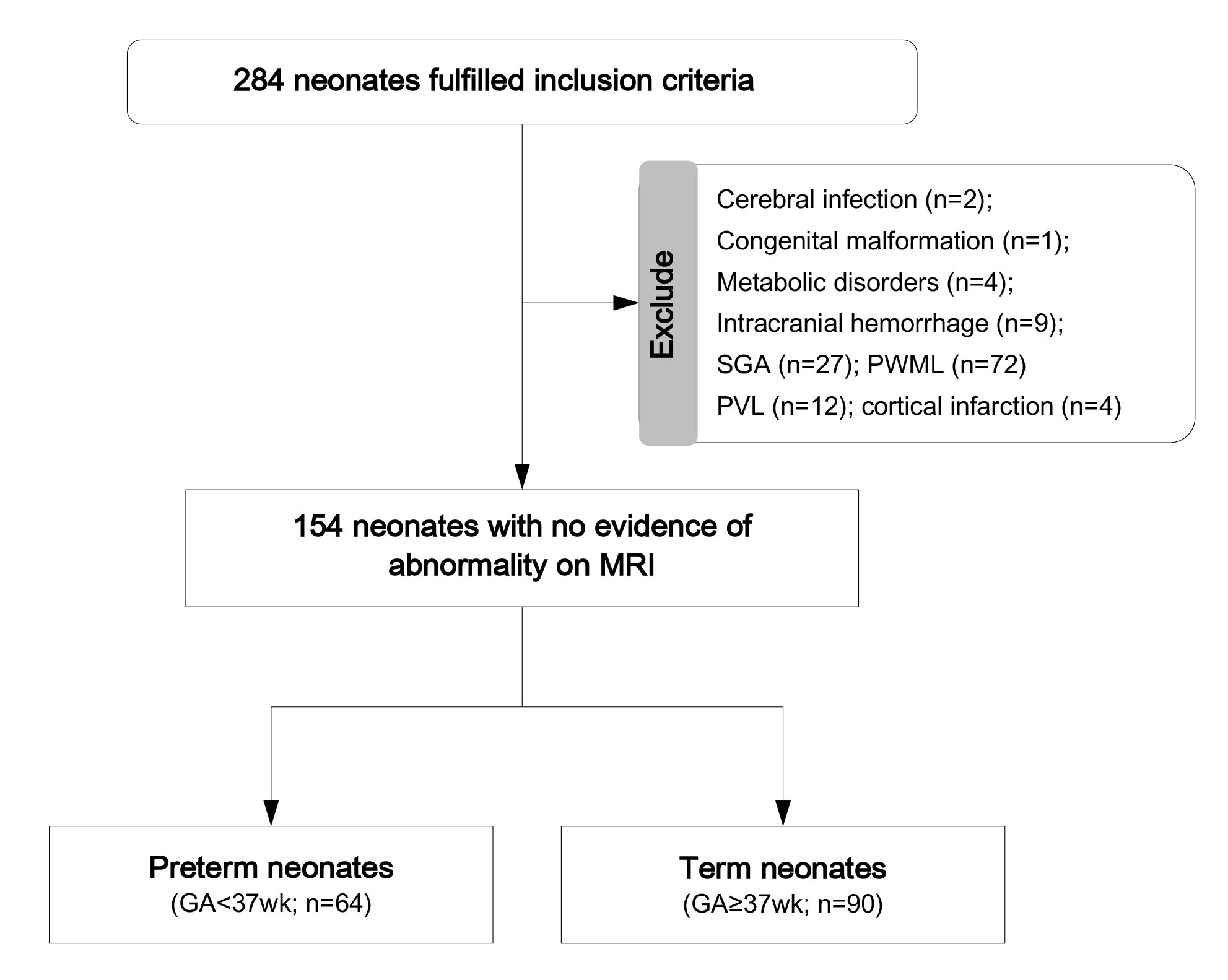
Fig.1 Flow chart for determining the research subject based on the inclusion and exclusion criteria