2361
Early diagnosis and prognosis of full-term neonatal hypoxic-ischemic encephalopathy using 3D arterial spin labeling perfusion imaging1Radiology, The First Affiliated Hospital of USTC, Hefei, China, 2GE Healthcare, Shanghai, China
Synopsis
Keywords: Neonatal, Pediatric
Motivation: Hypoxic ischemic encephalopathy (HIE) is the most important reason for morbidity and mortality in full-term infants.
Goal(s): This study aimed to investigate the clinical value of 3DASL perfusion imaging for the early diagnosis and prognosis of HIE in full-term neonates.
Approach: This study recruited 40 full-term HIE neonates and 40 healthy controls. Cerebral blood flow (CBF) values of bilateral frontal lobes, basal ganglia, thalamus were measured for comparison.
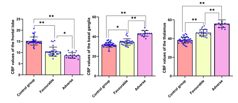
Results: The HIE groups showed significant decreased CBF values in the frontal lobes but significantly increased CBF values in basal ganglia and thalamus as compared to the control group.
Impact: We observed significant group differences between HIE neonates and healthy controls. 3DASL could reflect abnormal changes of cerebral perfusion in HIE infants and demonstrated the clinical value for early diagnosis and diagnosis of HIE.
Introduction
Hypoxic ischemic encephalopathy (HIE) is the most important reason for morbidity and mortality in full-term infants [1]. Understanding pathophysiology of the brain damage is essential for the early detection of HIE and development of strategies for their treatments. A novel quantitative cerebral perfusion imaging technique 3D arterial spin labeling (3DASL) has been widely used, which is especially suitable for pediatric imaging without the involvement of contrast agents [2]. This study aimed to investigate the clinical value of 3DASL technique for the early diagnosis and prognosis of HIE in full-term neonates.Methods
Our Institutional Review Board approved the study and written informed consent was obtained from all the participants’ parents. We retrospectively enrolled 40 full-term neonates clinically diagnosed with HIE in our hospital from January 2020 to September 2023. All patients were divided into favorable (26 patients) and adverse (14 patients) outcome groups. Another 40 healthy controls were recruited for comparison. Neonatal cerebral MRI was performed in natural sleep on a 3-T scanner (Discovery MR750w, GE Healthcare, WI) with an eight-channel head coil. We used the feed and bundle method to acquire MR imaging without sedation. The axial 3DASL scan was performed with the following parameters: TR/TE=4562/11ms, voxel size=0.8 × 0.8 × 3 mm3, post-labeling delay=1025ms, labeling duration=1450ms. Cerebral blood flow (CBF) was calculated using the vender provided workstation. An experienced radiologist manually delineated the regions of interest (ROI) including bilateral frontal lobes, basal ganglia and thalamus. Statistical analysis was conduct using SPSS. One-way ANOVA was performed to evaluate the group differences and post-hoc analysis was conducted to compare each pair of three groups. The group differences were considered significant when P < 0.05.Results
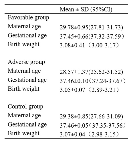
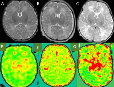
The demographic data showed no significant differences among three groups (Table 1). Figure 1 shows the T2-weighted images and CBF maps of a representative healthy control, two HIE patients with the favorable and adverse outcomes respectively. The HIE group with the adverse outcomes showed significant decreased CBF values in the frontal lobes as compared to the HIE group with favorable outcomes, which in turn showed significant deceased CBF values relative to the control group. The CBF values in the basal ganglia and thalamus showed the opposite pattern (Figure 2).Discussion and Conclusion
We observed significant group differences between HIE neonates and healthy controls. 3DASL could reflect abnormal changes of cerebral perfusion in HIE infants and demonstrated the clinical value for early diagnosis and diagnosis of HIE.Acknowledgements
No acknowledgement found.References
1. Yıldız EP, Ekici B, Tatlı B. Neonatal hypoxic ischemic encephalopathy: an update on disease pathogenesis and treatment. Expert Rev Neurother. 2017;17(5):449-459.
2. Jill B, Hendrikse J, Petersen ET, et al. Arterial spin-labelling perfusion MRI and outcome in neonates with hypoxic-ischemic encephalopathy. European Radiology. 2015:25(1): 113-121.
Figures