2355
Evaluation of the changes of GABA and T2 mapping in patients with post-stroke cognitive impairment1Department of Radiology, Hefei First People’s Hospital, Heifei, China, 2Philips Healthcare, Shanghai, China
Synopsis
Keywords: Stroke, Aging
Motivation: Exploring alterations in brain microstructure and neurotransmitter content in post-stroke cognitive impairment (PSCI).
Goal(s): To investigate the influence of T2 mapping and MEGA-PRESS on brain microstructure and neurotransmitter content in PSCI individuals.
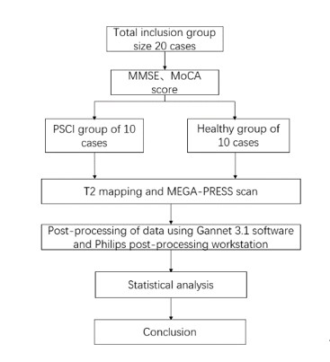
Approach: MRI scans were performed on 14 patients from the PSCI group and 11 healthy volunteers. GABA and Glx spectra were gathered, followed by T2 mapping.
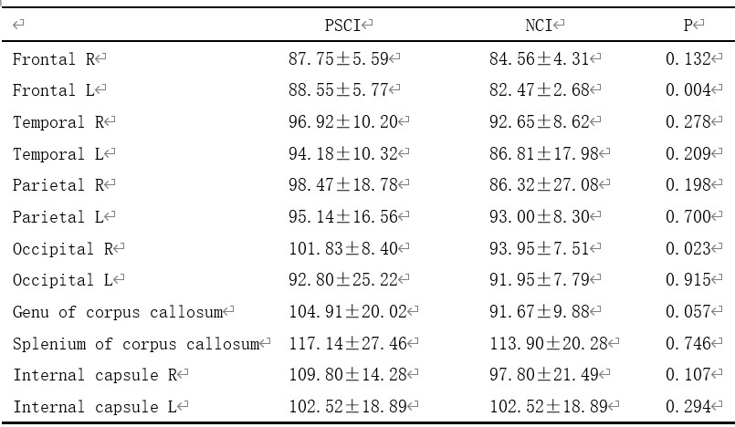
Results: The study found lower GABA+/Water ratios in the prefrontal cortex of the PSCI group than the healthy group. Additionally, the PSCI group showed significantly higher T2 values in the right frontal and left occipital lobes.
Impact: This study confirmed that the combined measurement of brain GABA content and relaxation using MEGA-PRESS and T2 mapping sequence has clinical value. This provides a clinical idea for exploring the occurrence and development of cognitive disorders such as PSCI.
Introduction
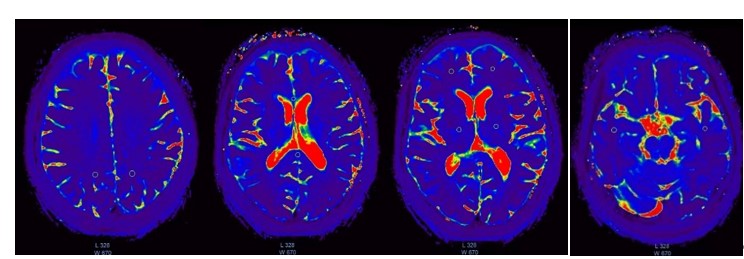
Cognitive impairment is a prevalent and debilitating outcome following an ischemic stroke[1]. The primary function of the prefrontal cortex (PFC) is cognitive function, which orchestrates psychological processes and actions in line with present objectives and future plans[2-3]. Research indicates that ischemia in the sub-frontal cortex is a critical factor in stroke damage[4]. The T2-mapping technique can identify the condition of diseased tissue at the microstructure level, reflecting the content and environment of water, protein, and other solutes in the tissue. It can also quantitatively analyze the tissue characteristics of organisms by measuring the transverse relaxation time (T2 value) of voxels, demonstrating high stability and repeatability[5-6]. The Mecher-Garwood dot discrimination spectral sequence (MEMA-PRESS) technique has been confirmed to detect the concentration of GABA, the primary inhibitory neurotransmitter. Abnormalities in GABA are implicated in a range of neuropsychiatric disorders[7-8]. This study aims to investigate the alterations in brain microstructure and neurotransmitters in cognitive impairment following cerebral infarction, using a combination of T2 mapping and MEGA-PRESS sequences.Methods
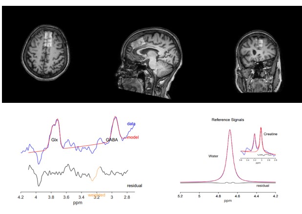
The scanning sequence included MEGA-PRESS and T2 mapping. MR scans were conducted on a 3.0T scanner (Ingenia Elition, Philips Healthcare) with a 32-channel head coil. The parameters for MEGA-PRESS were as follows: TR/TE 2000/68 ms, voxel size 2×4×2 cm3, approximately 10 minutes per acquisition. The regions of interest (ROIs) for MEGA-PRESS were set in the prefrontal cortex (PFC). The GABA and Glx signals in PSCI and NCI were quantified using the Matlab-based analysis toolkit Gannet 3.1 with the water signal as an internal reference (Figure 2). The GABA signal detected by MEGA-PRESS also includes co-edited signal from macromolecules and homocarnosine, hence it is referred to as GABA+ below. Only spectra with a relative fitting error (Fit-Error) of GABA+/Water and Glx/Water generated by Gannet smaller than 15% were included in the final statistical analysis. Differences in GABA+ and Glx levels between the two groups were analyzed using a T test. Fourteen brain regions of interest (bilateral frontal lobe, bilateral parietal lobe, bilateral occipital lobe, bilateral temporal lobe, knee part of corpus callosum, compression part of corpus callosum, posterior limb of internal capsule) were studied on mapping images, and the GABA data of PFC were processed by the Gannet3.1 program.Results
The research undertaken encompassed a total of 25 participants, comprising of 14 patients diagnosed with Post-Stroke Cognitive Impairment (PSCI) and 11 healthy control subjects. An in-depth analysis of the demographic and clinical data, including variables such as age and gender, showed no statistically significant discrepancies between the two cohorts, thereby ensuring a balanced and comparable study sample. Subsequent to rigorous statistical evaluation, a noteworthy finding was the lower Gamma-Aminobutyric Acid to Creatine (GABA+/Cr) ratio in the Prefrontal Cortex (PFC) of the PSCI group as opposed to the control group. Furthermore, the study also revealed a marked increase in the relaxation time within the right frontal lobe and left occipital lobe in the PSCI group when juxtaposed with the control group. This suggests a potential disruption in the neurophysiological functioning of these regions, which could be related to the cognitive deficits observed in PSCI patients.Discussions
PFC is a vital component in the orchestration of cognitive functions, serving as a hub for complex cognitive processes and executive functions. Predominantly composed of pyramidal efferent neurons and GABA intermediate neurons, the PFC plays a pivotal role in the modulation and management of cognitive operations. The brain's neurometabolite concentration, particularly the inhibitory GABA concentration, is subject to variability contingent on the severity and level of brain injury. A plethora of research studies have underscored the diminished activity of GABA in the brains of patients suffering from cognitive impairment, corroborating the findings of our research. Furthermore, the elongation of T2 relaxation time in the frontal lobe, as observed in various studies, could be indicative of the progression of cognitive impairment. This phenomenon may be attributable to alterations in the neurotransmitter concentration within the PFC, suggesting that damage to GABA neurons could precipitate changes in the water and protein content of the PFC. This line of inquiry points to a potential avenue for the development of therapeutic interventions targeting the restoration of GABA function to mitigate cognitive impairment.Conclusion
The results indicate that alterations in GABA levels and T2 relaxation time in the frontal pole cortex (FPC) of individuals with post-stroke cognitive impairment (PSCI) may be associated with cognitive dysfunction. This discovery could offer a fresh neuroanatomical foundation for understanding the mechanism of cognitive decline in patients with cognitive impairment and potentially serve as a theoretical basis for the treatment of PSCI patients.Acknowledgements
No acknowledgement found.References
[1].Ohlmeier, L., et al., Prevalence of, and risk factors for, cognitive impairment in lacunar stroke. International journal of stroke, 2023. 18(1): p. 62-69.
[2].Tang, W.K., et al., Association of Frontal Subcortical Circuits Infarcts in Poststroke Depression: A Magnetic Resonance Imaging Study of 591 Chinese Patients With Ischemic Stroke. Journal of Geriatric Psychiatry and Neurology, 2011. 24(1): p.44-49.
[3].Boto, J., et al., Cerebral Gray and White Matter Involvement in Anorexia Nervosa Evaluated by T1, T2, and T2 Mapping. Journal of neuroimaging, 2019. 29(5): p.598-604.
[4].Yan, H., et al., Assessment of cognitive impairment after acute cerebral infarction with T1 relaxation time measured by MP2RAGE sequence and cerebral hemodynamic by transcranial Doppler. Frontiers in Neurology, 2022.13(6): p1056423-1056438.
[5].Baeshen, A., et al., Test–Retest Reliability of the Brain Metabolites GABA and Glx With JPRESS, PRESS, and MEGA‐PRESS MRS Sequences in vivo at 3T. Journal of magnetic resonance imaging, 2020.51(4): p.1181-1191.
[6].Menon, V. and M. D'Esposito, The role of PFC networks in cognitive control and executive function. Neuropsychopharmacology (New York, N.Y.), 2022.47(1): p. 90-103.
[7].Wang, S., et al., The resting-state topological organization damage of language-related brain regions in post-stroke cognitive impairment. Brain Imaging and Behavior, 2022.16(6): p.2608-2617.
[8].Sood, A., et al., Glia: A major player in glutamate–GABA dysregulation‐mediated neurodegeneration. Journal of Neuroscience Research, 2021. 99(12): p. 3148-3189.
Figures