2351
Exploration of brain GABA levels alterations in patient with neurologic Wilson disease1Department of Neurology, Guizhou Provincial People's Hospital, Guiyang, China, 2Department of Radiology, Guizhou Provincial People's Hospital, Guiyang, China, 3Philips Healthcare, Beijing, China, 4Philips Healthcare, Chengdu, China
Synopsis
Keywords: Neurotransmission, Spectroscopy, GABA, Wilson disease, Copper Metabolism
Motivation: To better understand the effect of abnormal copper metabolism on the brain GABA level in patients with neurologic Wilson disease.
Goal(s): To explore brain GABA level alterations in patients with neurologic Wilson disease compared to healthy controls.
Approach: Five neurologic WD patients and five healthy controls underwent magnetic resonance spectroscopy (MRS) on a 3.0 scanner and GABA signal was acquired from left striatum and left thalamus using MEGA-PRESS.
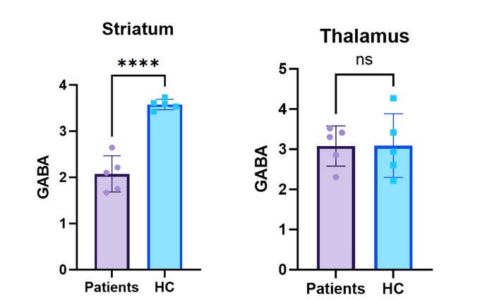
Results: The GABA level in the left striatum was significant reduced while in left thalamus it had no changes.
Impact: This study suggests that the abnormality of GABA energy system may exist in the pathogenesis of neurologic WD, and adjusting neurotransmission of γ-aminobutyric acid may be an important target for neuroprotection in WD.
Introduction
Wilson disease, also known as hepatolenticular degeneration, is a rare autosomal recessive copper metabolism disorder affecting 1 in 30,000-40,000 individuals [1][2]. Under normal physiological conditions, copper reversibly inhibits the response evoked by GABA by binding to GABA receptor protein[3]. When it comes to patients with Wilson disease (WD), the concentration of copper in the brain is 10-15 times higher than that of the normal control group[4][5], the effect of which on the level of GABA has not been reported yet. Therefore, this study was performed in order to explore the changes of GABA levels in the brains of patients with WD.Method
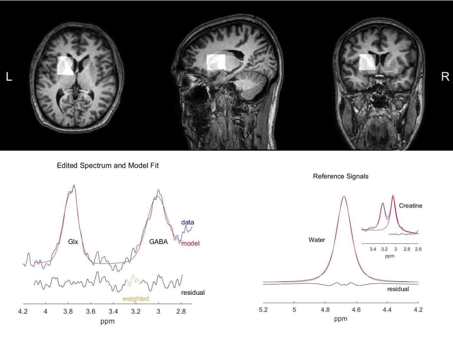
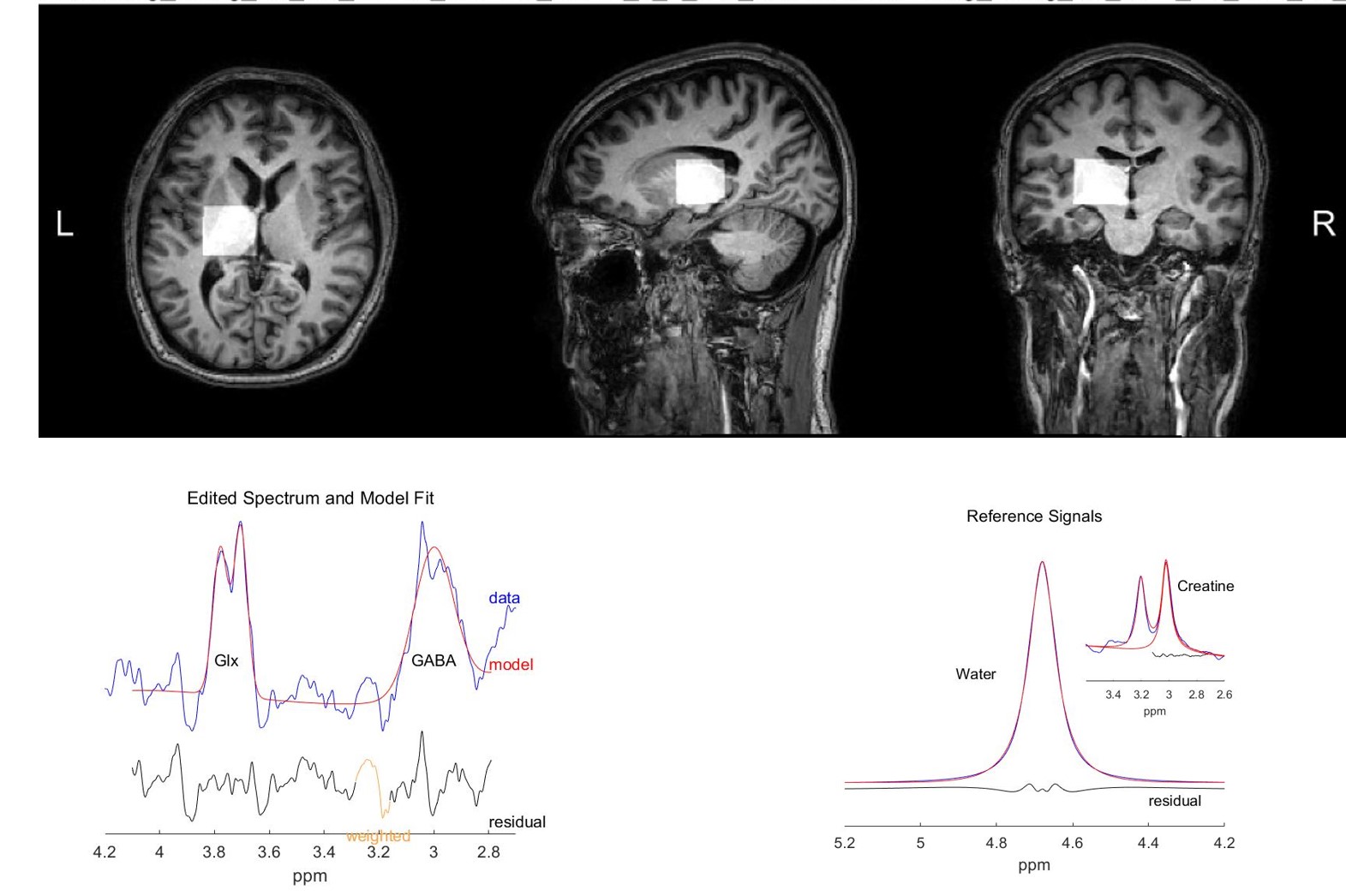
This study recruited a cohort of ten participants, with five individuals diagnosed with Wilson's disease (WD). The control group consisted of five healthy individuals matched for age and gender, each with no history of neurological or psychiatric conditions. All participants were scanned on a 3.0 Tesla scanner (Ingenia Elition, Philips Healthcare, Best, The Netherlands) equipped with a 32-channel head coil. Single-voxel MRS scans were performed using the MEGA-PRESS sequence, with foam padding utilized to minimize head movement to the greatest extent possible. The scanning protocol included the following parameters: TR/TE: 2000/68 ms, VOI (Volume of Interest): 3x3x3 cm^3 for both left striatum and left thalamus, spectral bandwidth = 2000 Hz, Acquisition time: 8 min and 56s per VOI. Before the MRS scan, a high-resolution anatomical image was acquired using a T1-weighted sequence to aid in the placement of the voxel. The GABA+ signal at 3.02 ppm and the Glx signal at 3.75 ppm were fitted using the Gannet 3.0 toolkit within MATLAB. A fitting error of GABA+ and Glx ≤ 15% was considered acceptable for inclusion in the statistical analysis. Levels of GABA+ between patients and healthy controls were analyzed using t-tests (GraphPad Prism 10.0 software).Result
The fitting errors for both GABA+ and Glx were below 15% for all participants. Figure 1 and Figure 2 illustrate typical volumes of interest (VOIs) in the striatum and thalamus, respectively, along with the corresponding spectral data. Figure 3 demonstrates a significant reduction in the levels of GABA+ (referenced to water) in the striatum of WD patients compared to healthy controls (HC), with no significant difference observed in the thalamic region.Discussion
The significance of the striatum in motor control and its dense GABAergic innervation make it a region particularly susceptible to the dysregulation of neurotransmission seen in WD. The reduced GABA+ levels in the striatum observed in this study could be reflective of alterations in both pre- and postsynaptic components of GABAergic neurotransmission as well as a decrease in the density of inhibitory neurons, which may be a consequence of copper toxicity. Copper is known to inhibit the activity of GABA receptors, and such inhibition could exacerbate axonal firing. Interestingly, the lack of significant difference in the thalamic GABA+ levels suggests that copper’s neurotoxic effects may have a region-specific impact on the inhibitory neurotransmission system. Future studies should explore the direct effects of copper chelation therapy on GABA metabolism and whether restoration of GABAergic tone can ameliorate the neurological deficits associated with WD.Conclusion
In conclusion, the observed decline in striatal GABA levels in Wilson's disease patients suggests that the abnormality of the GABA energy system may exist in the pathogenesis of neurologic WD, and adjusting neurotransmission of γ-aminobutyric acid may be an important target for neuroprotection in WD.Acknowledgements
No acknowledgement found.References
[1] Tamas Kozicz et al. Mitochondrial Etiology of Psychiatric Disorders: Is This the Full Story? JAMA Psychiatry, 2018
[2] Rebecca A. Muhle et al. The Emerging Clinical Neuroscience of Autism Spectrum Disorder: A Review. JAMA Psychiatry, 2018
[3] Nadia D'Ambrosi, Luisa Rossi. Copper at synapse: Release, binding and modulation of neurotransmission [J]. Neurochemistry International, 2015, 90(0197-0186): 36-45.
[4] Członkowska A, Litwin T, Dusek P, et al. Wilson disease [J]. Nature reviews Disease primers, 2018, 4(1): 21.
[5] Schilsky ML, Roberts EA, Bronstein JM, et al. A multidisciplinary approach to the diagnosis and management of Wilson disease: 2022 Practice Guidance on Wilson disease from the American Association for the Study of Liver Diseases [J]. Hepatology (Baltimore, Md), 2022.
Figures