2349
Voxel-based morphometry for evaluating nigrostriatal damage in unilateral 6-OHDA-induced Parkinson’s rat model1Department of NMR, All India Institute of Medical Sciences, New Delhi, India, 2Department of Computer Science, University of Delhi, Delhi, India, 3Department of Psychiatry, Yale School of Medicine, New Haven, CT, United States, 4Department of Physiology, All India Institute of Medical Sciences, New Delhi, India
Synopsis
Keywords: Parkinson's Disease, Segmentation
Motivation: Parkinson’s disease (PD) is a progressive neurodegenerative disorder characterized by loss of dopaminergic neurons in the substantia nigra pars compacta resulting in classical motor and nonmotor symptoms.
Goal(s): Our goal was to characterise the gray matter atrophy in 6-OHDA PD model as compared to sham control.
Approach: Structural MRI (3D T1W images) combined with behavioural techniques was used.
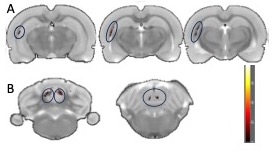
Results: Voxel based morphometry (VBM) revealed reductions of gray matter volume in Fimbria (Ipsi), hippocampal formation (Ipsi) and accumbens nucleus (contra) in 3rd week PD and cerebellum (Ipsi and contra both) in 7th week PD as compared to sham.
Impact: Identification of gray matter atrophy longitudinally helps in understanding the progression of structural alterations in response to dopaminergic cell loss.
Introduction
Parkinson’s disease (PD) is a progressive neurodegenerative disorder characterized by a gradual loss of dopaminergic neurons in the substantia nigra pars compacta1. We investigated the utility of magnetic resonance imaging (MRI) in detecting macroscopic structural brain changes in the unilaterally-lesioned 6-hydroxydopamine (6-OHDA) model.Methods
After the approval of Institute Animal Ethics Committee, adult male Wistar rats (n = 12, weighing 250–300 g) were divided into PD and Sham groups (n=6 in each). Two injections of 2 µL of 6-OHDA solution (7.5 µg/µL 6-OHDA dissolved in 0.2% ascorbate saline) were given in the right hemispheric striatum in the coordinates with reference to the bregma2. The sham-lesioned group went through the same procedure with the injection of saline (instead of 6-OHDA). Necessary postoperative care was provided and all procedures were performed as per the guidelines of the Committee for Control and Supervision of Experiments on Animals (CCSEA).The extent of the 6-OHDA lesioning was confirmed by the apomorphine-induced rotational test. Rats received a subcutaneous injection of apomorphine (0.1 mg/kg in 0.9% saline, E-Biomed GmbH, Heidelberg, Germany). After 5 min, the rat was placed in a hemispherical bowl for 30 minutes and monitored with the help of the tracking software (XT software, EthoVision, The Netherlands). The number of ipsiversive and contraversive full-body turns was recorded automatically over a period of 30 minutes.
High-resolution T1- weighted 3D gradient-echo MRI sequence (RF-spoiled fast low-angle shot (FLASH), repetition time (TR)/ echo time (TE) = 317.89/4.5ms, flip angle = 30°; voxel size = 0.0684 x 0.0684x0.8mm3; number of averages = 3.
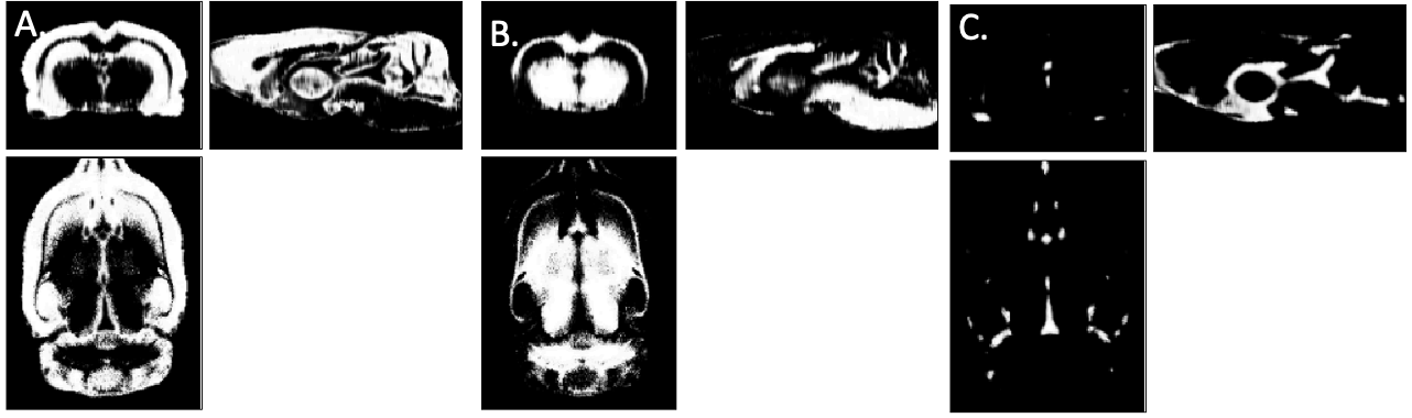
Each image was reconstructed using Bruker’s Paravision software, exported in DICOM format, and converted to NIfTI format, manually rotated to match the standard stereotaxic space in rat3, and resized by a factor of 10 to account for the brain size difference between rodents and humans; further aligned to the stereotaxic space by registering each image to the template image using “Coregister” tool in SPM12 and was resampled into 0.125-mm isotropic voxels and then segmented into probability maps of gray matter (GM), white matter (WM), and cerebrospinal fluid with rat tissue priors using SPM8 (Figure 1). The resulting GM and WM volumetric maps were then used in the second level analysis (paired t-test for intra-group comparisons and two-sample t-test for inter-group comparisons) to estimate the differences in atrophy between the groups.
Results
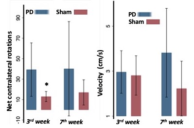
A contralateral rotational response to apomorphine was exhibited by the 6-OHDA PD model at 3rd and 7th week post-surgery (confirming the lesion) compared to sham control (Figure 2).VBM revealed widespread bilateral changes in GM volume on a topographic scale in the brains of 6-OHDA rats, compared to sham-operated model. A reductions of GM volume were observed in fimbria (ipsi), hippocampal formation (ipsi) and accumbens nucleus (contra) in 3rd week PD and cerebellum (ipsi and contra both) in 7th week PD as compared to sham. Additionally, GMV revealed a decreasing trend in the retrosplenial granular cortex (c-region) and cerebellum in contralateral hemisphere in the 7th week PD model as compared to 3rd week (p<0.001unc).
Discussion
Behavioural results revealed striatal dopaminergic cell loss and denervation in the striatum. GMV was decreased in cortical and subcortical regions of the ipsilateral (lesioned) and contralateral (unlesioned) hemisphere, including the site of the primary lesion following 6-OHDA lesioning. The reductions observed are similar to that reported in previous studies4,5 in the third week post-lesion, while the cerebellum exhibited changes in the seventh week post-lesion. This temporal dimension provides a deeper understanding of the progression of structural alterations in response to dopaminergic cell loss.Conclusion
Unilateral nigrostriatal 6-OHDA lesioning leads to widespread gray matter volume changes, which extend beyond the nigrostriatal system and resemble advanced Parkinsonism.Acknowledgements
We acknowledge the funding from Department of Health Research (DHR), Govt. of India, Women Scientist Scheme: R.12013/06/2023-HR (for SK) and Department of Health Research (DHR), Govt. of India, Grant No.R.11013/68/2021-GIA/HRReferences
1. Hatano T, Saiki S, Okuzumi A, et al. Identification of novel biomarkers for Parkinson’s disease by Metabolomic technologies. J. Neurol. Neurosurg. Psychiatry. 2016.
2. Paxinos G, Watson C. The rat brain in stereotaxic coordinates. London Acad. Press. 2009:456
3. Valdés-Hernández PA, Sumiyoshi A, Nonaka H, et al. An in vivo MRI Template Set for Morphometry, Tissue Segmentation, and fMRI Localization in Rats. Front Neuroinform. 2011; 24;5:26.
4. Heidi F, Elijah M, Russell J C, et al. Associations of hippocampal subfields in the progression of cognitive decline related to Parkinson's disease. NeuroImage: Clinical. 2017;14:37-42
5. Cools R, Lewis SJ, Clark L, et al. L-DOPA disrupts activity in the nucleus accumbens during reversal learning in Parkinson's disease. Neuropsychopharmacology. 2007;32(1):180-9.
Figures