2348
Whole Brain Amide Proton Transfer Weighted Imaging in Children with Obstructive Sleep Apnea1shenzhen children's hospital, shenzhen, China, 2Philips Healthcare, guangzhou, China
Synopsis
Keywords: Other Neurodegeneration, Neuro
Motivation: Investigations of amide proton signal changes in the brain of children with OSA may provide imaging markers for early diagnosis.
Goal(s): To explore the APTw signal changes in children with OSA and to evaluate the association of APTw signal in different brain regions with the cognitive impairment.
Approach: Forty-six children with mild to severe OSA and 20 HV underwent whole brain APTw imaging scan. Statistical analysis was performed for inter-group comparison and correlation analysis.
Results: The APTw signals in some brain regions in children with moderate-severe OSA were significantly lower than that of HV. The APTw signals were significantly associated with cognitive scores.
Impact: APTw imaging could be used for assessment of neurocognitive impairment in children with OSA.
Introduction
Obstructive sleep apnea (OSA) is a common respiratory disorder affecting 1.2% to 5.7% of pediatric population1, which is characterized by recurrent partial or complete upper airway obstruction. A growing body of evidence suggests that long-term untreated OSA is associated with neurocognitive dysfunction2, growth problems3, and adverse cardiovascular outcome4. Irreversible cognitive impairments may happen in some children with OSA5, which adversely affects their long-term development as children are in the golden period of nervous system maturation. Additionally, untreated childhood OSA considerably increases utilization of health resource6. All of these highlight the need for prompt recognition, diagnosis, and treatment of children with OSA. In this study, we aimed to explore the brain amide proton transfer weighted (APTw) signal changes in children with OSA and to evaluate the association of APTw signal in different brain regions with the cognitive impairment, provide imaging markers for early diagnosis.Methods
This prospective study was conducted from September 2021 to December 2022. Forty-six children with mild to severe OSA and 20 health volunteers (HV) of matched age and gender underwent whole brain APTw imaging scan (not including cerebellum). The APTw signals of 29 regions of the brain were compared between children in OSA group and HV with Fisher’s least significant differences post-hoc analysis or Kruskal–Wallis test with Steel-Dwass test. The correlation of the APTw signals of different brain regions with cognitive assessment scores was analyzed with Person’s correlation analysis or Spearman’s correlation analysis.Results
The APTw signals in the white matter of inferior frontal gyrus, angular gyrus and thalamus of children with mild OSA and HV were significantly lower than that of children with moderate-severe OSA. The APTw signals of the gray matter of supramarginal gyrus, the gray matter of lingual gyrus, corona radiata, and genu of corpus callosum in children with moderate-severe OSA were significantly lower than that of HV. The APTw signals of the frontal-parietal-temporal regions, hippocampus, and corona radiata were significantly associated with single or multiple cognitive assessment scores.Discussion
Children with OSA tend to have more severe cognitive decline7. Compared with HV, children in the PG had decreased FIQ, VIC and PSI, which was consistent with the findings of Zhao’s study of children (age ≥ 6 years) with OSA8. Chemical and structural cellular injury of the prefrontal cortex in patients with OSA has been suggested to be related to certain patterns of cognitive impairment, especially for executive dysfunction9. The development of the frontal-parietal-temporal network that includes both the cortex and the white matter is related to visuo-spatial working memory during childhood and the impairment of this network could cause decline of specific cognitive functions10,11. In the present study, the APTw signal intensity of the frontal-parietal-temporal regions was significantly related to the multiple cognitive assessment scores, which suggested that APTw signal may be a possible imaging biomarker for assessment of neurocognitive impairment in children with OSA. In addition, the APTw signals intensity of the hippocampus, and corona radiata were significantly associated with a few cognitive assessment scores. Hippocampus and para-hippocampal cortex are interconnected with multiple brain areas in the frontal-parietal lobes, which plays an important role in cognitive processes12. Hippocampus is also well-known for its vital role of declarative memory. However, we did not identify a correlation between the APTw signal intensity of the hippocampus with WMI or FIQ. A coronal section of the hippocampus would be better for measurement of the APTw signal, which was not performed in our study. This might be a reason for not detecting the association. Corona radiata is vulnerable to microvascular damage and the microstructural integrity of corona radiata has been shown to be associated with cognitive performance[33]. Our finding suggested that the damage of the corona radiata may be related to certain cognitive impairment of children with OSA.conclusion
APTw imaging could be used for assessment of neurocognitive impairment in children with OSA.Acknowledgements
No acknowledgement found.References
1. Marcus, C.L., et al., Diagnosis and management of childhood obstructive sleep apnea syndrome. Pediatrics, 2012. 130(3): p. 576-84. 2. Wu, Y., et al., Aberrant Awake Spontaneous Brain Activity in Obstructive Sleep Apnea: A Review Focused on Resting-State EEG and Resting-State fMRI. Front Neurol, 2020. 11: p. 768. 3. Salorio, C.F., et al., Learning, memory, and executive control in individuals with obstructive sleep apnea syndrome. J Clin Exp Neuropsychol, 2002. 24(1): p. 93-100. 4. Mansukhani, M.P., B.P. Kolla, and V.K. Somers, Hypertension and Cognitive Decline: Implications of Obstructive Sleep Apnea. Front Cardiovasc Med, 2019. 6: p. 96. 5. Gozal, D., Obstructive sleep apnea in children: implications for the developing central nervous system. Semin Pediatr Neurol, 2008. 15(2): p. 100-6. 6. Reuveni, H., et al., Health care services utilization in children with obstructive sleep apnea syndrome. Pediatrics, 2002. 110(1 Pt 1): p. 68-72. 7. Lo Bue, A., A. Salvaggio, and G. Insalaco, Obstructive sleep apnea in developmental age. A narrative review. Eur J Pediatr, 2020. 179(3): p. 357-365. 8. Zhao, J., et al., Association between mild or moderate obstructive sleep apnea-hypopnea syndrome and cognitive dysfunction in children. Sleep Med, 2018. 50: p. 132-136. 9. Beebe, D.W. and D. Gozal, Obstructive sleep apnea and the prefrontal cortex: towards a comprehensive model linking nocturnal upper airway obstruction to daytime cognitive and behavioral deficits. J Sleep Res, 2002. 11(1): p. 1-16. 10. Pearson, J., The human imagination: the cognitive neuroscience of visual mental imagery. Nat Rev Neurosci, 2019. 20(10): p. 624-634. 11. Klingberg, T., Development of a superior frontal-intraparietal network for visuo-spatial working memory. Neuropsychologia, 2006. 44(11): p. 2171-7. 12. Opitz, B., Memory function and the hippocampus. Front Neurol Neurosci, 2014. 34: p. 51-9.Figures
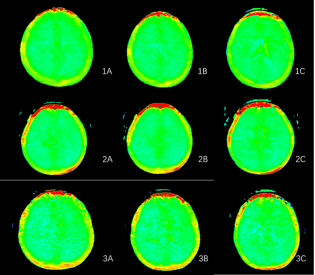
Figure.1 False-color images of APT frontotemporal lobe(1A), radiation crown(1B) and occipital lobe(1C) in healthy volunteer
Figure.2 False-color images of APT frontotemporal lobe(2A), radiation crown(2B) and occipital lobe(2C) in mild OSA children group
Figure.3 False-color images of APT frontotemporal lobe(3A), radiation crown(3B) and occipital lobe(3C) in severe OSA children group