2346
Identifying Brain Calcifications in Down Syndrome Patients Using ZTE-Derived Pseudo-CT Imaging1Department of Neuroimaging, Institute of Psychiatry Psychology and Neuroscience, King's College London, London, United Kingdom, 2Forensic & Neurodevelopmental Sciences, Institute of Psychiatry, Psychology and Neuroscience, King's College London, London, United Kingdom, 3South London and Maudsley NHS Foundation Trust, London, United Kingdom
Synopsis
Keywords: Aging, Aging, ZTE pseudo-CT calcification
Motivation: Patients with Down Syndrome may undergo nearly ten times as many medical imaging procedures in their childhood alone compared to the general population, exposing this already high-risk group to elevated levels of radiation.
Goal(s): We want to explore the potential of Zero Echo-Time (ZTE) MRI as a radiation-free alternative to CT scans for imaging cortical bone and calcifications in Down's Syndrome patients.
Approach: We employed a published method for tissue segmentation, assigning Standard Hounsfield Units for air and soft tissue, while a continuous linear mapping technique was used for bone.
Results: The images obtained resemble true CT images and calcifications are also visible.
Impact: ZTE to pseudo-CT image conversion offers a one-stop-shop solution for imaging patients without ionising radiation. The calcifications found and their potential clinical implications in ageing, or as markers for other conditions, warrant further investigation.
Introduction
Zero echo-time (ZTE) MRI is an innovative imaging approach which enables the visualization of short-T2 tissues before decay by minimizing the delay between radiofrequency excitation and data acquisition, effectively reducing it to zero. This approach offers rapid, artifact-resistant imaging, ideal for cortical bone examination without ionizing radiation, serving as a safe alternative to CT scans and providing a comprehensive solution for imaging both hard and soft tissues. By eliminating the need for multiple scans, ZTE MRI streamlines the imaging process and offers a convenient "one-stop-shop" solution. This study used ZTE to obtain CT-like images from ZTE MRI in a Down’s Syndrome (DS) study and identify calcifications within the choroid plexus, basal ganglia, and pineal gland which are known to occur from CT [1].Methods
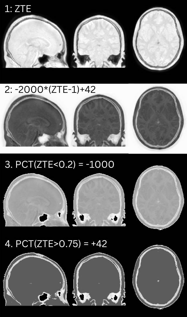
Pseudo-CT images were generated from proton density weighted ZTE images acquired from 39 patients (36.19 ± 11.80) and 19 healthy controls. (37.17 ± 9.59). One patient was discounted due to a brain shunt, and 4 patients due to incorrigible motion artifacts. 6 patients had additional scans 6 months after the first. All ZTE images were obtained with a matrix size of 200x200, a voxel size of 1.5x1.5x1.5 mm3, a TR of 1.08 ms and a bandwidth of ±62.5 kHz. After bias field correction, normalization, and registration to MNI space, the images were segmented into three categories: bone, soft tissue, and air. Following Wiesinger et al., standard Hounsfield Units were assigned for air (-1000 HU) and soft tissue (+42 HU), and a continuous linear mapping technique was used for bone (Fig.1) [2]. The analysis further involved ROI masking, voxel counting, and statistical analysis. We used masks from the Automated Talairach Atlas project for the basal ganglia (ATAG), the probabilistic atlas for the pineal gland derived by Razavi et al. [3] and the Harvard Oxford Lateral Ventricle atlas from FSL for the choroid plexus. Voxel counting was performed with a threshold of 43.Results
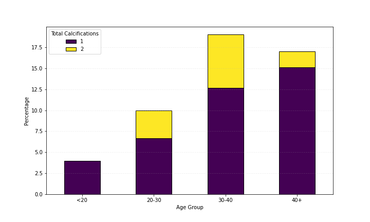
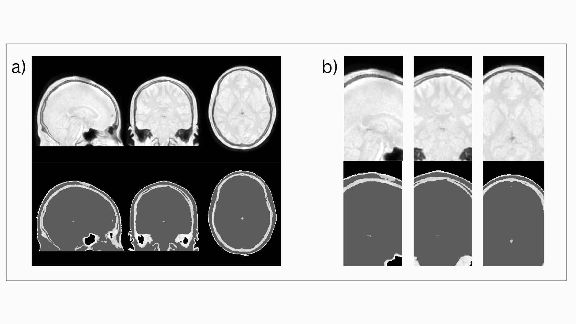
The obtained ZTE-derived pseudo-CT images resemble true CT images and calcifications, where present, are visible (Fig.3). A potential association between increasing age and a rise in calcification frequency is illustrated in Figure 2. Using a generalised linear model regression, we found that an increase in age corresponds to an increase in pineal gland calcifications, when both controls and patients are considered (p-value=0.02). We observed pineal gland calcifications in 37% of the patients and 21% in the control group (p = 0.24, z-test). Choroid plexus calcifications were present in 17% of the patients (p = 0.07) and in none of the healthy control. No study participants displayed any detectable anomalies within the basal ganglia. One patient for which longitudinal data was available presented with choroid calcifications at the second scan session, where none appeared in the first scan.Discussion
The results indicate a higher incidence of pineal gland and choroid plexus calcifications in the patient group. However, a comparison with a previous CT study of Down's syndrome, reveals some discrepancies in the location and frequency of calcifications. Specifically, the CT study reported a high incidence (10.7%) of bilateral calcification of basal ganglia in Down's syndrome, which was not detected in our pseudo-CT images [1]. Potential factors contributing to this disparity include variations in imaging modalities, insufficient sensitivity in the ZTE-pCT sequence, differences in patient age groups, or the different methods of calcification detection and quantification. We postulate that the insignificant p-values are heavily influenced by the smaller size of the healthy control group for which data collection is ongoing.Conclusion
The method described enables MR to pseudo-CT image conversion in DS patients, in a robust, and fast manner, allowing potential calcification detection. However, the location, frequency and clinical significance of calcifications, warrants further investigation and validation with larger and more diverse samples.Acknowledgements
The authors would like to thank Florian Wiesinger for his insightful advice on the ZTE to pseudo-CT conversion pipeline.References
[1] Ieshima, A., et al. “A morphometric CT study of Down's syndrome showing small posterior fossa and calcification of basal ganglia” Neuroradiology (1984). doi: 10.1007/BF00342687
[2] Wiesinger, F., et al. " Zero TE-based pseudo-CT image conversion in the head and its application in PET/MR attenuation correction and MR-guided radiation therapy planning” British Journal of Radiology (2018). doi:10.1259/bjr.20220059
[3] Razavi, F., et al. “A Probabilistic Atlas of the Pineal Gland in the Standard Space” Frontiers in Neuroinformatics (2021). doi:10.3389/fninf.2021.554229
Figures