2343
Discrimination between white and gray matter in the cervical spinal cord using the Myelin-Sensitive Inversion Recovery (MySIR)1Department of Radiology, Chiba University Hospital, Chiba-shi, Japan, 2Department of Diagnostic Radiology and Radiation Oncology, Graduate School of Medicine, Chiba-shi, Japan, 3Philips Japan, Tokyo, Japan, 4Philips Canada, Mississauga, ON, Canada
Synopsis
Keywords: Spinal Cord, Neuro, Myelin
Motivation: Discrimination between gray and white matter of the spinal cord remains challenging.
Goal(s): To investigate imaging techniques that best discriminate gray matter from white matter.
Approach: Inhomogeneous magnetization transfer (ihMT) and phase-sensitive inversion recovery (PSIR) have been reported effective in discriminating between gray and white matter. Myelin-sensitive inversion recovery (MySIR) was compared with ihMT and PSIR for evaluating the discriminability between gray and white matter.
Results: MySIR had the highest contrast ratio compared to ihMT and PSIR.
Impact: MySIR was superior to ihMT and PSIR in discriminating gray matter from white matter, and MySIR can be useful as a new myelin-specific MR imaging technique for spinal cord segmentation.
Introduction
The development of myelin-specific MR imaging techniques remains an active area of research where new approaches are being sought. Discrimination between gray matter (GM) and white matter (WM) in cervical MRI remains challenging due to small structure size and motion artifacts from swallowing, breathing, and blood flow. Previous studies have reported promising imaging techniques in this area, including inhomogeneous magnetization transfer (ihMT)1), short-TR adiabatic inversion recovery (STAIR)2), and phase-sensitive inversion recovery (PSIR)3). The STAIR technique is a short TR IR technique, and it is possible to image only myelin with a short T2 value by setting the null point for magnetization of intracellular/extracellular water with a long T2 value.The PSIR technique is a fast gradient echo technique to obtain an intensity image with T1 contrast after TI and a low flip angle reference image just before the next IR pulse. Myelin-sensitive inversion recovery (MySIR) is an imaging technique that combines the STAIR technique, which is an IR technique with a short TR, and the PSIR technique, which reconstructs a corrected real image by acquiring an IR image and a reference image, allowing only myelin to be depicted. MySIR is expected to provide better myelin-specific MR imaging than conventional methods. T this study aimed to compare ihMT, PSIR, and MySIR in terms of contrast ratio and to investigate their ability to discriminate between GM and WM in the cervical spinal cord.
Methods
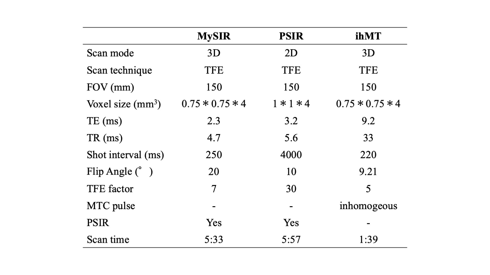
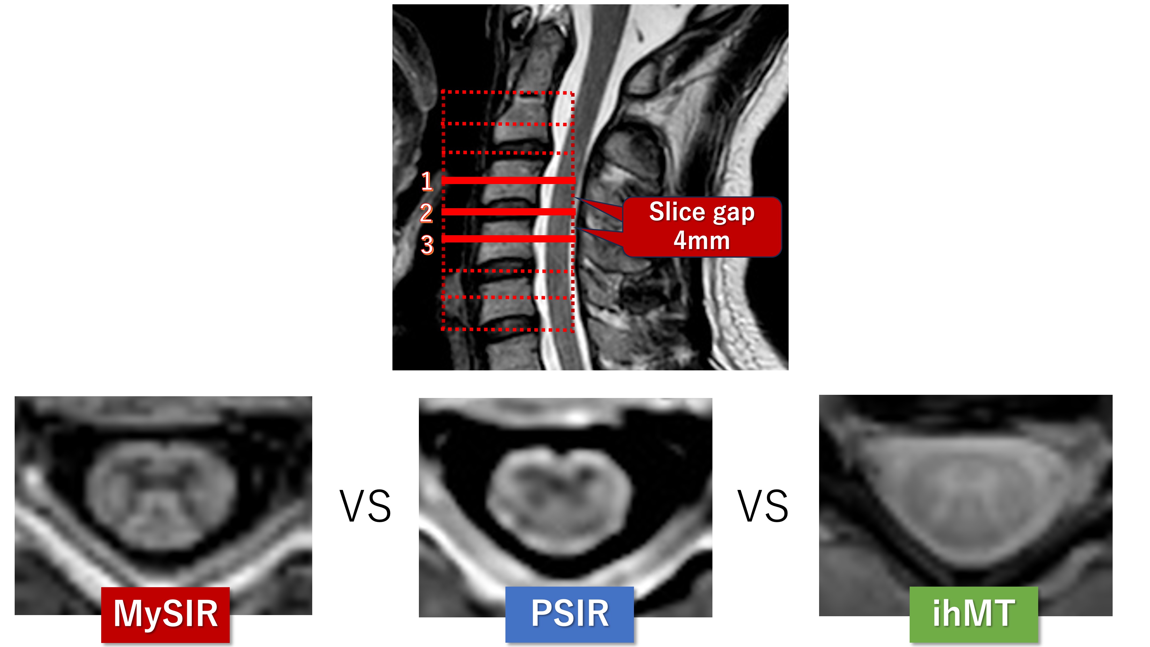
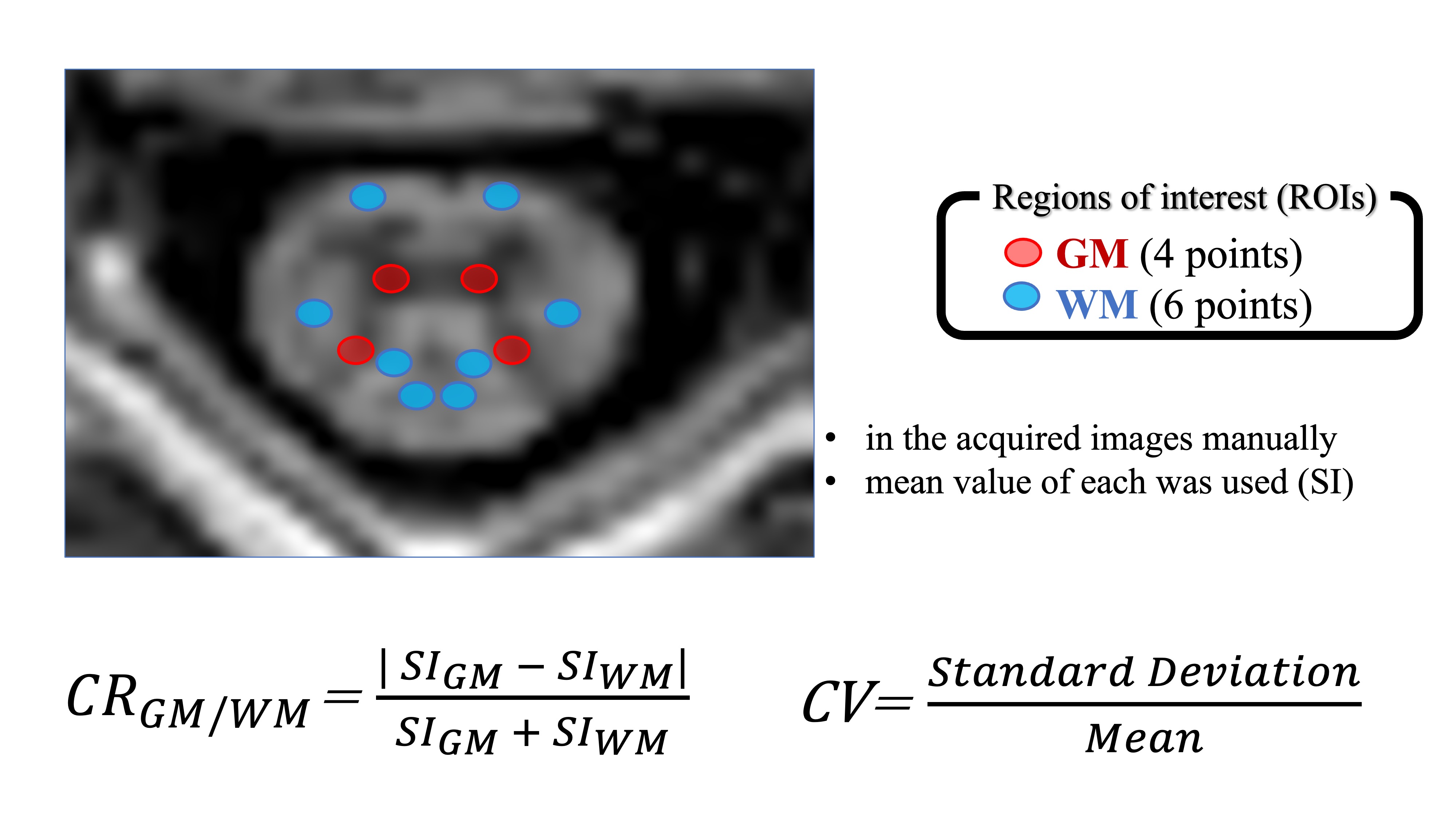
MRI examination: ten healthy subjects (mean age 27.0 ± 3.4 years, nine males) underwent an MRI examination of the cervical spinal cord using a 3.0T scanner (Ingenia, Philips Healthcare). The scan parameters are summarized in Figure. 1.Evaluation: for each healthy subject, a total of three slices (n=30) were selected and evaluated based on sagittal T2-weighted images: the C3-C4 disc level and two slices above and below it (Figure. 2). Regions of interest (ROIs) were manually set at GM (4 points) and WM (6 points) in the acquired images, and the mean and standard deviation of each were measured (Figure. 3).
1. The contrast ratio (CR) within GM and WM was calculated from the mean values as follows
CRGM/WM = | SIGM – SIWM | / SIGM + SIWM
2. Coefficients of variation for GM and WM (CVGM, CVWM) were calculated from the standard deviation and mean values as follows
CV= Standard Deviation/Mean
Statistical analyses: the Kurskal-Wallis test was used to compare CR, CVGM, and CVWM for each sequence, respectively. Statistical significance was considered P < 0.05
Results
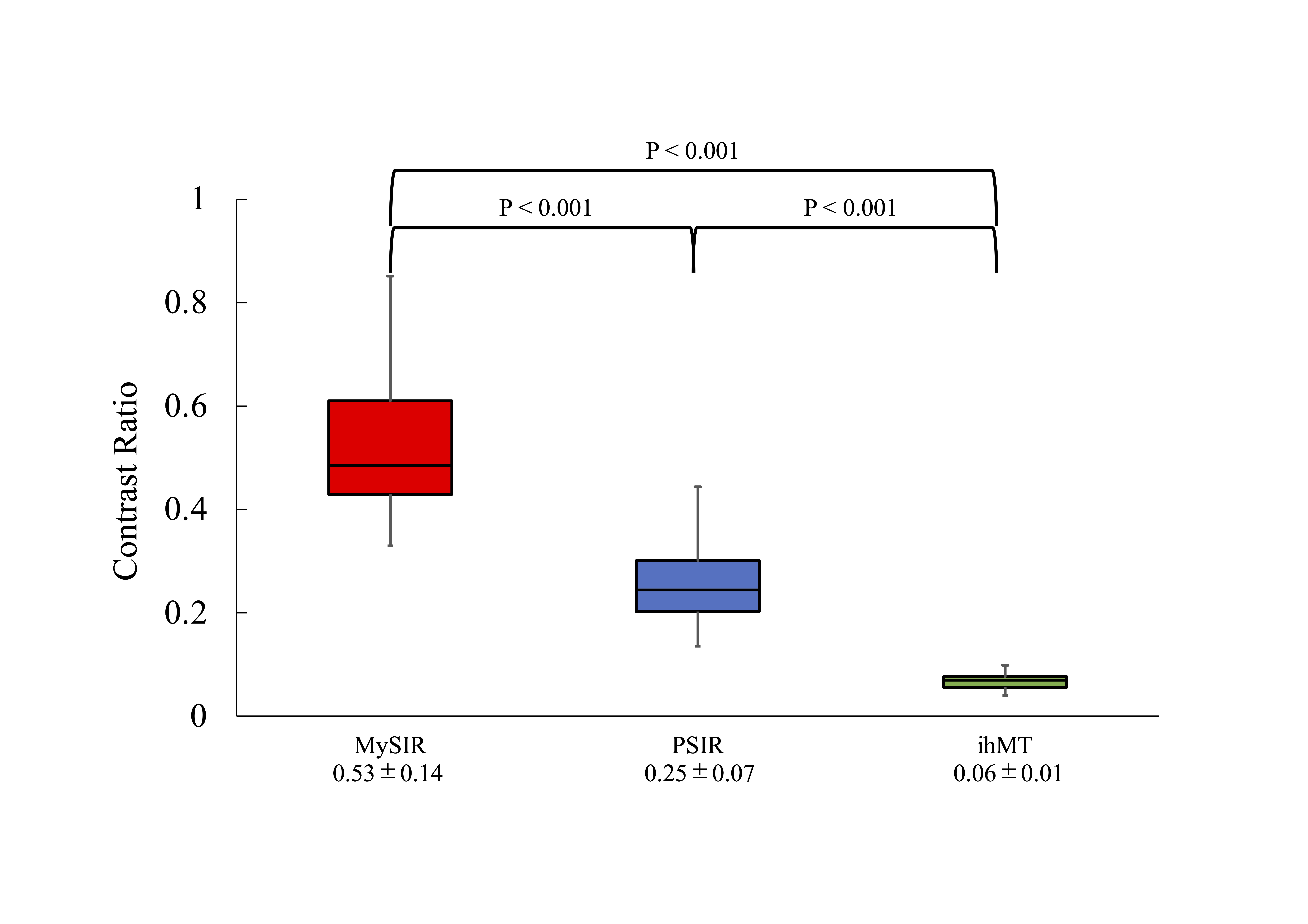
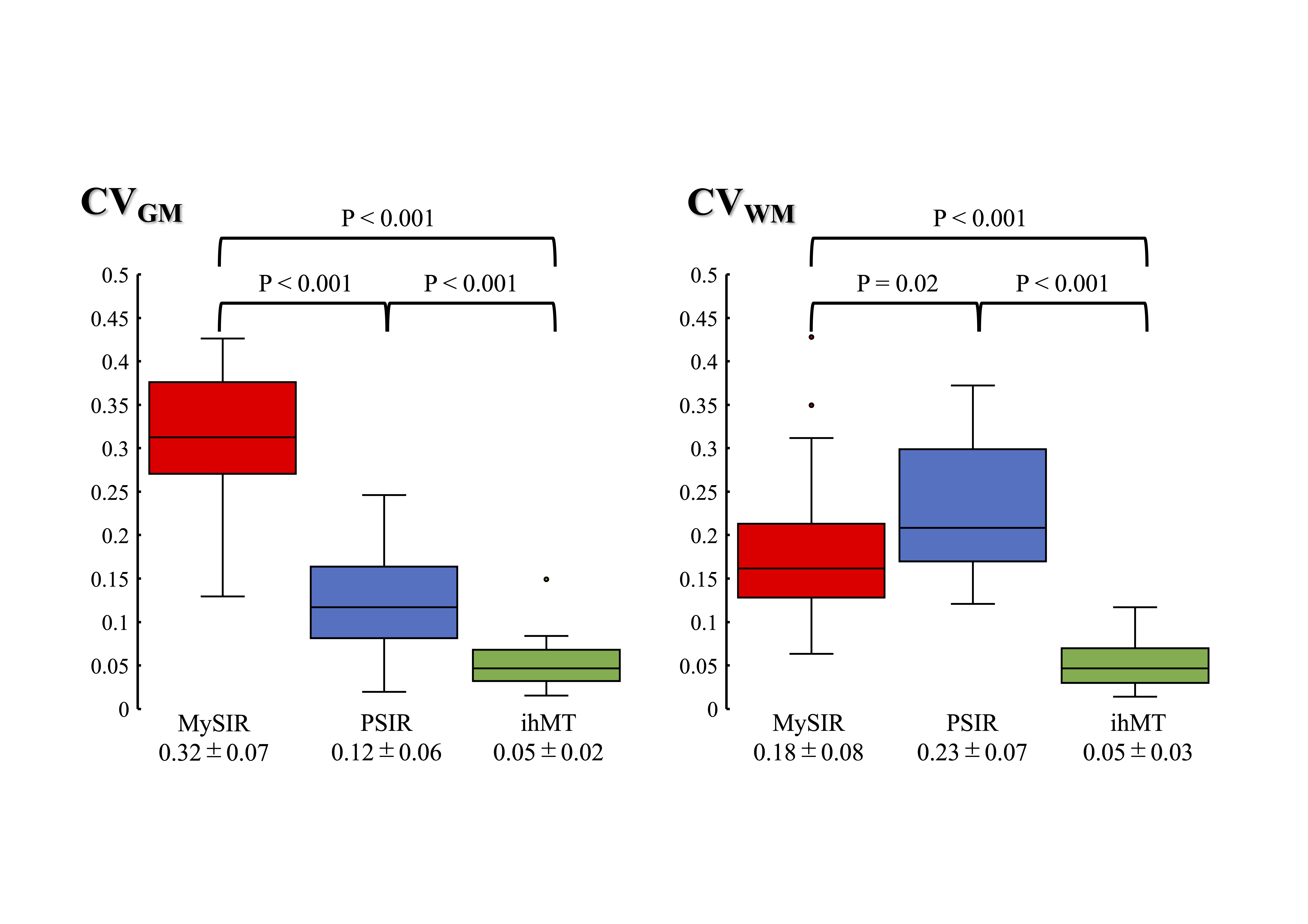
Figure. 4 shows box-and-whisker plots of CR between GM and WM for each sequence, with CRs of MySIR: 0.53 ± 0.14, PSIR: 0.25 ± 0.07, and ihMT: 0.06 ± 0.01. The CRs for each sequence showed significant differences, respectively. Figure. 5 shows box plots of CVGM and CVWM for each sequence. CVGM was MySIR: 0.32 ± 0.07, PSIR: 0.12 ± 0.06, and ihMT: 0.05 ± 0.02. CVWM was MySIR: 0.18 ± 0.08, PSIR: 0.23 ± 0.07, and ihMT: 0.05 ± 0.03. CVGM and CVWM for each sequence showed statistically significant differences.Discussion
The ability of MySIR to discriminate GM and WM evaluated by CR was the highest. The PSIR and ihMT techniques have been reported to discriminate GM and WM in the cervical spinal cord. However, the results of this study showed that MySIR outperformed both PSIR and ihMT in discriminating GM and WM. Therefore, it is suggested that MySIR is effective as a new myelin-specific MR imaging agent. However, the coefficient of variation of MySIR was higher than that of ihMT. MySIR delineates myelin; signals from other tissues are suppressed. In some subjects, the low signals from GM were insufficient. Therefore, it is assumed that the coefficient of variation of MySIR was higher than that of ihMT. The high coefficient of variation of MySIR is a point for improvement. Another issue is that MySIR requires a longer imaging time than the ihMT. Although this study was conducted in young, healthy subjects and the effect of motion was considered small, further deterioration of image quality is a concern in actual clinical practice4). Therefore, it is necessary to adjust the imaging acquisition time and resolution, and evaluate the lesion detectability in actual clinical practice.Conclusion
MySIR effectively differentiated between white and gray matter in the cervical spinal cord. MySIR is expected to be a new myelin-specific MR imaging technique.Acknowledgements
No acknowledgement found.References
1. Rasoanandrianina H, et al. Region-specific impairment of the cervical spinal cord (SC) in amyotrophic lateral sclerosis: A preliminary study using SC templates and quantitative MRI (diffusion tensor imaging/inhomogeneous magnetization transfer). NMR Biomed. 2017 Dec;30(12).
2. Ma YJ, et al. Myelin water imaging using a short-TR adiabatic inversion-recovery (STAIR) sequence. Magn Reson Med. 2022 Sep;88(3):1156-1169.
3. Papinutto N, et al. 2D phase-sensitive inversion recovery imaging to measure in vivo spinal cord gray and white matter areas in clinically feasible acquisition times. J Magn Reson Imaging. 2015 Sep;42(3):698-708.
4. Li G, et al. Improving the robustness of 3D turbo spin echo imaging to involuntary motion. Magn Reson Mater Physics, Biol Med 2015;28:329–45.
Figures