2340
Magnetization Transfer Ratio Shows Selective Spinal Tracts Damage in Patients with Degenerative Cervical Myelopathy1Federal Neurosurgical Center, Novosibirsk, Russian Federation, 2Novosibirsk State University, Novosibirsk, Russian Federation
Synopsis
Keywords: Spinal Cord, Magnetization transfer
Motivation: Quantitative spinal cord imaging is essentially helpful in various pathological conditions, such as degenerative cervical myelopathy (DCM). However, the role of myelin-sensitive techniques in DCM seems to be underestimated.
Goal(s): Assess demyelination within different spinal tracts in patients with DCM.
Approach: Spinal cord 3T MRI with magnetization transfer ratio (MTR) technique was performed with subsequent post-processing in Spinal Cord Toolbox (v5.5).
Results: We revealed structural impairment of the spinocerebellar, rubrospinal, and reticulospinal tracts, as well as fasciculus cuneatus, in patients with DCM compared to the control group. The associations between JOA score and MTR values within spinal cord white matter were also found.
Impact: Our results could help to better understand the pathophysiology of DCM. Further studies in this field could be dedicated toassociations of MTR values with surgical recovery, links with supraspinal structural changes, or comparisons with other quantitative techniques.
Introduction
Degenerative cervical myelopathy (DCM) is the most common cause of spinal cord dysfunction in developed countries, and its prevalence is increasing due to the increase in life expectancy [1]. However, we are still far from complete understanding of the pathobiology of the disease, and we have limited ability to monitor the development of DCM and predict the potential for recovery with existing neuroimaging methods [2]. Previously published studies showed great potential for the magnetization transfer ratio (MTR) technique [3], [4] under this condition but were carried out with small sample sizes and did not selectively evaluate each tract. Thus, the main objective of our study was to understand the role of changes in the MTR signal in the evaluation of the structural integrity of the spinal cord white matter in patients with clinically significant DCM and disability.Methods
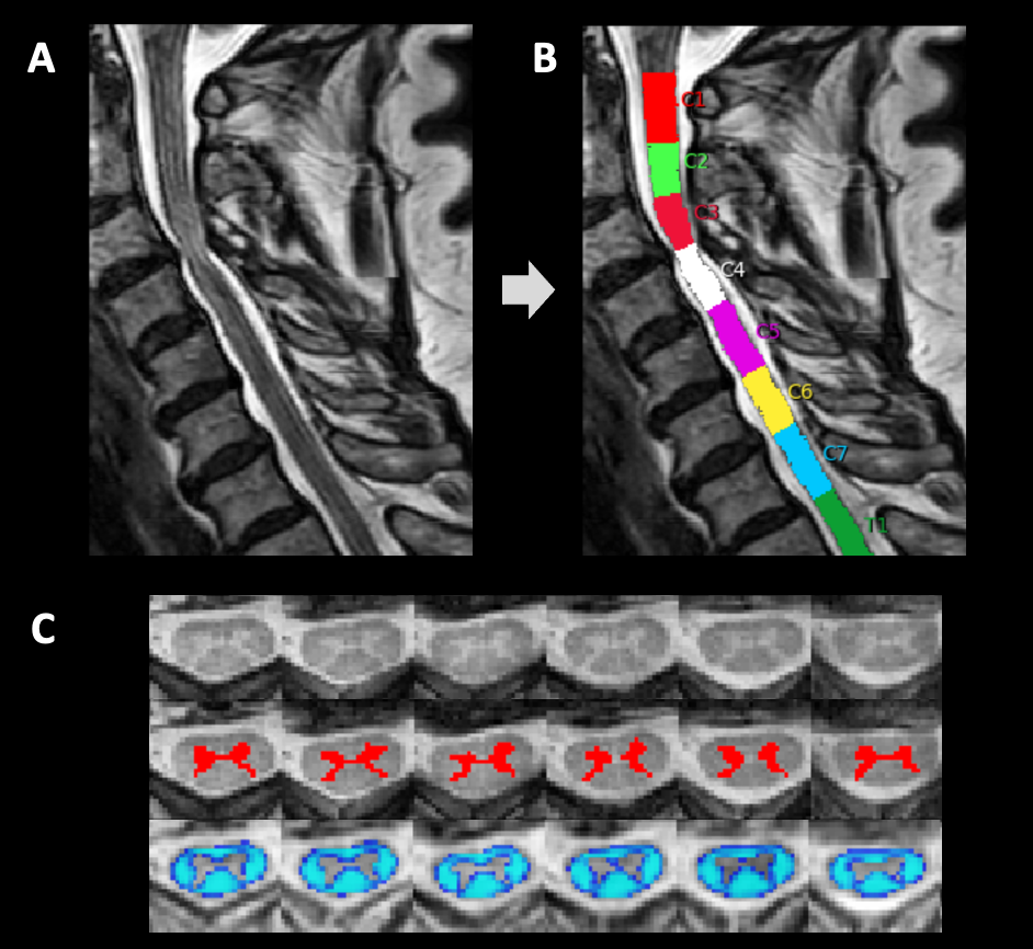
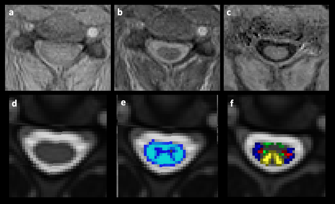
Fifty-three patients with DCM and 41 patients without spinal cord compression (with cervical radiculopathy syndrome) were evaluated using high-resolution cervical spinal cord magnetic resonance imaging (MRI), as previously described, which included the magnetization transfer technique (as described in [5]). 3T MRI data were analysed with the Spinal Cord Toolbox (v5.5) [6], with automatic gray and white matter segmentation (Figure 1); magnetization transfer ratio (MTR) values in each spinal tract were calculated (Figure 2). Covariance analyses (ANCOVAs) were performed and included the group (DCM, radiculopathy) as the factor between subjects, the MTR values within different regions of the spinal cord as the dependent variable and the age and sex of the patient as covariates. Correlations between MTR data and the clinical disability rate of the patients(according to the JOA score) were also evaluated in patients with DCM with Pearson’s correlation test.Results
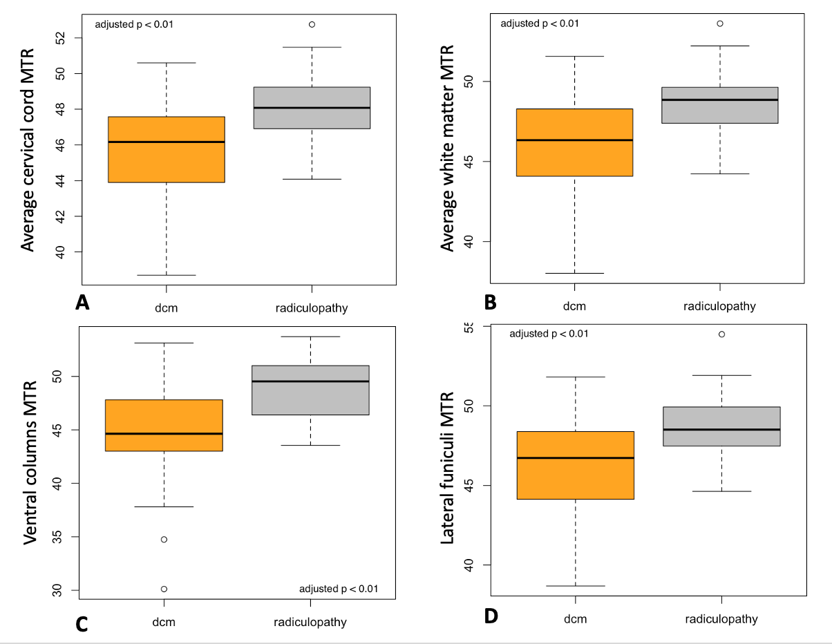
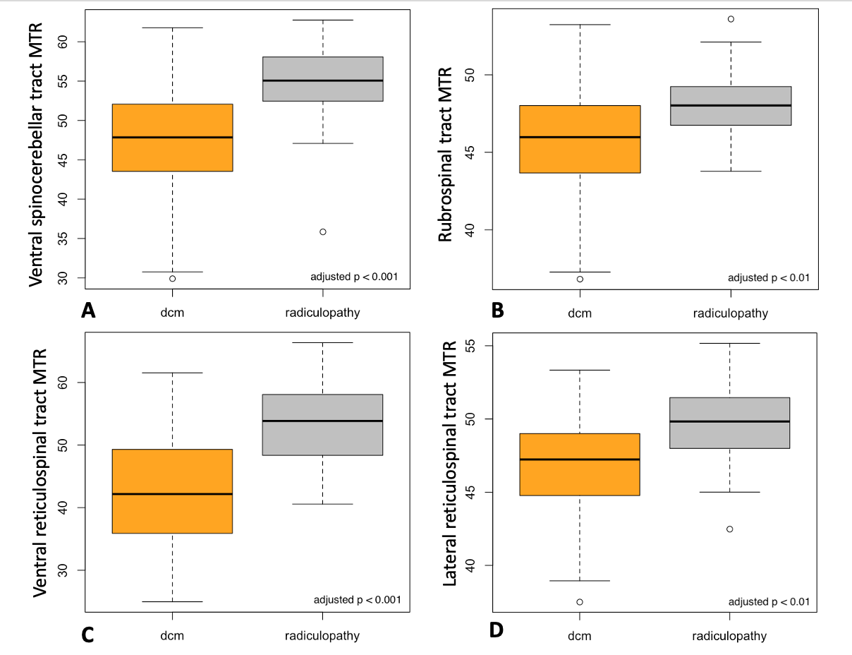
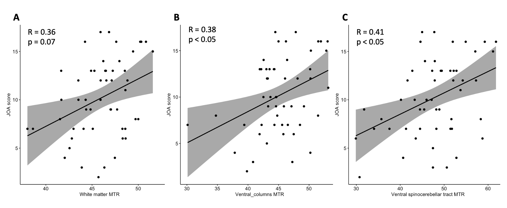
Statistically significant reductions in the average MTR values of the spinal cord white matter, as well as the MTR values of the ventral columns and lateral funiculi, were revealed in the DCM group compared to the radiculopathy group (p < 0.01 in all cases, FDR corrected; Figure 3). Furthermore, reductions in MTR values of the fasciculus cuneatus, spinocerebellar, rubrospinal andreticulospinal tracts were found in patients with DCM (p < 0.01, p < 0.001, p < 0.01, and p < 0.01, respectively, FDR corrected; Figure 4). Furthermore, a trend towards a decrease in MTR values within the ventral corticospinal tract was found (p = 0.087, FDR corrected). Positive correlations between the JOA score and the MTR values within the ventral columns of the spinal cord (R = 0.38, adjusted p < 0.05; Figure 5b) and the ventral spinocerebellar tract (R = 0.41, adjusted p < 0.05; Figure 5c) were revealed, as well as a trend towards a positive association between the JOA score and the average white matter MTR values (R = 0.36; adjusted p = 0.06; Figure 5a).Discussion
Chronic spinal cord compression in DCM is believed to lead to demyelination and axonal destruction [7], and the ventral areas are more prone to these changes [4]. In general, our results confirm the existing data. However, we revealed that some spinal tracts are more susceptible to damage than others, regardless of anatomic location or blood supply. Specifically, structural impairmentsare more prominent within the spinocerebellar, rubrospinal, and reticulospinal tracts, as well as the fasciculus cuneatus. To our knowledge, this is the first attempt to evaluate differences in the vulnerability of spinal tracts to damage in DCM with a relatively large sample size. However, more studies with follow-up data and the application of other quantitative techniques are needed to prove our results and evaluate their utility in patient care.Conclusion
Our results show that the spinal tracts have selective vulnerability to demyelination in patients with DCM.Acknowledgements
The authors thank Aria Nouri (Switzerland) for inspiration to this study and providing valuable advices.References
[1] R. Yurac et al., “[Degenerative cervical myelopathy],” Rev Med Chil, vol. 150, no. 3, pp. 339–352, Mar. 2022, doi: 10.4067/S0034-98872022000300339.
[2] E. Filimonova, I. Vasilenko, Y. Kubetsky, O. Prokhorov, M. Abdaev, and J. Rzaev, “Brainstem and subcortical regions volume loss in patients with degenerative cervical myelopathy and its association with spinal cord compression severity,” Clin Neurol Neurosurg, vol. 233, Oct. 2023, doi: 10.1016/J.CLINEURO.2023.107943.
[3] M. Paliwal et al., “Magnetization Transfer Ratio and Morphometrics of the Spinal Cord Associates with Surgical Recovery in Patients with Degenerative Cervical Myelopathy,” World Neurosurg, vol. 144, p. e939, Dec. 2020, doi: 10.1016/J.WNEU.2020.09.148.
[4] M. B. Cloney, Z. A. Smith, K. A. Weber, and T. B. Parrish, “Quantitative magnetization transfer MRI measurements of the anterior spinal cord region are associated with clinical outcomes in cervical spondylotic myelopathy,” Spine (Phila Pa 1976), vol. 43, no. 10, p. 675, May 2018, doi: 10.1097/BRS.0000000000002470.
[5] J. Cohen-Adad et al., “Open-access quantitative MRI data of the spinal cord and reproducibility across participants, sites and manufacturers,” Scientific Data 2021 8:1, vol. 8, no. 1, pp. 1–17, Aug. 2021, doi: 10.1038/s41597-021-00941-8.
[6] B. De Leener et al., “SCT: Spinal Cord Toolbox, an open-source software for processing spinal cord MRI data,” Neuroimage, vol. 145, pp. 24–43, Jan. 2017, doi: 10.1016/J.NEUROIMAGE.2016.10.009.
[7] F. Akter et al., “The pathophysiology of degenerative cervical myelopathy and the physiology of recovery following decompression,” Front Neurosci, vol. 14, p. 507668, Apr. 2020, doi: 10.3389/FNINS.2020.00138/BIBTEX.
Figures