2326
Characterizing Cerebral Neurometabolic Changes in Diaschisis from Stroke using Fast 3D High-Resolution MRSI
Ziyu Meng1, Tianyao Wang2, Hong Zhou3, Chang Xu1, Bin Bo1, Yibo Zhao4,5, Yudu Li4,6, Wen Jin4,5, Zhi-Pei Liang4,5, and Yao Li1
1School of Biomedical Engineering, Shanghai Jiao Tong University, Shanghai, China, 2Radiology Department, Renji Hospital, Shanghai Jiao Tong University of Medicine, Shanghai, China, 3Department of Radiology, The First Affiliated Hospital of South China of University, South China of University, Hengyang, China, 4Beckman Institute for Advanced Science and Technology, University of Illinois at Urbana-Champaign, Urbana, IL, United States, 5Department of Electrical and Computer Engineering, University of Illinois at Urbana-Champaign, Urbana, IL, United States, 6The National Center for Supercomputing Applications, University of Illinois at Urbana-Champaign, Urbana, IL, United States
1School of Biomedical Engineering, Shanghai Jiao Tong University, Shanghai, China, 2Radiology Department, Renji Hospital, Shanghai Jiao Tong University of Medicine, Shanghai, China, 3Department of Radiology, The First Affiliated Hospital of South China of University, South China of University, Hengyang, China, 4Beckman Institute for Advanced Science and Technology, University of Illinois at Urbana-Champaign, Urbana, IL, United States, 5Department of Electrical and Computer Engineering, University of Illinois at Urbana-Champaign, Urbana, IL, United States, 6The National Center for Supercomputing Applications, University of Illinois at Urbana-Champaign, Urbana, IL, United States
Synopsis
Keywords: Stroke, Stroke
Motivation: Understanding metabolic changes in diaschisis is essential for stroke rehabilitation.
Goal(s): Our goal was to investigate the neurometabolite alterations in the disconnected white matter (WM) tracts and the downstream cortical gray matter (GM) regions, and their associations in early stroke outcome.
Results: Our findings reveal a trend for decreased cortical GM NAA as the WM NAA in the diaschitic hemisphere decreases, both associated with early stroke outcome.
Impact: Using 3D high-resolution 1H-MRSI, we found a potential involvement of neuronal mitochondria metabolism in the metabolic changes observed in diaschisis from stroke. It may offer valuable prognostic biomarkers in stroke patients management.
Introduction
Diaschisis is a frequent outcome of cerebral infarction and notably impacts stroke recovery. It usually occurs when strokes impair neuronal synaptic functions in brain areas distant from the lesion site. Characterizing the neurometabolic changes in diaschisis is crucial for the development of stroke rehabilitation strategies and the prediction of stroke outcomes. Proton MR spectroscopic imaging (1H-MRSI) provides a unique tool for mapping neurometabolic changes, including reduced N-acetylaspartate (NAA) levels indicating potential axonal metabolic impairment in diaschisis 1. However, conventional MRSI technology is limited in spatial resolution and partial brain coverage, unable to provide precise measurements of tissue-specific neurometabolites concentrations. Therefore, the relationship between white matter (WM) neurometabolic changes and resulting gray matter (GM) metabolite abnormalities in diaschisis remains elusive. In this work, we characterized the cerebral neurometabolic changes in GM and WM post stroke using a fast high-resolution 3D 1H-MRSI technique, called SPICE (SPectroscopic Imaging by exploiting spatiospectral CorrElation). The NAA alterations in the disconnected WM tracts as well as in the downstream cortical GM regions were investigated, which provided complementary information about neuronal dysfunction and contributed to an understanding of the physiological changes associated with diaschisis in stroke.Methods
A total of 105 ischemic stroke patients from the Fifth People’s Hospital of Shanghai (n = 55), Renji Hospital (n = 13), and the First Affiliated Hospital of South China University (n = 37), participated in this study. Patients with hemorrhage or non-stroke lesions on structural MRI, and contraindication for MRI were excluded. All patients were assessed using the NIHSS score at admission. Fifty-five patients had their NIHSS scores at discharge. This study was approved by the local hospital IRBs with written informed consents provided. Demographics are detailed in Table 1.MRI scans were conducted using 3T Siemens Prisma or Skyra scanners (Siemens Healthcare, Erlangen, Germany). The image acquisition protocol included: 3D MRSI using the SPICE sequence (TR/TE = 160/1.6 ms, resolution = 2.0×3.0×3.0mm3, FOV = 240×240×72mm3, scan time = 8min), MPRAGE (TR/TE/TI = 2400/2.13/1100ms, resolution = 1.0×1.0×1.0mm3, FOV = 256mm), DWI (TR/TE = 4300/[74,125]ms, resolution = 1.3×1.3×4.0mm3, FOV = 240mm, b = 0 and 1000 s/mm2), and FLAIR (TR/TE/TI = 9000/89/2500ms, resolution = 0.5×0.5×2.0mm3, FOV = 240mm). Neurometabolite maps were obtained using the standard processing pipeline of SPICE 2–4.
Neurometabolite maps (NAA, choline, creatine) and lesion masks (manually delineated by an experienced neuroradiologist) were nonlinearly registered to standard MNI152 space. All images were co-registered to the MPRAGE images using an affine transformation in ANTs 5. Structural disconnection maps resulting from each lesion were estimated using Lesion Quantification Toolkit 6. The affected WM tracts were identified by thresholding the disconnection maps. Affected GM was the connected area to the affected WM tracts. Contralateral GM regions were defined as the mirroring counterparts along the midline in MNI152 space. Paired t-tests were conducted to compare relative NAA levels (normalized over contralateral neurometabolites) in both hemispheres. Pearson’s correlation analyses were used to examine the relationships between NAA levels in affected WM and GM regions. Linear regression analysis, using leave-one-out cross-validation, was employed to compare predictive models for patient NIHSS scores at discharge. All statistical analyses were performed using MATLAB.
Results
Representative high-resolution 3D NAA maps, along with the spectra and DWI images in lesion and normal-appearing layers, are shown in Fig.1. Reduced NAA levels were observed in the lesion region compared to the contralateral normal area. This NAA decrease persisted even in regions that appeared normal on DWI of the ipsilateral hemisphere. Figure 2 illustrates the comparisons of NAA levels in ipsilateral and contralateral WM and GM regions, respectively. NAA levels in ipsilateral WM or GM were significantly lower than their contralateral counterparts (WM: p = 0.041; GM: p = 0.008). As displayed in Fig.3, the ipsilateral WM NAA levels were strongly correlated with ipsilateral cortical GM NAA levels (r = 0.436, p < 0.001), but not with the affected cortical GM volume (r = - 0.165, p = 0.092). We further explored the prediction performance for patients' NIHSS score at discharge using NAA levels in ipsilateral WM and GM acquired at the patients' admission. As illustrated in Fig.4, both NAA levels in disconnected WM (r = 0.613) and in affected GM (r = 0.587) showed promising prediction performance for patients' functional scores.Conclusion
Using 3D high-resolution 1H-MRSI, we found a potential involvement of neuronal mitochondria metabolism in the metabolic changes observed in diaschisis from stroke. It may offer valuable prognostic biomarkers in stroke patients management.Acknowledgements
This work was supported by Shanghai Pilot Program for Basic Research—Shanghai Jiao Tong University (21TQ1400203), the Program for Professor of Special Appointment (Eastern Scholar) at Shanghai Institutions of Higher Learning; Key Program of Multidisciplinary Cross Research Foundation of Shanghai Jiao Tong University (YG2021ZD28, YG2023ZD22), and New Faculty Start-up Foundation of Shanghai Jiao Tong University (23X010501992).References
- Chu WJ, Mason GF, Pan JW, et al. Regional cerebral blood flow and magnetic resonance spectroscopic imaging findings in diaschisis from stroke. Stroke. 2002;33(5):1243-1248.
- Liang Z.P. Spatiotemporal imaging with partially separable functions. In: Proceedings of the 4th IEEE International Symposium on Biomedical Imaging: From Nano to Macro. ; 2007:988-991.
- Lam F, Ma C, Clifford B, Johnson CL, Liang Z.P. High-resolution 1H-MRSI of the brain using SPICE: Data acquisition and image reconstruction. Magn Reson Med. 2016;76(4):1059-1070.
- Li Y, Lam F, Clifford B, Liang Z.P. A subspace approach to spectral quantification for MR spectroscopic imaging. IEEE Trans Biomed Eng. 2017;64(10):2486-2489.
- Avants BB, Tustison N, Song G. Advanced normalization tools (ANTS). Insight j. 2009;2(365): 1-35.
- Griffis JC, Metcalf NV., Corbetta M, Shulman GL. Lesion Quantification Toolkit: A MATLAB software tool for estimating grey matter damage and white matter disconnections in patients with focal brain lesions. NeuroImage Clin. 2021;30:102639.
Figures
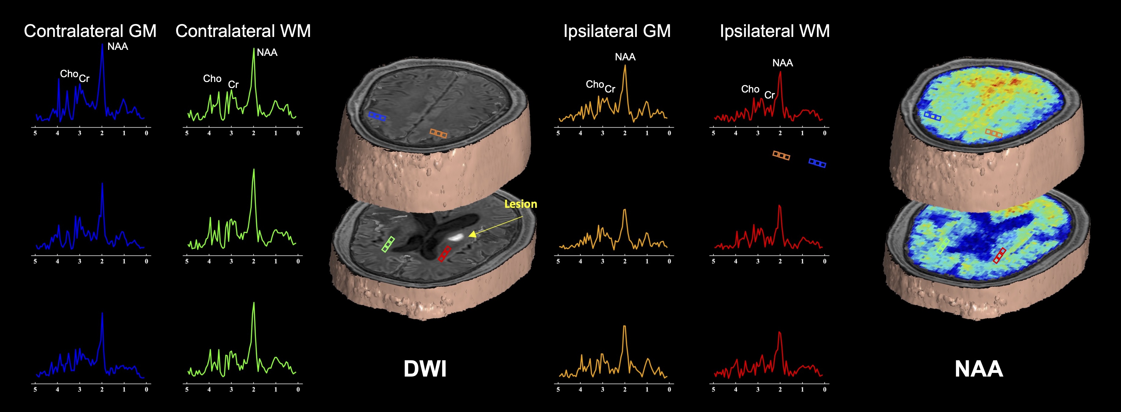
Figure 1. Representative high-resolution 3D NAA maps, along with the spectra and DWI images in lesion and normal-appearing layers obtained from an acute stroke patient.
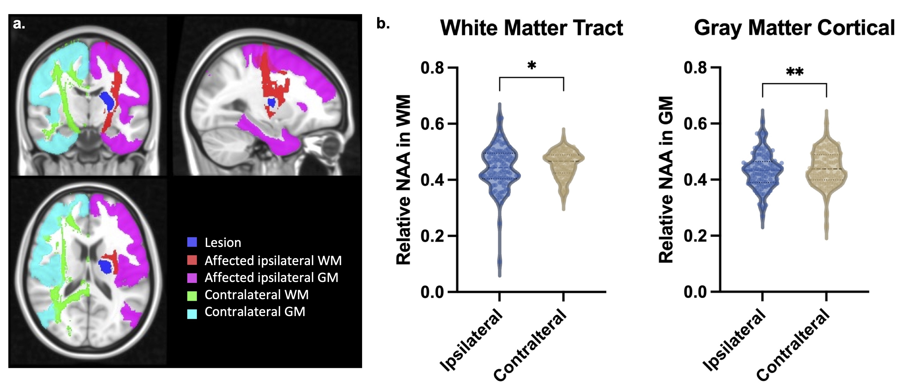
Figure 2. (a) Illustration of regions of interest. (b) Comparison between relative NAA levels in white matter tracts and cortical gray matter of ipsilateral and contralateral hemispheres.
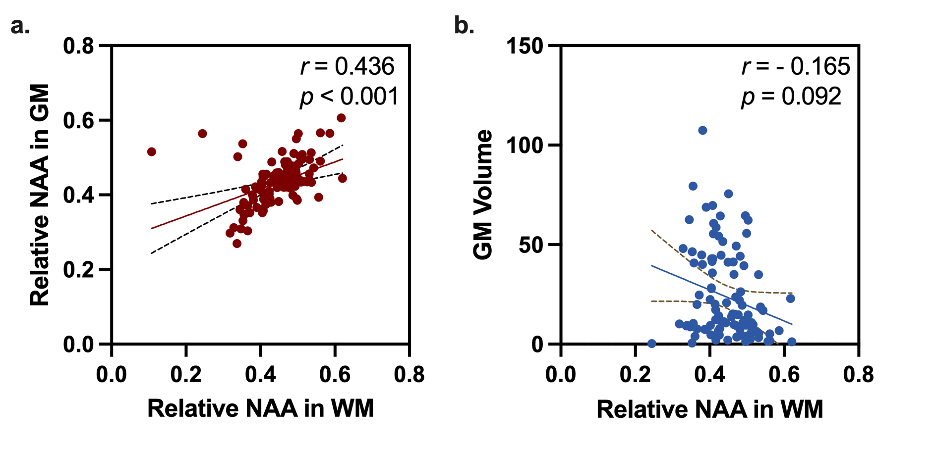
Figure 3. Correlation between relative NAA levels in ipsilateral white mater (WM) and (a) ipsilateral cortical gray matter (GM) NAA levels, and (b) ipsilateral affected GM volume. Each dot represents one subject.
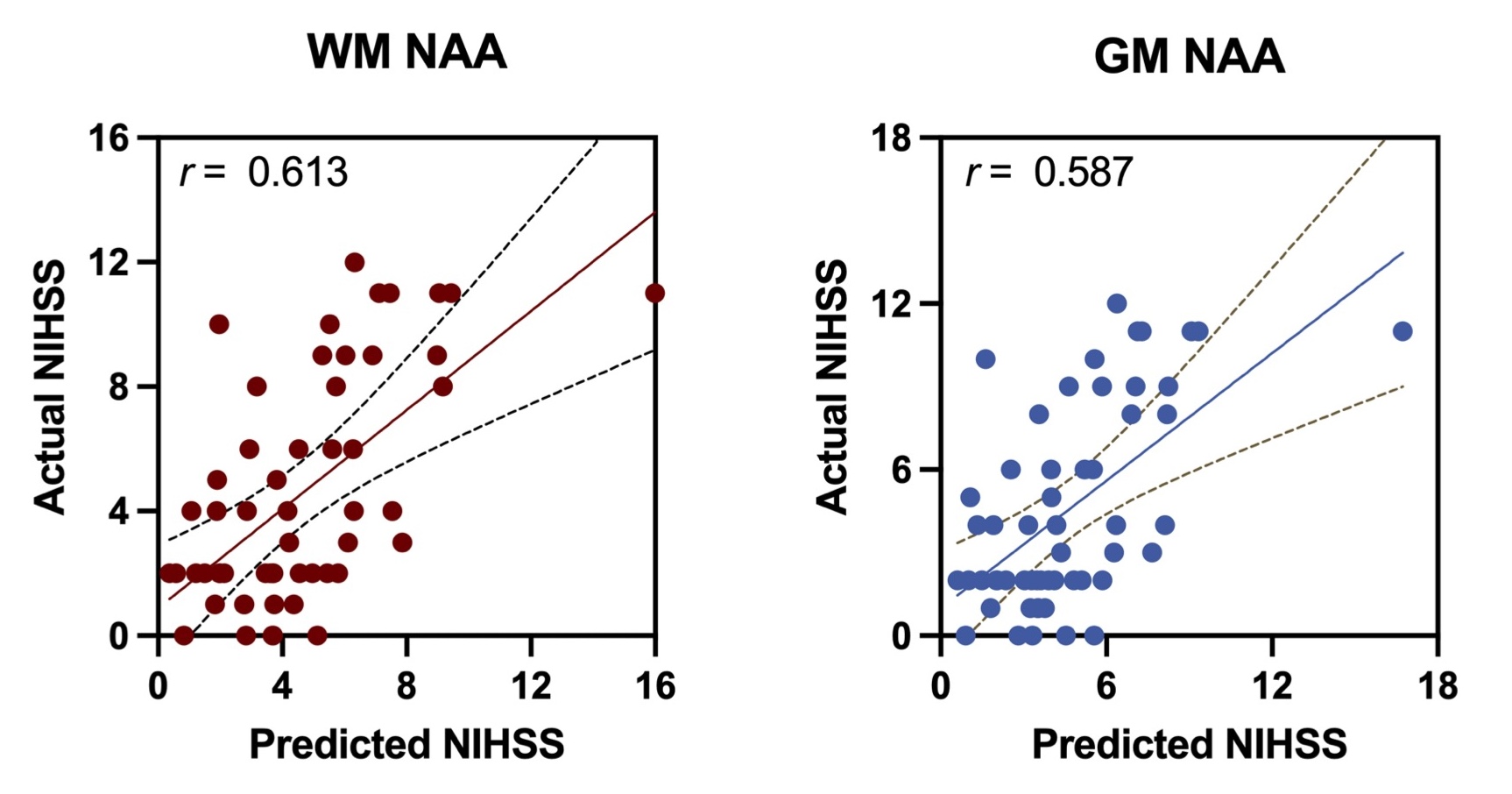
Figure 4. Predictions of NIHSS scores at discharge using NAA in ipsilateral white matter (WM) and cortical gray matter (GM), respectively.
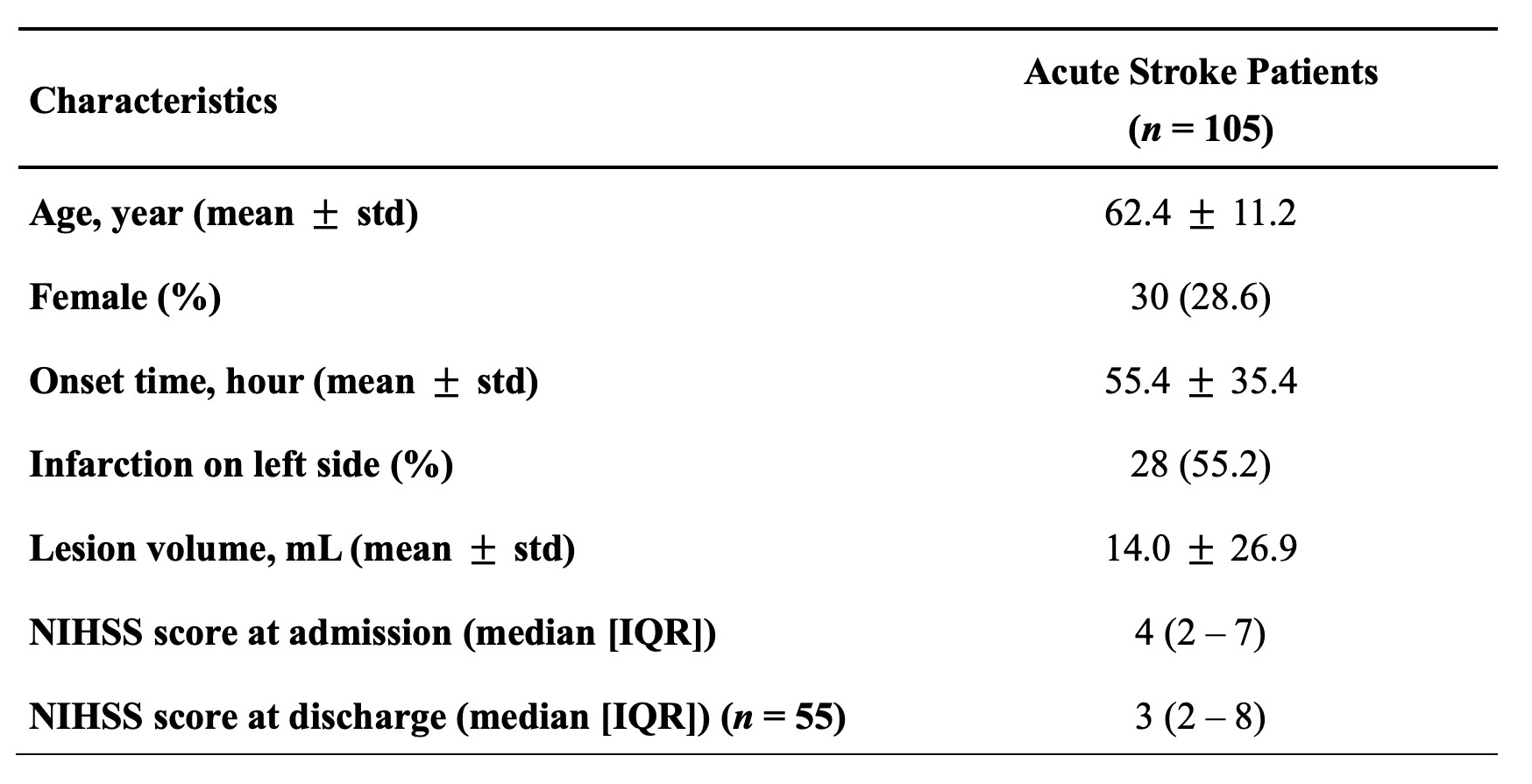
Table 1. Demographic and clinical characteristics of participants
DOI: https://doi.org/10.58530/2024/2326