2323

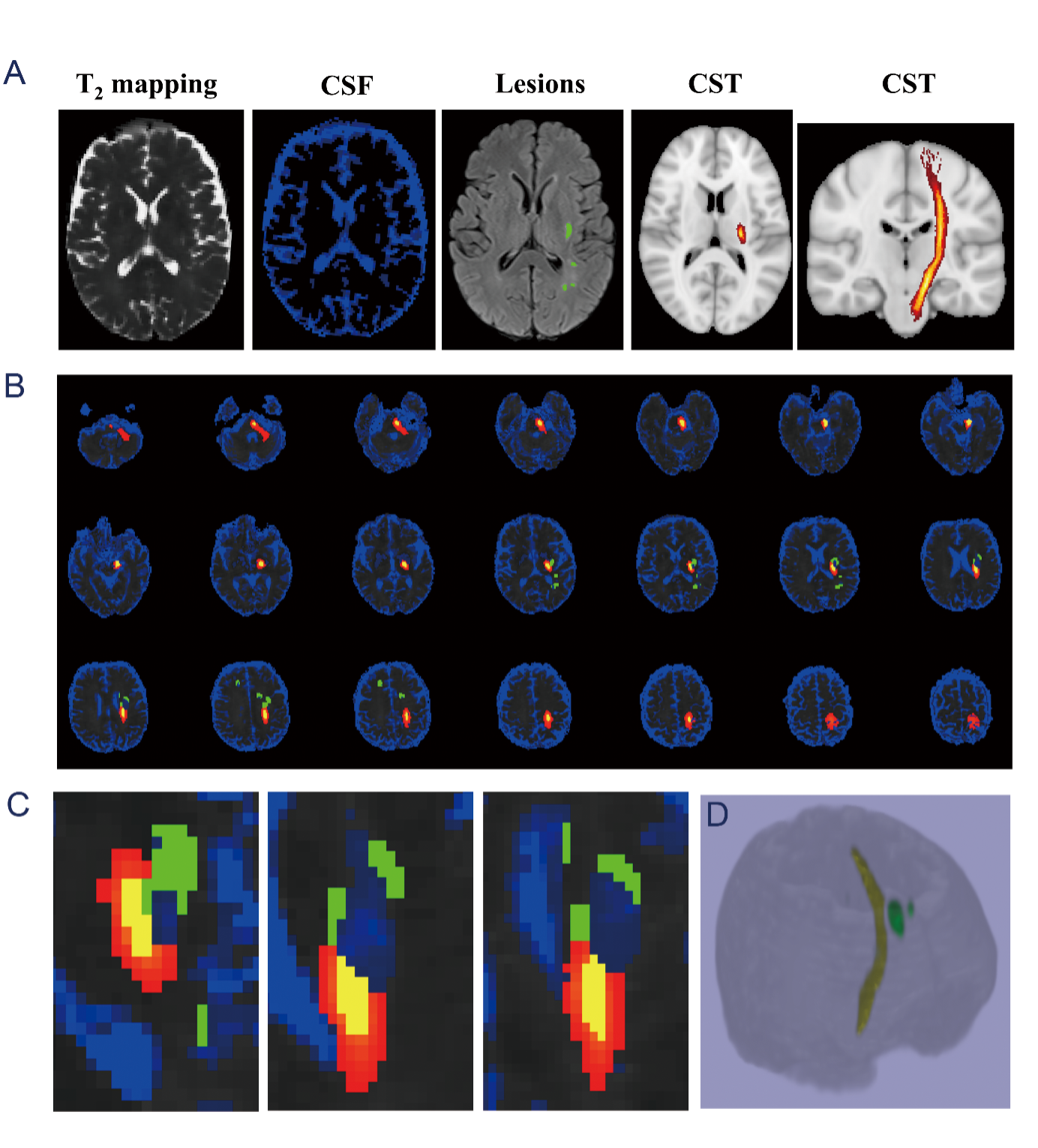
Increased T2 value suggest the NA-CST degeneration following stroke proved by multiple overlapping-echo detachment qMRI
Jianfeng Bao1, Xiao Wang1, Ming Ye2, Qinqin Yang2, Congbo Cai2, Shuhui Cai2, Andrey Tulupov3, Yanbo Dong4, Liangjie Lin5, Yong Zhang1, Zhong Chen2, and Jingliang Cheng1
1Functional Magnetic Resonance and Molecular Imaging Key Laboratory of Henan Province, Department of Magnetic Resonance Imaging, the First Affiliated Hospital of Zhengzhou University, Zhengzhou University, Zhengzhou, China, 2Department of Electronic Science, Fujian Provincial Key Laboratory of Plasma and Magnetic Resonance, School of Electronic Science and Engineering, National Model Microelectronics College, Xiamen University, Xiamen, China, 3The Laboratory «MRT TECHNOLOGIES», The Institute International Tomography Center of the Russian Academy of Sciences, Novosibirsk, Russian Federation, 4Pingdingshan College, Pingdingshan, China, 5Philips Healthcare, Beijing, China

Synopsis

Keywords: Stroke, Stroke

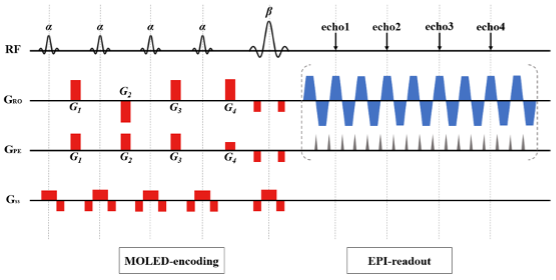
Motivation: Stroke patients commonly face challenges during clinical magnetic resonance imaging (MRI) examinations due to loss of consciousness and involuntary movements. This study aims to address these challenges using a self-developed ultra-fast, multiple overlapping-echo detachment (MOLED) quantitative magnetic resonance technology.

Goal(s): Through this technology, we seek to quantitatively detect potential damage to the motor-related normal-appearing corticospinal tract (NA-CST) following stroke.

Approach: Total 79 patients underwent routine scan and MOLED. A deep learning network was utilized for quantitative image reconstruction.

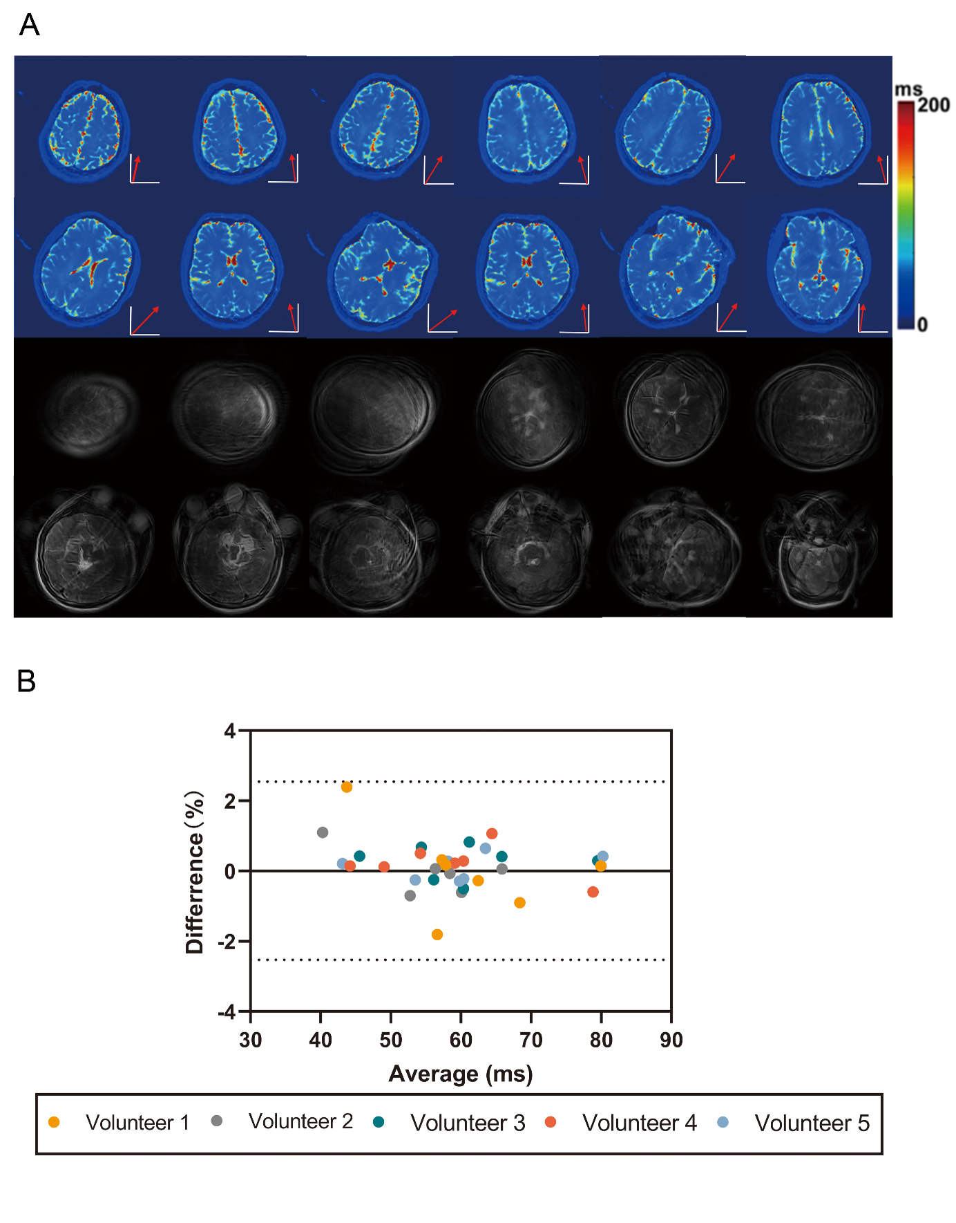
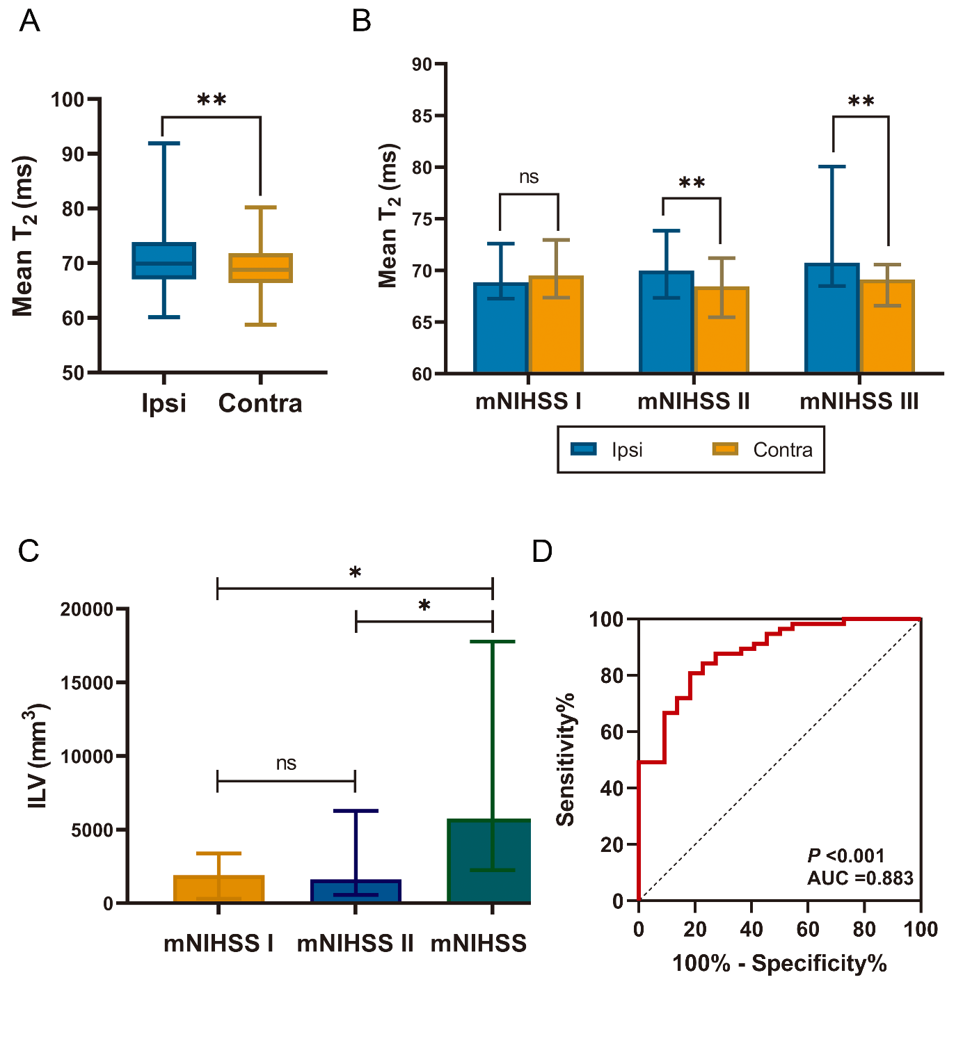
Results: MOLED T2 imaging showed high accuracy and repeatability, was unaffected by head motion, correlated with motor dysfunction severity, and predicted motor impairment post-stroke.

Impact: The MOLED technique quickly and accurately quantifies imaging in stroke patients with involuntary movements and helps monitor post-stroke motor impairment progression.

Stroke patients commonly face challenges during clinical MRI examinations due to loss of consciousness and involuntary movements. This study aims to address these challenges using a self-developed ultra-fast, multiple overlapping-echo detachment (MOLED) quantitative magnetic resonance technology. Through this technology, we seek to quantitatively detect potential damage to the motor-related normal-appearing corticospinal tract (NA-CST) following stroke. A total of 79 patients underwent 3 T MRI exam, which included routine scan and MOLED technique. A deep learning network was utilized for quantitative image reconstruction. The MOLED technique's accuracy, reliability, and resistance to motion were validated on phantoms and five healthy volunteers. Subsequently, we assessed motor dysfunction severity, ischaemic lesion volume, quantitative T2 values of the bilateral NA-CST, and the T2 ratio (rT2) between the ipsilesional and contralesional of the NA-CST in the 79 patients.Results: MOLED-derived T2 showed high accuracy (P < 0.001) and excellent repeatability, with a mean coefficient of variation (CoV) of 1.11%, and accessed reliable quantitative results even under head movement: Meandiff = 0.28%, SDdiff = 1.34%. Moreover, the mean T2 value of the NA-CST on the ipsilesional was significantly higher than the contralesional (P < 0.001), and a positive correlation was observed between rT2 and the severity of motor dysfunction (rs = 0.575, P < 0.001). Moreover, rT2 was able to predict post-stroke motor impairment, with the area under the curve (AUC) was 0.883. The fast quantitative MOLED technique provides substantial benefits for the quantitative imaging of stroke patients who exhibit involuntary movements. Furthermore, T2 mapping derived from MOLED can detect NA-CST degeneration after a stroke, assisting in monitoring the progression of stroke-induced motor impairments.

Acknowledgements

No acknowledgement found.

References

No reference found.

Figures

Figure 1. MRI pulse sequence diagram of MOLED. The first four RF pulses with a flip angle α are excitation pulses, while the fifth pulse with a flip angle β is a refocusing pulse. G1-G4 are echo-shifting gradients along the phase coding dimension and the frequency coding dimension, which is used to shift spin echoes from the center of the k-space to certain locations.

Figure 2. A. The comparison between T2 MOLED and T2 MESE in the same slice on No.3 volunteer are presented under motion. Coordinate axes and red arrows indicate the space and direction of motion of the slices. B. Bland-Altman plots of mean ROI T2 values for five repeat scans without motion and T2 values for ROIs under motion (7 ROIs per volunteer). The Bland-Altman plots also show the mean differences (solid lines) and the 95% limits of agreements (dashed lines). MOLED = multiple overlapping-echo detachment imaging; MESE = multi-section multi-echo spin-echo; ROIs = regions of interest.

Figure 3: Extraction of the NA-CST. A. Left 1. The T2 mapping obtained by deep learning reconstruction. Left 2. The extracted CSF mask. Middle. The lesions mask, consisting of ischaemic lesion and WMH. Right 1.2. The canonical CST tract template B. Brain atlas showing the distribution of lesions and the anatomical location of the CST. C. The local enlargements of B in the z-direction. D. 3D image of NA-CST. Green represents lesions, Red represents the anatomical distribution of CST; Yellow represents the NA-CST after removing lesions and the CST with probability values less than 20.

Figure 3: A. Box plot shows the comparison of mean T2 values between ipsilesional and contralesional in patients. B. The bar graph shows the mean T2 of ipsilesional and contralesional in patients with different severity of motor dysfunction. C. The bar graph shows the comparison of ILV in patients with different severity of motor dysfunction. D. The ROC analysis based on rT2 parameters for distinguishing the onset of movement disorders. ILV = ischaemic lesion volume; Ipsi = ipsilesional; Contral = contralesional; *P<0.05, **P<0.01.

Proc. Intl. Soc. Mag. Reson. Med. 32 (2024)
2323
DOI: https://doi.org/10.58530/2024/2323