2320
Predicting the prognosis of Single Subcortical Infarction using lenticulostriate artery features and subacute perfusion in its region1West China Hospital of Sichuan University, chengdu, China
Synopsis
Keywords: Stroke, Vessels, lenticulostriate artery;perfusion
Motivation: Due to the limitations of standard imaging tools, few studies have directly reserched infarct lesion or its corresponding arteries to predict prognosis. Now, the morphology of lenticulostriate artery can be seen via VWI1, and perfusion of infarct lesion can be determined via PWI2
Goal(s): To evaluate the effect of LSA features and quantitative perfusion on SSI prognosis
Approach: The patients were categorized into groups with poor and good prognoses based on the modified Rankin scale (mRS) score. Each group's LSA morphological parameters and perfusion status were compared.
Results: Poorer results are correlated with shorter LSA and lower rCBF
Impact: The prognosis of the SSI can be assessed by analyzing the morphology of the LSA and the subacute perfusion. Our findings provide evidence in support of early therapy intervention.
Introduction
Single Subcortical Infarction (SSI) is an important type of ischemic stroke. Improving the clinical outcome of these patients can be achieved by research on risk factors. The LSA is the main artery supplying blood to the basal ganglia and surrounding cerebral area. The recent development of intracranial vascular wall imaging (VWI) allows detailed evaluation of the lumen of the LSA. It's unclear whether LSA feature has effect on prognosis. Meanwhile, magnetic resonance perfusion-weighted imaging (PWI) can detect perfusion deficits in patients with SSIs and reflect perfusion status of the supply area of the LSA 3. Previous studies focused on the acute phase or super phase4,5. However, cerebral blood perfusion is highly dynamic in the first few days of infarction, and cerebral perfusion dynamics and risk factors after infarction remain unclear. The quantitative perfusion of cerebral in the subacute stage was required because some patients failed the acute stage perfusion examination. Exploring PWI and VWI as a way to predict prognosis, which may be better than methods based on traditional imaging modalities. In this study, we investigated the predictive value of LSA features and subacute cerebral perfusion status on the prognosis of patients with SSI.Methods
Patients: From September 2021 to May 2022, 25 patients with acute SSI were enrolled and underwent head MRI examinations within 7 days of symptom onset. Function outcome was assessed by the mRS score at 90 days: A score of 0 to 1 is defined as good prognosis, and a score of 2 to 5 is defined as poor prognosis.Imaging parameters: This study was performed on a 3T MR scanner with a dedicated 32-channel head coil (Siemens Medical Systems, Trio, Erlangen, Germany), including conventional T1-weighted, T2-weighted, T2-FLAIR, DWI, 3D time-of-flight MRA, whole-brain VWI, and dynamic susceptibility contrast-PWI. Parameters of whole-brain VWI were: TR/TE=900/14ms; FOV=170 ×170;a total of 240 slices with a slices thickness of 0.53mm;scan time=8min. Parameters of perfusion were: repetition time, 1500 ms; echo time, 32 ms; flip, 90; FOV= 220 × 220 mm; matrix size=128 × 128; A total of 19 slices with a thickness of 5 mm ; scan time=83s.
Data processing: All images were reviewed and analyzed by two experienced radiologists blinded to the clinical data. Raw images were processed by using the IntelliSpace Portal (version 9.05, Philips Healthcare, Netherlands). Through the registration of DWI and PWI images, a region of interest (ROI) covering the hyperintense area of SSI in the DWI map (ROI 1) was manually outlined, and was mirrored to the contralateral unaffected hemisphere (ROI 2). Then the relative CBF, CBV, MTT, and TTP values of the infarct lesion were calculated as the ratio of ROI 1 to ROI 2(Figure 1).
LSA features on the symptomatic side were measured on MinIP with 10–20 mm thickness on WB-VWI. LSAs were manual tracked using a workstation (RadiAnt DICOM viewer). The morphological parameters included the number of stems,branches, total length and average length(Figure 2).
Statistical analysis: SPSS 29.0 software was used for statistical analysis. Multivariable logistic regression models were used to identify independent predictors of the two groups, including all variables with P < 0.05 on univariate analysis. Receiver operating characteristic (ROC) curve was used to analyze the diagnostic efficacy of the indicators between two groups.
Results
The poor prognosis group was associated with significantly shorter average LSA length (21.27 ± 4.01mm versus 25.28 ± 3.41 mm, p = 0.016). There was no significant differences in the total length of LSAs, numbers of stems and branches. And there was no significant difference about the rCBV, rMTT, and rTTP. The poor prognosis group had lower rCBF compared with the good prognosis group (0.76±0.24 versus 0.98±0.20, p = 0.027). Multivariable logistic regression showed that the average LSA length is independent risk factor of predicting the prognosis (p<0.05)( Table 1). The diagnostic effect in combination with rCBF and average LSA length proved effective, with an area under the ROC curve of 0. 867 (95% CI: 0.724 -1.010).Acknowledgements
No acknowledgement found.References
1. Pan YT, Tsai YH, Lee JD, et al. Evaluation of clinical relevance and underlying pathology for hemodynamic compromise in acute small subcortical infarction using MRI-based neuroimaging markers. Biomed J. 2023 Apr;46(2):100529.
2. Jiang S, Cao T, Yan Y, et al.. Lenticulostriate artery combined with neuroimaging markers of cerebral small vessel disease differentiate the pathogenesis of recent subcortical infarction. J Cereb Blood Flow Metab. 2021 Aug;41(8):2105-2115.
3. Jiang S, Cui JY, Yan Yy et al. Association of compromised cerebral perfusion with lenticulostriate artery impairments in the subacute phase of branch atheromatous disease. Ther Adv Neurol Disord. 2022 Jul 4;15:17562864221109746.
4. Yamada M, Yoshimura S, Kaku Y, et al. Prediction of neurologic deterioration in patients with lacunar infarction in the territory of the lenticulostriate artery using perfusion CT. AJNR Am J Neuroradiol. 2004 Mar;25(3):402-8.
5. Pan YT, Tsai YH, Lee JD, et al. Evaluation of clinical relevance and underlying pathology for hemodynamic compromise in acute small subcortical infarction using MRI-based neuroimaging markers. Biomed J. 2023 Apr;46(2):100529. doi: 10.1016/j.bj.2022.03.014. Epub 2022 Mar 30. PMID: 35367449; PMCID: PMC10267958.
Figures
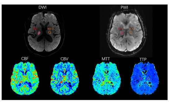
Figure 1. One representative patients of single subcortical infarction patients. An ROI was defined on the infarction in the DWI map (ROI 1) and mirrored to the contralateral unaffected hemisphere (ROI 2),and then through the registration of DWI and PWI images, each ROI was automatically co-registered onto the perfusion maps of cerebral blood flow (CBF), cerebral blood volume (CBV), mean transit time (MTT), and time to peak (TTP).
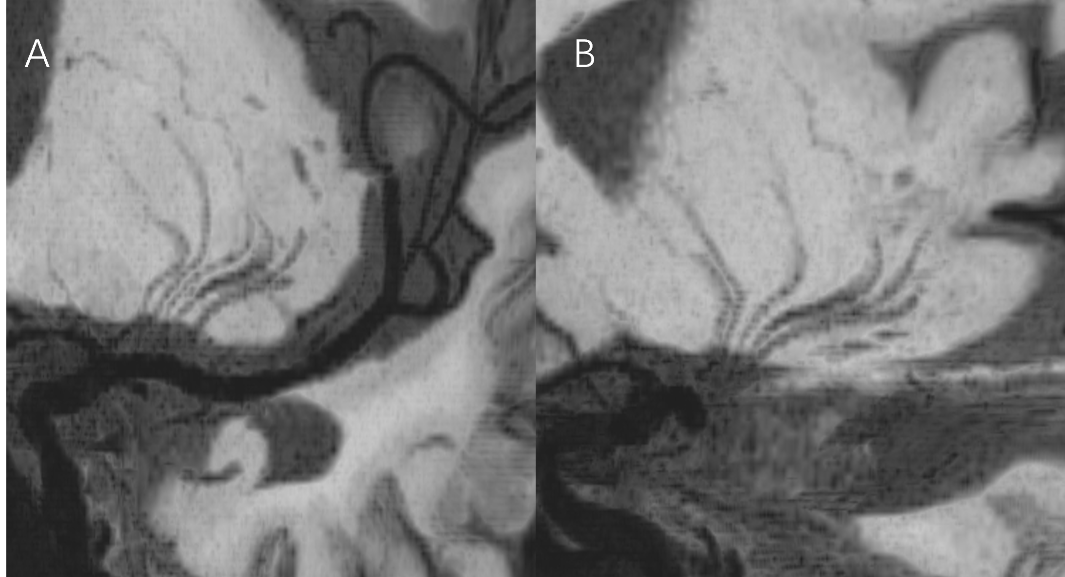
Figure 2. (A) Manual tracing of the LSA on coronal MinIP image alignment. (B) Corresponding tracing of LSA on sagittal MinIP image.

Table 1. Main LSA characteristics and radiological data of patients with Good prognosis and Poor prognosis group