2316
Predictive Value of PET/MR Indicators for Early Detection of Ischemic Brain Changes Following carotid artery stenting1Xuanwu Hospital Capital Medical University, Beijing, China
Synopsis
Keywords: Stroke, Inflammation
Motivation: Aiming to bridge the diagnostic gap in predicting perioperative ischemic lesions post-CAS, crucial for preempting recurrent strokes.
Goal(s): To pinpoint predictive markers on DWI for new ischemic brain lesions within 3 days post-CAS.
Approach: Employed a retrospective analysis of preoperative PET/MR imaging in 47 CAS patients, correlating with postoperative DWI outcomes.
Results: Identification of high PET SUVmax, SCAIL scores, and complex plaques as significant predictors of new ischemic lesions, offering a strategic tool for early high-risk patient detection.
Impact: This study aids clinicians in stratifying stroke risk, influencing patient management and treatment tailoring. It opens avenues for precision medicine in stroke prevention and underscores the need for advanced imaging in preoperative assessments.
Background and Objective
Carotid Artery Stenting (CAS) is an established treatment for carotid artery stenosis. New ischemic brain lesions on diffusion-weighted Images(DWI) appearing perioperatively are one of the common postoperative imaging manifestations, which may be related to the preoperative morphology of the vessel, plaque characteristics, and inflammatory uptake. Therefore, early identification and prediction of new ischemic brain lesions on DWI that may occur within the perioperative period (within 3 days) after CAS stenting are crucial for predicting high-risk patients for recurrent ischemic stroke1.Methods
In this retrospective study, 47 patients who underwent CAS were included. All participants received carotid PET/MR examination before surgery and routine brain MR examination after treatment. Experienced clinicians determined the responsible carotid artery based on symptoms and record reviews. Lumen and outer wall boundaries were segmented on hrVWI, as well as atherosclerotic components, and complicated carotid artery plaques ( AHA-lesion type VI ) were defined by multi-sequence contrast enhancement. The standardized uptake value and the tissue-to-background ratio quantified the 18F-FDG uptake, and the validated symptomatic carotid atheroma inflammation lumen-stenosis(SCAIL) score2 included stenosis severity and inflammation measured by PET. The primary outcome was new ischemic brain lesions on DWI, and a multivariate model was established to evaluate the relationship between imaging biomarkers and new infarction changes.Results
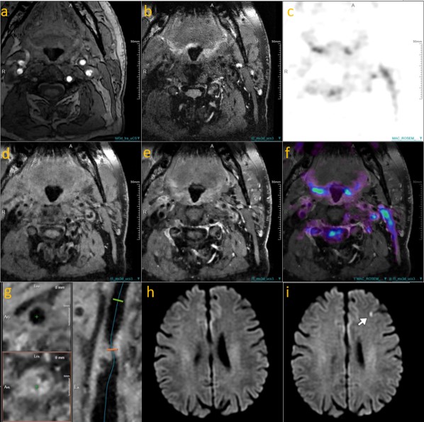
A total of 47 participants were included in the study (mean age, 65.2 years ± 6.85 [SD]; 44 were male, 20 of whom had symptomatic carotid stenosis). Of these 47 participants, 32 (68%) exhibited new ischemic brain changes. The new ischemic brain lesions on DWI were located in (28%, 13/47) and/or beyond (40%, 19/47) the territory of the territory of the treated artery. Higher PET SUVmax (>2.86) was positively associated with necrosis, intraplaque hemor-rhage, ulceration, and calcification (P =0.04 to P 0.001). In the adjusted model , higher SUVmax (>2.86) ,high SCAIL score(>2), complicated plaques was an independent predictor of new ischemic brain lesions, with an odds ratio (OR) of 9.58 [95% CI: 1.12, 82.06, p=0.039], 4.02[95% CI:1.05-15.42,p=0.043], 6.5[95% CI:1.26,33.58,p=0.025]. A post hoc analysis demonstrated A new indicator combined with high SCAIL score(>2) and complicated plaques(+) was a better predictor(aOR 15.0, 95%CI 1.5-149.68, p=0.021) of new ischemic brain lesions.Discussion
The study’s findings illuminate the significant role of imaging biomarkers in predicting ischemic brain lesions post-CAS. High PET SUVmax, elevated SCAIL scores, and the presence of complex plaques were correlated with new ischemic events, aligning with current literature that underscores the relevance of inflammatory processes in stroke pathogenesis. The predictive power of these markers suggests a paradigm shift towards more individualized patient risk assessments, potentially refining perioperative care. Furthermore, the novel indicator combining SCAIL scores and plaque complexity could spearhead more targeted interventions3. However, larger-scale studies are warranted to substantiate these preliminary observations and solidify their clinical utility.Conclusion
Carotid PET/MR-related indicators can effectively predict the risk of new ischemic brain lesions on DWI during the perioperative period after carotid artery stenting, providing important references for early identification of high-risk patients for recurrent ischemic stroke after surgery. Further large-scale randomized controlled studies are necessary to validate the clinical application value of these indicators.Acknowledgements
We extend our gratitude to the patients who participated in this study and their families for their invaluable contribution to medical research. Special thanks to the radiology and neurology departments for their collaboration and expertise in image analysis and clinical evaluation. We also appreciate the technical support provided by the imaging technicians. This work was supported in part by National Natural Science Foundation of China(82130058). Finally, we acknowledge the insightful comments of our peer reviewers, which have greatly enhanced the quality of this work.References
1.Hou Z, Jing J, Yan L, et al. New Diffusion Abnormalities Following Endovascular Treatment for Intracranial Atherosclerosis. Radiology. 2023;307(4):e221499.
2.Camps-Renom P, McCabe J, Martí-Fàbregas J, et al. Association of Plaque Inflammation With Stroke Recurrence in Patients With Unproven Benefit From Carotid Revascularization. Neurology. 2022;99(2):e109-e118.
3.Kopczak A, Schindler A, Sepp D, et al. Complicated Carotid Artery Plaques and Risk of Recurrent Ischemic Stroke or TIA. J Am Coll Cardiol. 2022;79(22):2189-2199.
Figures