Jiali Sun1, Yue Chen1, Min Lv1, Wei Wang1, Jianxiu Lian2, Sicong Huang2, and Yuefei Ma2
1First Affiliated Hospital of Harbin Medical University, Harbin, China, 2Philips Healthcare, Beijing, China, Beijing, China
Synopsis
Keywords: Stroke, Brain, Cerebral ischemia, NODDI, Vic, ODI
Motivation: It is currently unclear whether neurite orientation dispersion and density imaging (NODDI) can reflect the microstructure in early cerebral hypoperfusion.
Goal(s): Therefore, this study aims to detect the cerebral microstructure using NODDI in rabbits’ cerebral ischemia model.
Approach: The orientation dispersion index (ODI) and intracellular volume fraction (Vic) were calculated for comparing among groups, including pre-operative and post-operative of rabbit models, and control group.
Results: The results showed that Vic of bilateral cerebral hemispheres in post-operative state was higher than that of pre-operative and control groups (all P < 0.05). NODDI could be used to reflect the mild injury in early cerebral ischemia.
Impact: Early
recognition of injury in brain tissue microstructure in patients with cerebral
ischemia is helpful to guide clinical treatment, and NODDI will be able to
identify early brain injury.
Introduction
Cerebral ischemia caused by decreased cerebral blood flow (CBF) is an important cause of acute cerebral infarction (ACI)[1]. Studies have shown that the probability of ACI in TIA patients with persistent brain hypoperfusion is 4%-10% within 7 days[2]. Moreover, the risk of recurrence with cerebral ischemia symptoms within 3 months is higher than that in patients with normal perfusion[3]. Cerebral perfusion weighted imaging is the most commonly employed imaging technique to evaluate CBF, but it can solely depict hemodynamics alterations and is not suitable for evaluating the microstructure changes in pathological state. Researchers demonstrated that it is feasible to evaluate ischemic stroke with NODDI, which exhibits greater sensitive in characterizing the microstructure changes of brain tissue when compared with DTI and DKI [4]. Therefore, the purpose of this study is to explore the microstructure changes under hypoperfusion state by using NODDI.Materials and methods
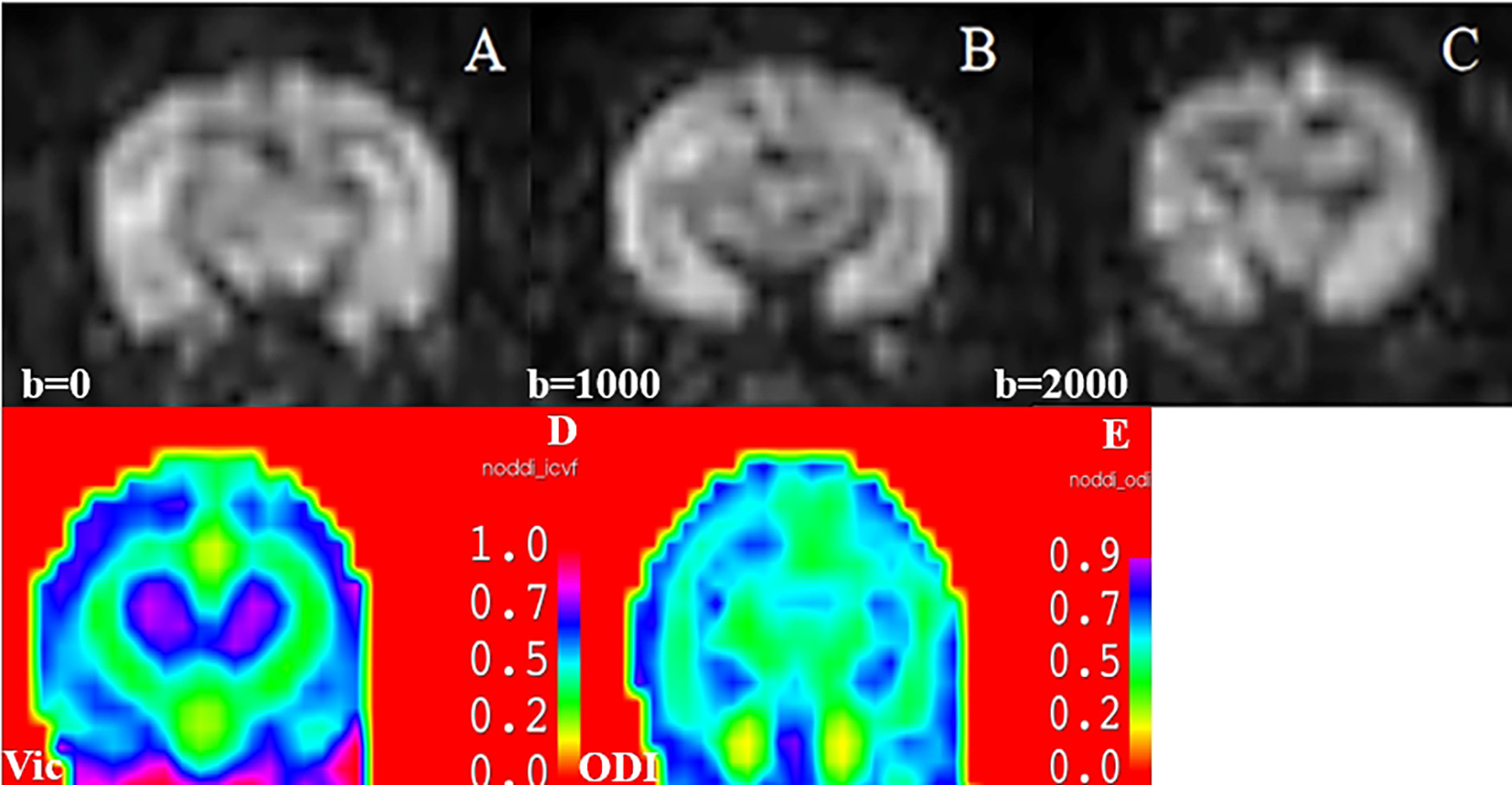
There were 10 Japanese big-eared white rabbits selected in this study (2.5±0.5 kg), which were raised in accordance with standard animal laboratory conditions. They were randomly divided into control group (n=3) and test group (n=7). The test group underwent bilateral carotid artery occlusion surgery (2VO) to establish a model of cerebral hypoperfusion and the control group underwent sham operation. All rabbits were scanned with 3.0T MRI (Ingenia Elition; Philips Healthcare, Best, the Netherlands) scanner and 8-channel elbow coil. The scanning sequence included pseudo-continuous arterial spin labeling imaging(pCASL), NODDI and 3D-T1WI. All rabbits underwent a baseline MRI scan. In the test group, the second MRI scan was performed after 1 hour of 2VO operation, and CBF of both hemispheres was measured by pCASL sequence. The model was considered successfully when the CBF after operation reduced by 30% compared with that before operation [5]. IntelliSpace Medicina Scientia (ISMS, v2.7.0, Philips Healthcare, China) software provided by Philips platform was used to process NODDI DICOM images, and Vic and ODI images were generated (Figure1), and then 3D-Slicer (v5.2.2) software were utilized to measure the quantitative parameters of bilateral cerebral hemispheres. The selected measurement section is the lateral ventricle level and its upper and lower adjacent levels, and the average value would be the final measurement result. Paired t test was used to compare the differences of Vic and ODI between pre-operation (normal perfusion period) and post-operation (hypoperfusion period). The differences of Vic and ODI between the control group and the test group were compared by independent sample t test.Results
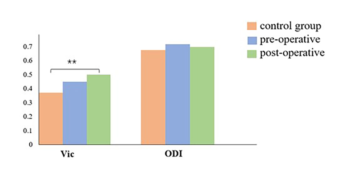
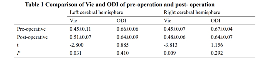
The results of this study showed that the CBF in test group decreased obviously after operation. Vic of the left and right hemispheres in post-operation was higher than pre-operation (P = 0.031, P = 0.009), and there was no significant difference in ODI between pre-operation and post-operation (Table 1). There was no statistical difference between the control group and the pre-operation in Vic and ODI (P > 0.05). The Vic in the control group was lower than that in the post-operation (P = 0.01), and there was no statistical difference in ODI between the two groups (Figure 2).Discussion
This study simulated the early stage of human cerebral ischemia by establishing rabbit hypoperfusion model. The results showed that the Vic increased during hypoperfusion, but the ODI did not exhibit significantly changes. NODDI is an advanced non-Gaussian biophysical diffusion model, which can specifically characterize and quantify the microstructure characteristics of brain tissue through obtain indicators of unique directional dispersion index (ODI) and intracellular volume fraction (Vic) [6]. Vic represents the intracellular volume fraction [7]. Hypoperfusion is a consequence of the reduced CBF, brain tissue would experience mild hypoxia and metabolic obstruction during this period. As a result, the cellular volume may increase, then leading to an elevation in Vic value. ODI is an index to measure the dispersion of nonparallel axons around the central direction, which can reflect the integrity and complexity of axons in white matter and dendrites in gray matter[8]. In this study, it was found that there was no significant increase in the ODI value under the condition of hypoperfusion, which indicates that the brain tissue may primarily undergo pathological changes at the level of nerve cells, without affecting the morphological changes of axons and dendrites. According to the imaging findings of this study, further histological examination is still needed to confirm, which is also the main task in our further work.Conclusion
The microstructure changes of brain tissue in hypoperfusion state may have occurred before ACI by NODDI technique, which may be helpful to find the pathological changes in the early stage of cerebral ischemia.Acknowledgements
No acknowledgement found.References
[1] Huang HT, Tung TH, Lin M, et al. Characterizing spatiotemporal progression and prediction of infarct lesion volumes in experimental acute ischemia using quantitative perfusion and diffusion imaging. Appl Radiat Isot. 2021; 168:109522. doi:10.1016/j.apradiso.2020.109522 [2] Kelly, P. J., Albers, G. W., Chatzikonstantinou, A., De Marchis, G. M., Ferrari, J., George, P., et al. (2016). Validation and comparison of imaging-based scores for prediction of early stroke risk after transient ischaemic attack: a pooled analysis of individual-patient data from cohort studies. Lancet Neurol. 15, 1238–1247. doi: 10.1016/S1474-4422(16)30236-8 [3] Wang Y, Liang H, Jin L, Wang S. Power of Hypoperfusion in Predicting Recurrent Transient Ischemic Attacks: Protocol of a Prospective Cohort Study. Front Hum Neurosci. 2021; 15:654383. Published 2021 Jun 24. doi:10.3389/fnhum.2021.654383 [4] Wang Z, Zhang S, Liu C, et al. A study of neurite orientation dispersion and density imaging in ischemic stroke. Magn Reson Imaging. 2019;57:28-33. doi: 10.1016/j.mri.2018.10.018 [5] Jiang T, Luo J, Pan X, et al. Physical exercise modulates the astrocytes polarization, promotes myelin debris clearance and remyelination in chronic cerebral hypoperfusion rats. Life Sci. 2021; 278:119526. doi:10.1016/j.lfs.2021.119526 [6] Zhang H, Schneider T, Wheeler-Kingshott CA, Alexander DC. NODDI: practical in vivo neurite orientation dispersion and density imaging of the human brain. Neuroimage. 2012;61(4):1000-1016. doi:10.1016/j.neuroimage.2012.03.072 [7] Timmers I, Roebroeck A, Bastiani M, Jansma B, Rubio-Gozalbo E, Zhang H. Assessing Microstructural Substrates of White Matter Abnormalities: A Comparative Study Using DTI and NODDI. PLoS One. 2016;11(12):e0167884. Published 2016 Dec 21. doi:10.1371/journal.pone.0167884 [8] Wang ZX, Zhu WZ, Zhang S, Shaghaghi M, Cai KJ. Neurite Orientation Dispersion and Density Imaging of Rat Brain Microstructural Changes due to Middle Cerebral Artery Occlusion at a 3T MRI. Curr Med Sci. 2021;41(1):167-172. doi:10.1007/s11596-021-2332-3