2311
Microstructural Alterations in Language-Related White Matter Tracts in Post Basal Ganglia Stroke: A DSI study1The First Hospital of Jilin University, Changchun, China, 2MR Research Collaboration, Siemens Healthineers, Beijing, China
Synopsis
Keywords: Stroke, Diffusion/other diffusion imaging techniques
Motivation: Post-stroke basal ganglia aphasia is common in clinical practice, and it is necessary to explain the causes of aphasia after basal ganglia infarction from imaging perspectives.
Goal(s): To obtain white matter fiber bundles associated with the occurrence of basal ganglia aphasia.
Approach: To apply DSI studio and use deterministic fiber-tracking algorithm to reconstruct language-related fiber bundles and measurequantitative anisotropy (QA) of each white matter tracts.
Results: The damage to language related white matter fiber bundles such as corpus callosum fibers may be related to the occurrence of basal ganglia aphasia after stroke.
Impact: By studying the integrity of white matter fiber bundles of basal ganglia aphasia after stroke, it provides an auxiliary role for clinical analysis of its pathogenesis.
Introduction
Post stroke aphasia (PSA) standsasan acquired language impairmentresultingfrom cerebralcortex or disruption within the language-related brain network due tocerebrovascular diseases1. Classical language theorydelineates the transmission oflinguistic information from the Wernicke region through the arcuate tract to the Broca's region, where encoding transpires before manifesting as language functions.Thus, the language network in the cerebral cortex serves as the main anatomical basis of language processing. However, emerging evidence has suggested the potential of subcortical lesions in aphasia2,3. Notably, post-stroke basal ganglia aphasia, a frequent clinical presentation, show milder language deficits, accelerated recovery and improved prognosis compared with cortical aphasia, hinting at different mechanisms underlaying anatomical structure damage and subsequent functional rehabilitation4.This study aims to observe changes in microstructural integrity of language-related white matter tracts in patients with basal ganglia aphasia within 7 days after onset by diffusion spectrum imaging (DSI), in order to obtain white matter fiber bundles related to language function damage in basal ganglia aphasia. Quantitative anisotropy (QA) is a measure of anisotropy of a diffusion process in a biological tissue. In the central nervous system, QA value can reflect the integrity of axons or myelin sheath5.Methods
Eight aphasia patients with basal ganglia aphasia within 7 days of onset and eight healthy controls(gender, age and years of education matched)were enrolled in this study (Table 1). MRI examinations were performed on a 3T system (MAGNETOM Vida, Siemens Healthineers, Erlangen, Germany). The DSI mapping parameters were: TR/TE = 3000/93ms; FOV = 220×220mm; voxel size = 2.2×2.2×4.0mm3; bmax = 3000s/mm2; acquisition direction = 128; acquisition time = 7min 35 sec. The diffusion data were reconstructed using generalized q-sampling imaging in DSI-studio. Eight aphasia patients underwent the Chinese Aphasia Battery (ABC) to evaluate language function. Aphasia Quotient (AQ) refers to the language ability of patients with aphasia. The normal range of AQ values is 98.4~99.6, and AQ<93.8 can be considered as aphasia. And we measured quantitative anisotropy (QA) of white matter tractsof bilateral arcuate fasciculus (AF), superior longitudinal fasciculus (SLF), inferior fronto-occipital fasciculus (IFOF), inferior longitudinal fasciculus (ILF), uncinate fasciculus (UF), and corpus callosum (CC). The independent t-test was used to compare the differences in QAvalues between the aphasia group and HCgroups.The correlation between QA values of different language related fiber bundles and ABC scale scores was analyzed using DSI based correlation tractography. Correlation Tracography is a research method based on fiber tracing that tracks the correlation between anisotropy (such as QA) and research variables in population studies.Results
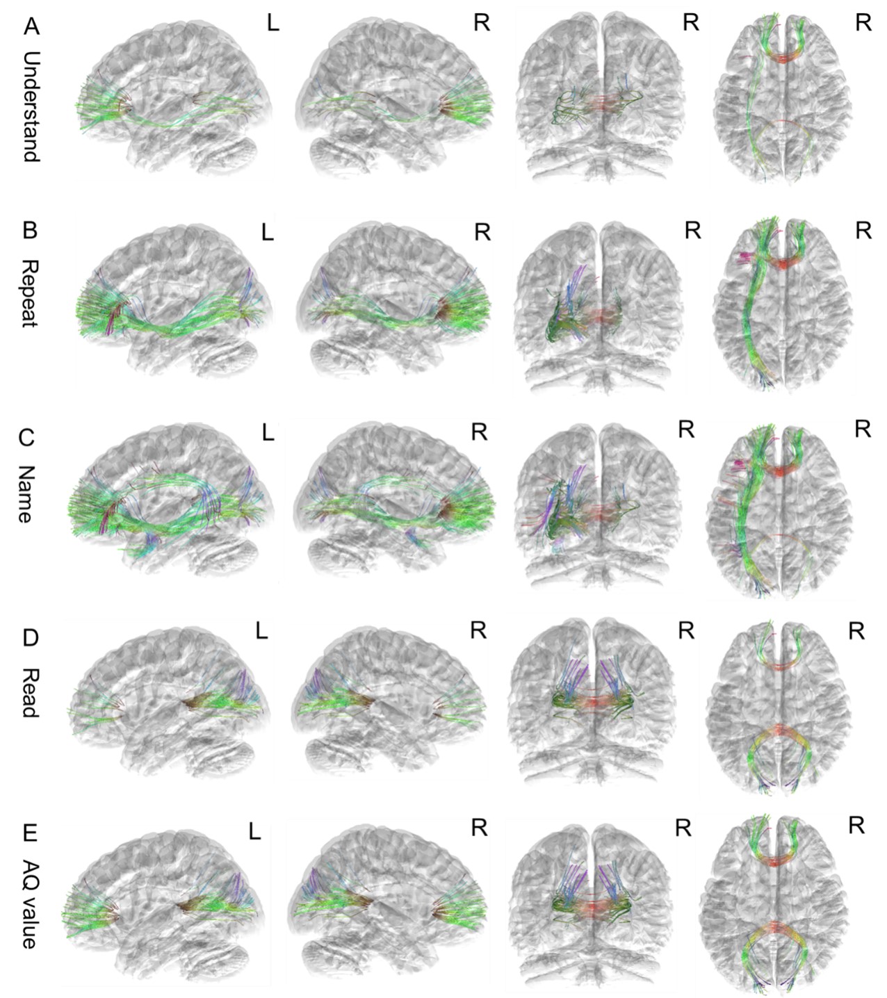
The comparison results of ABC scale scores between the aphasia group and the control group are shown in Table 2. Within 7 days after onset, the fluency, understanding, naming, reading, and AQ of the ABC scale in the aphasia group were lower than those in the control group (P<0.001), and the differences were statistically significant. The QA values of different white matter fibers related to languages between Aphasic patients and healthy controls are shown in Table 3. Compared with the healthy controls, the QA values statistically decreased in the aphasia group incorpus callosum body, left AF, left SLF II, left SLF III, left IFOF, and left UF (P<0.05). The results of the correlation between QA values of language related white matter tracts and clinical language scales in patients with aphasia within 7 days of onset is showed in Figure 1. The understanding, repeating, naming, reading, and AQ values were positively correlated with the QA values of the corpus callosum forceps minor or corpus callosum forceps major.Discussion
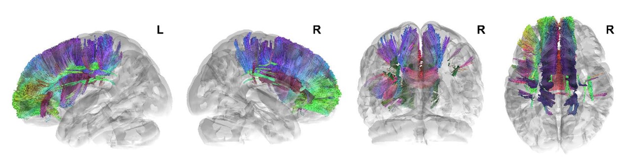
In this study, we found that the clinical features of basal ganglia aphasia after stroke are moderate retelling ability, impaired fluency, understanding, naming, and reading abilities.Secondly, the QA value of language related white matter fiber bundles such as corpus callosum body decreases, indicating that their integrity is disrupted, suggesting that the occurrence of basal ganglia aphasia may be related to the destruction of these fiber bundles(Figure 2). Finally, we found that understanding, repeating, naming, reading, and AQ values were positively correlated with the QA values of the corpus callosum forceps minor or corpus callosum forceps major. This may indicate that the integrity of the corpus callosum forceps minor and corpus callosum forceps major is closely related to the language function of patients with aphasia.Conclusion
In this study, we applied DSI technology to study the changes in white matter fiber bundles in basal ganglia aphasia after stroke. We found that the integrity of the corpus callosum, left AF, left SLF II, left SLFIII, left IFOF, and left UF was disrupted.Acknowledgements
No acknowledgement found.References
[1]Klingbeil, J., et al., Resting-state functional connectivity: An emerging method for the study of language networks in post-stroke aphasia. Brain Cogn, 2019. 131: p. 22-33.
[2]Démonet, J.-F., et al., “Subcortical” aphasia: Some proposed pathophysiological mechanisms and their rCBF correlates revealed by SPECT. Journal of Neurolinguistics, 1991. 6(3): p. 319-344
[3] Radanovic, M. and V.N. Almeida, Subcortical Aphasia. Curr Neurol Neurosci Rep, 2021. 21(12): p. 73.
[4] Kang, E.K., et al., Severity of post-stroke aphasia according to aphasia type and lesion location in Koreans. J Korean Med Sci, 2010. 25(1): p. 123-7.
[5] Yeh FC, Zaydan IM, Suski VR, Lacomis D, Richardson RM, Maroon JC, et al. Differential tractography as a track-based biomarker for neuronal injury. Neuroimage. 2019;202:116131.
Figures
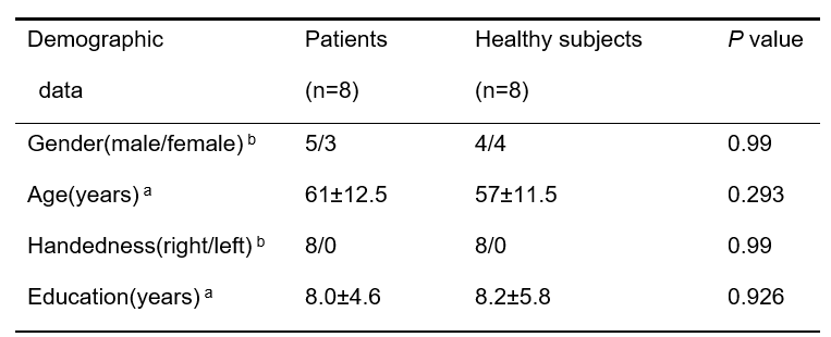
Table 1 Subject demographic information
aP values for the Chi-squared test; bP values for the independent t-test.
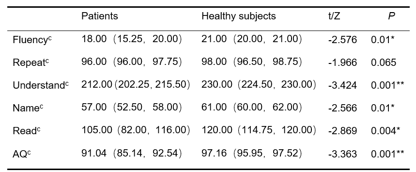
Table 2 Comparison of ABC clinical scale scores between the aphasia group and the control group within 7 days after onset
AQ indicates aphasia quotient; cP values for the Non parametric rank sum test. *P<0.05,**P<0.001.
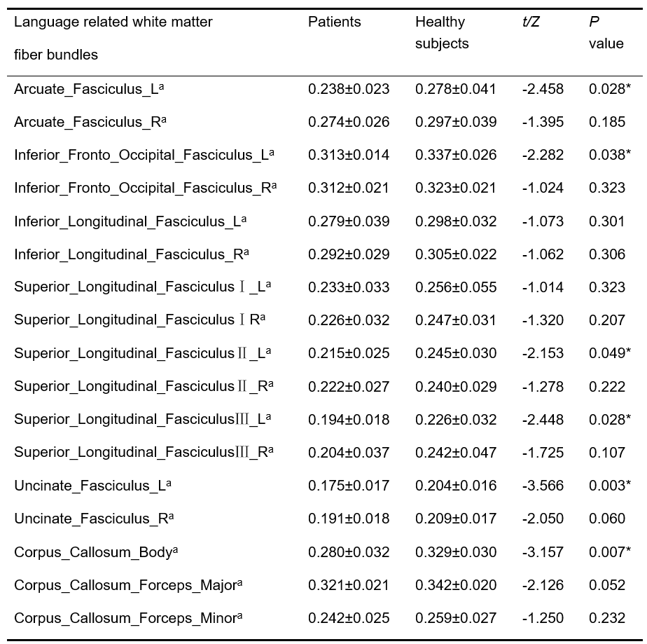
Table 3 Comparison of QA values of white matter tracts between the aphasia group and the control group within 7 days after onset
QA indicates quantitative anisotropy; aP values for the independent t-test. *P <0.05.