2305
Application of High-resolution MR reconstruction technique, Precise IQ Engine (PIQE) for Flow Sensitive Black Blood (FSBB) Imaging1Department of Diagnostic Imaging and Nuclear Medicine, Graduate School of Medicine, Kyoto University, Kyoto, Japan, 2Department of Advanced Imaging in Medical Magnetic Resonance, Graduate School of Medicine, Kyoto University, Kyoto, Japan, 3MRI Systems Division, Canon Medical Systems Corporation, Otawara, Japan
Synopsis
Keywords: Stroke, Neuro
Motivation: To apply the Precise IQ Engine (PIQE), a new high-resolution MR reconstruction technique, for Flow Sensitive Black Blood (FSBB) imaging.
Goal(s): To evaluate the usefulness of PIQE comparing the super-resolution FSBB images reconstructed from low-resolution FSBB images with short acquisition time using PIQE, low-resolution FSBB images, and high-resolution FSBB images.
Approach: On three FSBB images, cerebral microbleed (CMB) was identified and image quality was assessed for 121 patients who underwent FSBB images for CMB detection.
Results: PIQE made CMBs, blurred due to low resolution, clearly visible. PIQE-FSBB has image quality comparable to high-resolution FSBB even with shorter scan time.
Impact: PIQE, a technique that reconstructs high-resolution images from low-resolution images, was applied for FSBB imaging. The super-resolution FSBB reconstructed from low-resolution FSBB with short acquisition time had good quality, and was comparable to high-resolution FSBB for cerebral microbleeds detection.
INTRODUCTION:
High-resolution MR imaging (HR-MRI) is desirable for detailed evaluation which may lead to precise diagnosis. However, HR-MRI has a drawback of long acquisition time, which often prevents its clinical application. To overcome this drawback, super-resolution image reconstruction is gaining attention1, and a new reconstruction method, called Precise IQ Engine (PIQE), has been recently developed. PIQE reconstructs high-resolution images from low-resolution images using a technique included denoising part and zero-padding interpolation (ZIP) and the assist of neural network technology2. Susceptibility weighted MRI such as Flow Sensitive Black Blood (FSBB) enhances vessel contrast without the drawbacks of excessive T2* decay. FSBB employs very weak motion probing gradients in T2*-weighted sequences, making it sensitive to susceptibility and slow-flowing vessels3,4. Although FSBB needs a longer scan time compared to conventional T2*-weighted sequences, FSBB is clinically useful for conditions like cerebral microbleeds (CMB), brain injury, stroke, vascular malformations, and venous disease5-9. The purpose of this study was to compare the super-resolution FSBB images reconstructed from low-resolution FSBB images with short acquisition time using PIQE (PIQE-FSBB), low-resolution FSBB images with short acquisition time (LR-FSBB), and high-resolution FSBB images with long acquisition time (HR-FSBB) for evaluation of usefulness of PIQE.METHODS:
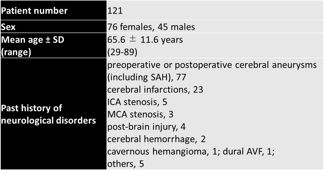
SubjectsOne hundred twenty-one patients who had undergone both LR-FSBB and HR-FSBB for CMB detection between January 2023 and May 2023 were enrolled under IRB approval. The demographics of all participants are shown in Figure 1.
Image Acquisition
The two FSBB sequences (HR-FSBB and LR-FSBB) were performed using a 3T-MR system (Canon Medical Systems Corporation, Vantage Centurian) with a 32-channel head coil. PIQE was adapted to FSBB as Work-In-Progress. Parameters of image sequence were as follows. HR-FSBB: TR/TE, 29/20ms; flip angle, 20°; acquisition matrix size, 240×320; FOV, 200×220mm; slice thickness, 1mm; number of slices 128; band width, 89Hz/pixel; number of acquisition, 1; acceleration factor, Speeder 2.0×1.5; acquisition time, 5min 28sec. LR-FSBB: TR/TE, 29/20ms; flip angle, 20°; acquisition matrix size, 120×320; FOV, 200×220 mm; slice thickness, 1mm; number of slices 128; band width, 89Hz/pixel; number of acquisition, 1; acceleration factor, Speeder 2.0×1.6; acquisition time, 2min 44sec.
Super-resolution Image Reconstruction
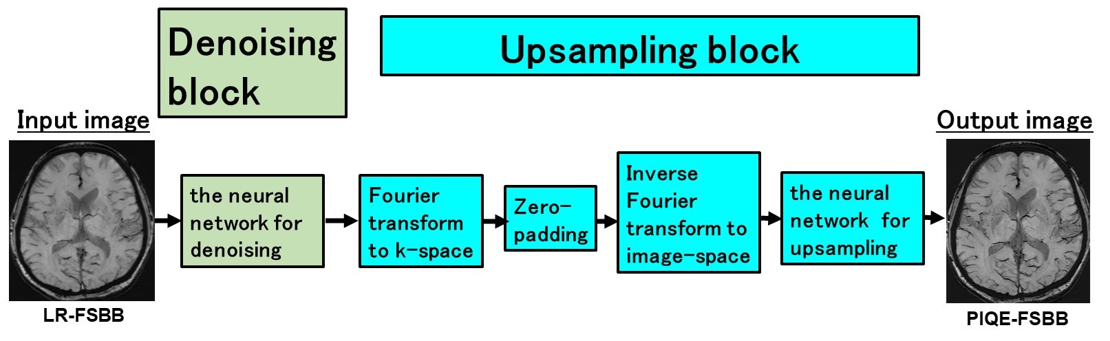
PIQE-FSBB was reconstructed from LR-FSBB using PIQE (matrix, 3×) (Figure 2).
Image Analysis
MinIP (minimum intensity projection) images of three FSBB images (PIQE-FSBB, LR-FSBB, and HR-FSBB) were evaluated. A board-certified radiologist, blinded to image type and patient information, identified and counted CMB. CMB was defined as small, round low signal intensities on FSBB that could not be followed on consecutive slices like blood vessels, 2–10 mm in size 10. Overall image quality, structural conspicuity, and artifact were assessed qualitatively using a 5-point Likert scale. Structural conspicuity was mainly evaluated for vessel visibility.
Statistical Analysis
The intraclass correlation coefficient (ICC) was calculated to determine CMB detection agreement among three FSBB images. The scores were compared among three FSBB images using the Friedman test followed by pairwise comparisons with Bonferroni correction. P value less than 0.05 was considered statistically significant.
RESULTS:
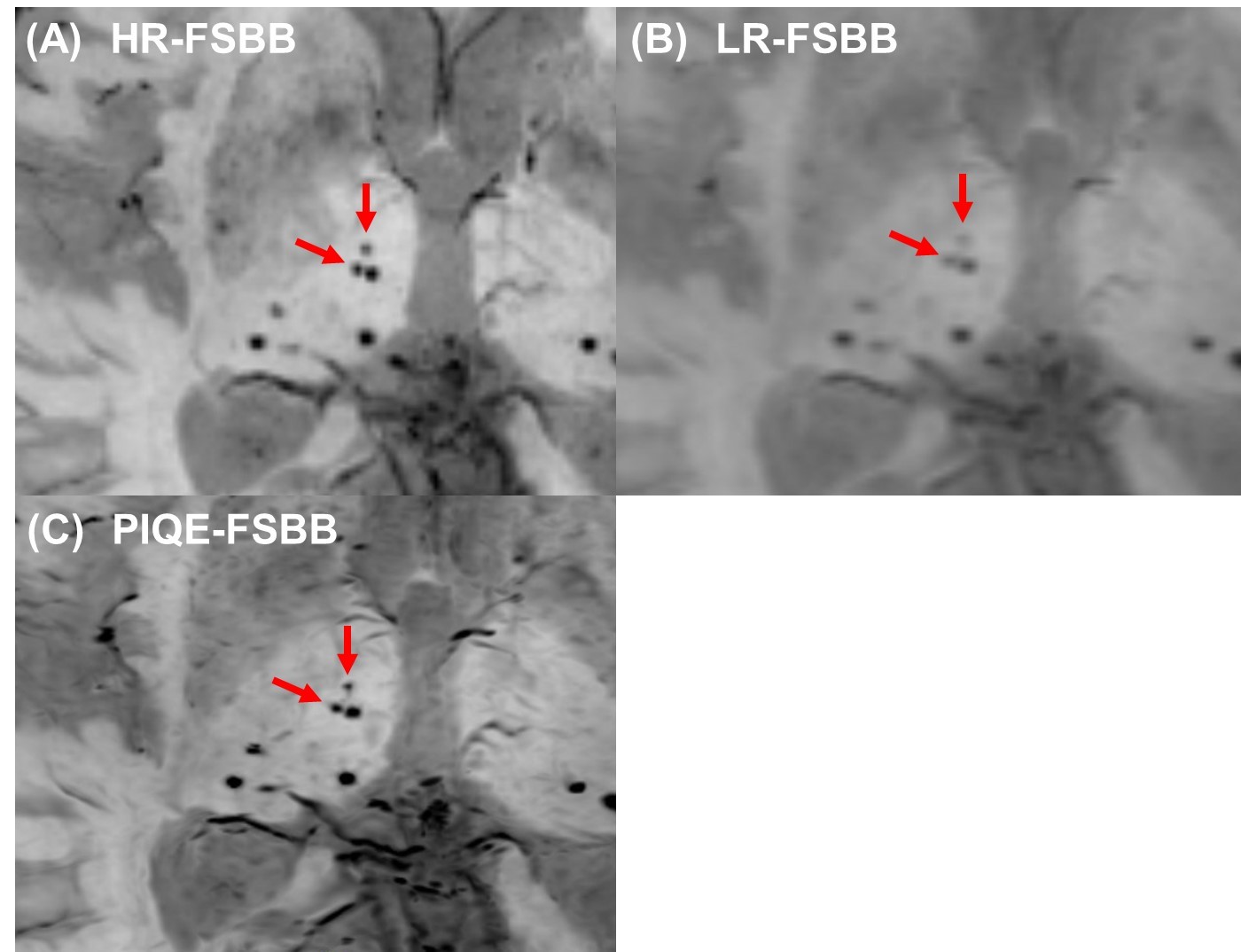
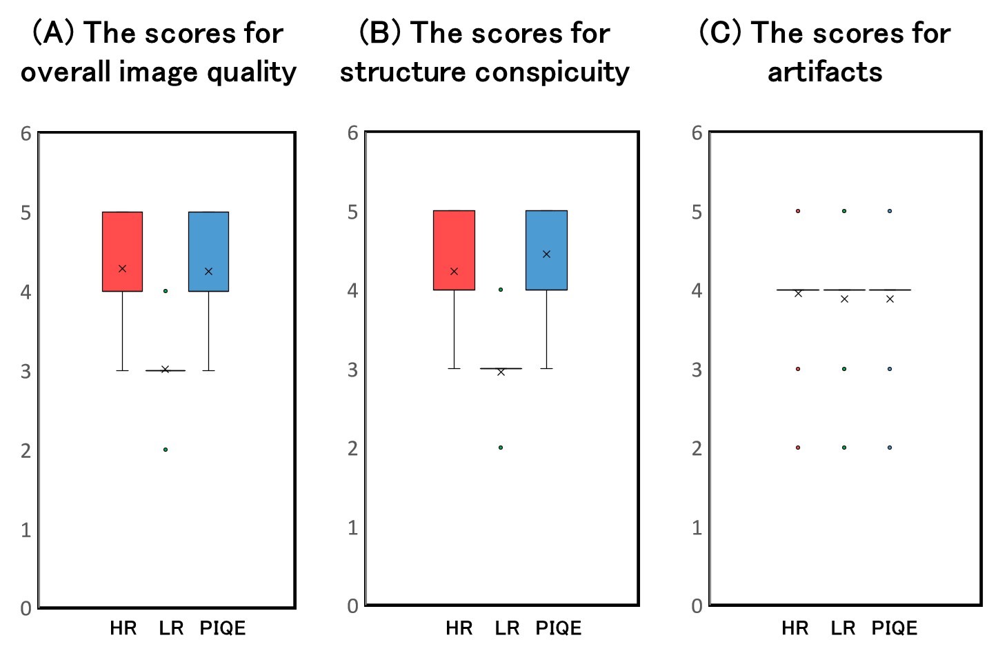
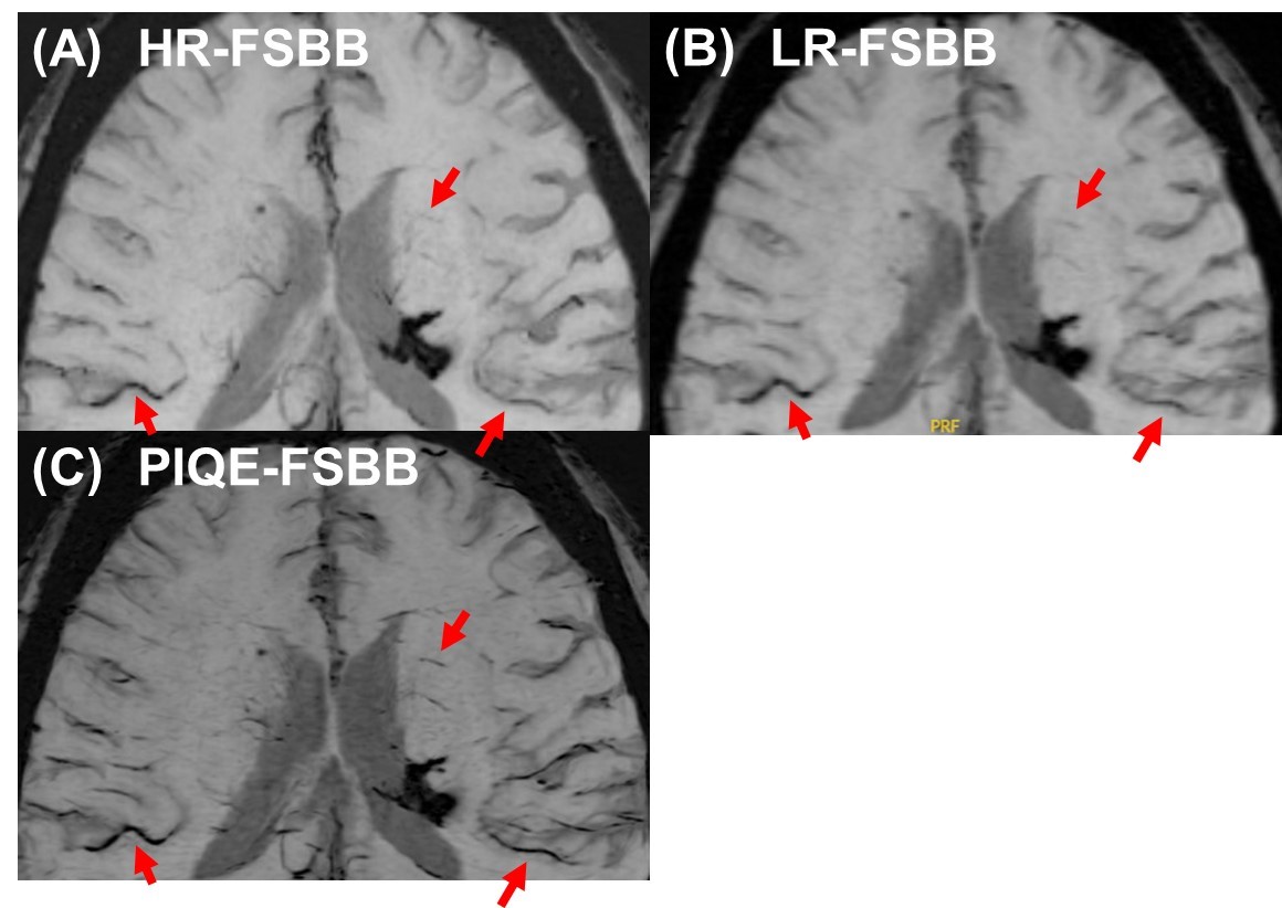
Out of 121 patients, 30 patients had 1-5 CMBs, 4 patients had 6-10 CMBs, 12 patients had 11-20 CMBs, 3 patients had >20 CMBs, and 72 patients had no CMB. ICC for CMB detection was 1.000 between HR-FSBB and PIQE-FSBB, 0.994 between LR-FSBB and HR-FSBB and between LR-FSBB and PIQE-FSBB. One CMB was detected on HR-FSBB but not on PIQE-FSBB. In contrast, 26 CMBs in 11 patients were detected on HR-FSBB but not on LR-FSBB (Figure 3). The scores for overall image quality were better in HR-FSBB and PIQE-FSBB than in LR-FSBB (P<0.001), and those were not significantly different between HR-FSBB and PIQE-FSBB (Figure 4A). The scores for structure conspicuity were the best in PIQE-FSBB, and better in HR-FSBB than in LR-FSBB (P<0.001) (Figure 4B and Figure 5). The scores for artifact were better in HR-FSBB than in PIQE-FSBB and LR-FSBB (P=0.02) (Figure 4C).DISCUSSION:
There was only one CMB that was detected by HR-FSBB but not by PIQE-FSBB, whereas there were many CMBs detected on HR-FSBB and PIQE-FSBB but not on LR-FSBB. These results showed that PIQE makes CMBs, which are blurred due to low resolution, clearly visible. The scores for overall image quality were almost equal both in HR-FSBB and PIQE-FSBB, which indicated PIQE-FSBB has image quality comparable to HR-FSBB even with shorter scan time. The scores for structure conspicuity were better in PIQE-FSBB than HR-FSBB, probably due to deep learining based denoising function of PIQE.CONCLUSION:
PIQE-FSBB had a good quality image and was comparable to HR-FSBB for CMB detection. PIQE is a clinically useful technique for CMB detection due to high-resolution image with shorter acquisition time.Acknowledgements
We are grateful to Mr. Nobuyasu Ichinose for his support.References
1. Wicaksono KP, Fujimoto K, Fushimi Y, et al. Super-resolution application of generative adversarial network on brain time-of-flight MR angiography: image quality and diagnostic utility evaluation. Eur Radiol 2023; 33(2): 936-46.
2. Kutsuna H, Uematsu S, Shinoda K. High Resolution MR Reconstruction with Functionally Separate Neural Networks. Proceedings of 2023 ISMRM & ISMRT Annual Meeting; 2023; Toronto, Canada; 2023. p. P. 2922
3. Kimura T, Ikedo M, Furudate N, Takemoto S. Flow-Sensitive Susceptibility Weighted Imaging. Proceedings of 2007 ISMRM Annual Meeting; 2007; 2007. p. p.3015.
4. Tsuchiya K, Tateishi H, yoshida M, et al. Flow-Sensitive Susceptibility-weighted Imaging of the Brain: Initial Experience in Ischemic Lesions. Proceedings of 2007 ISMRM Annual Meeting; 2007; 2007. p. p.3016.
5. Gotoh K, Okada T, Miki Y, et al. Visualization of the lenticulostriate artery with flow-sensitive black-blood acquisition in comparison with time-of-flight MR angiography. J Magn Reson Imaging 2009; 29(1): 65-9.
6. Mittal S, Wu Z, Neelavalli J, Haacke EM. Susceptibility-weighted imaging: technical aspects and clinical applications, part 2. AJNR Am J Neuroradiol 2009; 30(2): 232-52.
7. Okuchi S, Okada T, Ihara M, et al. Visualization of lenticulostriate arteries by flow-sensitive black-blood MR angiography on a 1.5 T MRI system: a comparative study between subjects with and without stroke. AJNR Am J Neuroradiol 2013; 34(4): 780-4.
8. Okuchi S, Okada T, Fujimoto K, et al. Visualization of lenticulostriate arteries at 3T: Optimization of slice-selective off-resonance sinc pulse-prepared TOF-MRA and its comparison with flow-sensitive black-blood MRA. Acad Radiol 2014; 21(6): 812-6.
9. Funaki T, Fushimi Y, Takahashi JC, et al. Visualization of periventricular collaterals in moyamoya disease with flow-sensitive black-blood magnetic resonance angiography: preliminary experience. Neurol Med Chir (Tokyo) 2015; 55(3): 204-9.
10. Wicaksono KP, Fushimi Y, Nakajima S, et al. Two-Minute Quantitative Susceptibility Mapping From Three-Dimensional Echo-Planar Imaging: Accuracy, Reliability, and Detection Performance in Patients With Cerebral Microbleeds. Invest Radiol 2021; 56(2): 69-77.
Figures