2304
Integrated PET/MR Imaging Using Synthetic MRI for Improved Stroke Lesion Characterization and Metabolism Prediction1Department of Radiology and Nuclear Medicine, Xuanwu Hospital Capital Medical University, Beijing, China, 2Institute of High Energy Physics, CAS, Beijing, China, 3GE Healthcare, MR Research China, Beijing, China
Synopsis
Keywords: Stroke, Stroke
Motivation: To explore synthetic MRI's potential for improved stroke lesion characterization and metabolic activity prediction.
Goal(s): To Enhance stroke lesion visualization and to estimate regional metabolism via quantitative relaxation values in synthetic MRI.
Approach: 10 stroke patients underwent integrated PET/MR scanning. We compared tissue contrast in synthetic tailored contrast-enhanced composite images with conventional T2 FLAIR. Relaxometry values were used to build predictive models for PET SUV.
Results: Composite images significantly improved stroke lesion visibility compared to traditional methods. Relaxometry values successfully predicted metabolic activity within the lesion.
Impact: This study demonstrates the potential of synthetic MRI in stroke patients, offering improved
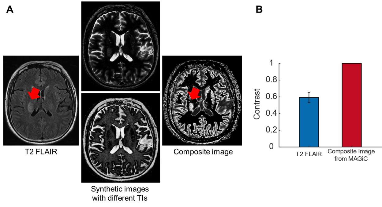
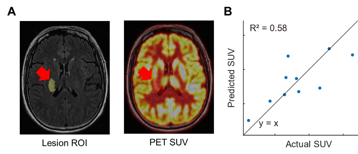
Methods: We conducted a pilot study on ten stroke patients. Each patient underwent Conventional T2-FLAIR, MAGiC and PET scanning with a 3.0 T scanner (SIGNA PET/MR, GE Healthcare). MAGiC offers flexibility in adjusting parameters such as repetition time (TR), echo time (TE), and inversion time (TI) to optimize imaging contrast for specific targets. In this study, we extracted the T1 value of the stroke lesion and surrounding tissue from the T1 map generated from MAGiC. Then, based on T1 measurements, two inversion recovery (IR) images were generated using different TI values to suppress stroke lesion (TI=800ms) and surrounding tissue (TI=500ms), respectively (Figure 1A). The two IR images were merged to create a composite image that enhanced stroke lesion contrast while maintaining anatomical context. These images were compared to traditional T2 FLAIR images for tissue contrast. Additionally, we leveraged the quantitative relaxation values from the lesion areas to build predictive models for PET Standardized Uptake Values (SUV), enabling the prediction of metabolic activity within the stroke lesion region (Figure 2A).
Results: The MAGiC-generated images provided significantly higher tissue contrast compared to conventional T2 FLAIR images (p<0.01, Figure 1B), allowing for improved lesion visualization. This increased contrast facilitated more accurate delineation of stroke lesions, enhancing their visibility for clinical diagnosis and research purposes. Additionally, quantitative relaxation values derived from the lesion areas in the T1, T2, and PD maps were utilized to establish a predictive model for PET Standardized Uptake Values (SUV). This model effectively predicted regional metabolic activity within the stroke lesion area (Figure 2B).
Discussion: The findings of this study demonstrate the potential of synthetic MRI using the MAGiC sequence in stroke patients. The improved tissue contrast provided by the generated images enhances the visualization of stroke lesion areas. Additionally, the prediction model based on the relaxation values offers a non-invasive method to estimate the metabolic level in the stroke regions. These findings contribute to the understanding and management of stroke patients, potentially leading to improved diagnostic accuracy and treatment strategies. However, further validation and clinical exploration with larger patient cohorts are still required.
Acknowledgements
No acknowledgement found.References
1. Feigin V L, Norrving B, Mensah G A. Global burden of stroke[J]. Circulation research, 2017, 120(3): 439-448.
2. Matsuda M, Tsuda T, Ebihara R, et al. Enhanced masses on contrast‐enhanced breast: differentiation using a combination of dynamic contrast‐enhanced MRI and quantitative evaluation with synthetic MRI[J]. Journal of Magnetic Resonance Imaging, 2021, 53(2): 381-391.
3. Ma Y J, Moazamian D, Cornfeld D M, et al. Improving the understanding and performance of clinical MRI using tissue property filters and the central contrast theorem, MASDIR pulse sequences and synergistic contrast MRI[J]. Quantitative Imaging in Medicine and Surgery, 2022, 12(9): 4658.
Figures