2303
Evaluation of iron deposition in unilateral middle cerebral artery stenosis patients using automatic brain regional susceptibility analysis1Department of Radiology, Zhongshan Hospital, Fudan University, and Shanghai Institute of Medical Imaging, Shanghai, China, 2GE Healthcare, Beijing, China
Synopsis
Keywords: Stroke, Stroke
Motivation: Iron deposition is associated with brain injury. Exploring alterations of iron deposition in cerebral regions caused by cerebral artery stenosis may help to predict degree of brain injury.
Goal(s): This study aimed to quantify iron alterations in both cortex and deep GM nuclei in patients with long-term unilateral MCA severe stenosis.
Approach: Using structural imaging and QSM, we employed automatic brain regional susceptibility analysis.
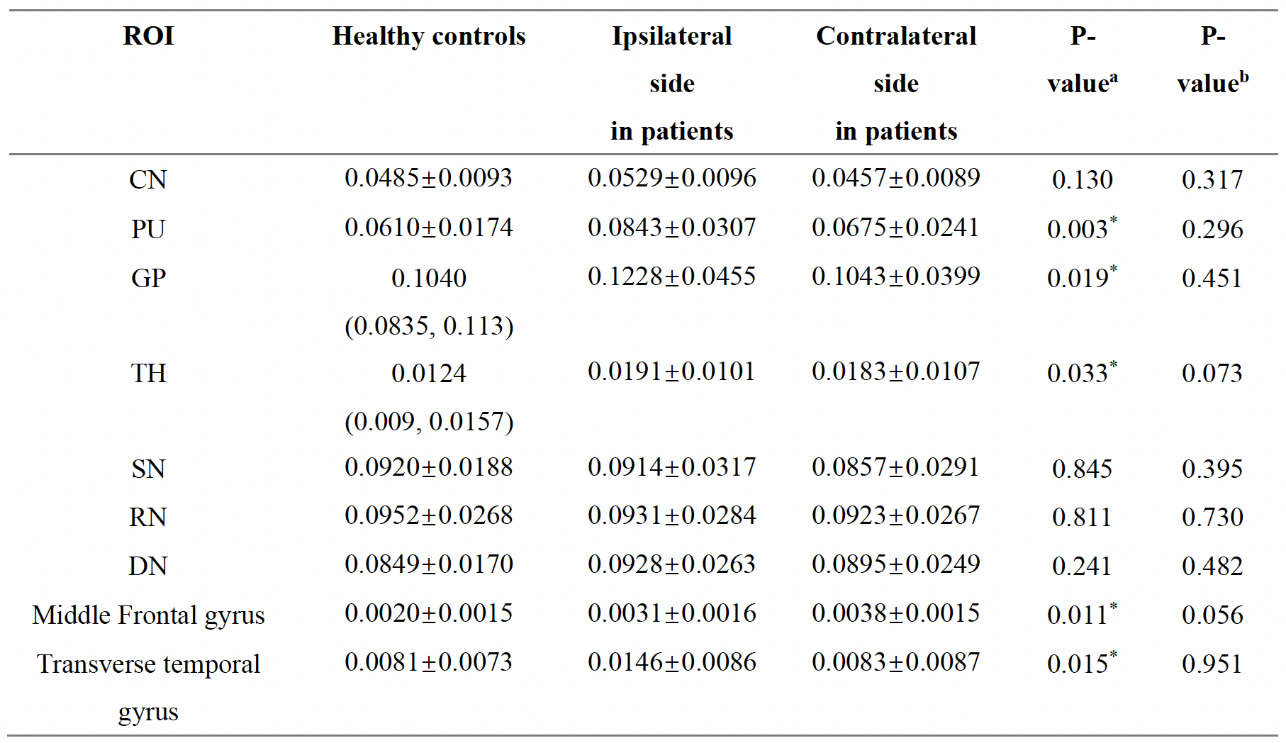
Results: We found that patients had higher susceptibility in ipsilateral CN, PU, GP, and transverse temporal gyrus than contralateral side, and patient group had higher susceptibility in PU, GP, TH, middle frontal gyrus, and transvers temporal gyrus than HCs.
Impact: By brain regional susceptibility analysis, this study demonstrated abnormal iron accumulation in deep GM nuclei, middle frontal gyrus, and transvers temporal gyrus after chronic MCA severe stenosis. Therefore, iron deposition may be a potential biomarker for evaluating long-term cerebral ischemia.
Introduction
Ischemic stroke stands as a leading cause of adult disability and mortality1, with intracranial artery stenosis, particularly middle cerebral artery (MCA) stenosis or occlusion, identified as the predominant instigator2. The occurrence of unilateral MCA stenosis or occlusion precipitates ischemia and hypoxia in the corresponding brain regions, causing neuronal injury. This process unfolds alongside a series of pathophysiological reactions, prominently featuring oxidative stress induced by iron accumulation3. Numerous neurological disorders have been linked to disrupted brain iron metabolism4,5, with iron deposits observed in affected regions, particularly in ischemic stroke models following unilateral MCA occlusion6. However, the regularity of iron deposition in cerebral ischemia patients has not been revealed clearly. Quantitative susceptibility mapping (QSM) has emerged as a promising MRI technique for accurately measuring the spatial distribution of magnetic susceptibility in biological tissues, enabling quantitative evaluation of iron levels7. Previous study had found increased iron deposition in the deep GM nuclei of patients with long-term cerebral ischemia8. However, to our knowledge, no studies have systematically investigated iron deposition throughout the cerebral cortex in ischemia patients. Therefore, based on high-resolution structural imaging and QSM, we first applied automatic segmentation of brain subregions to quantitatively evaluate iron alterations both in the cortex and deep GM nuclei for patients with long-term unilateral MCA severe stenosis.Materials and Methods
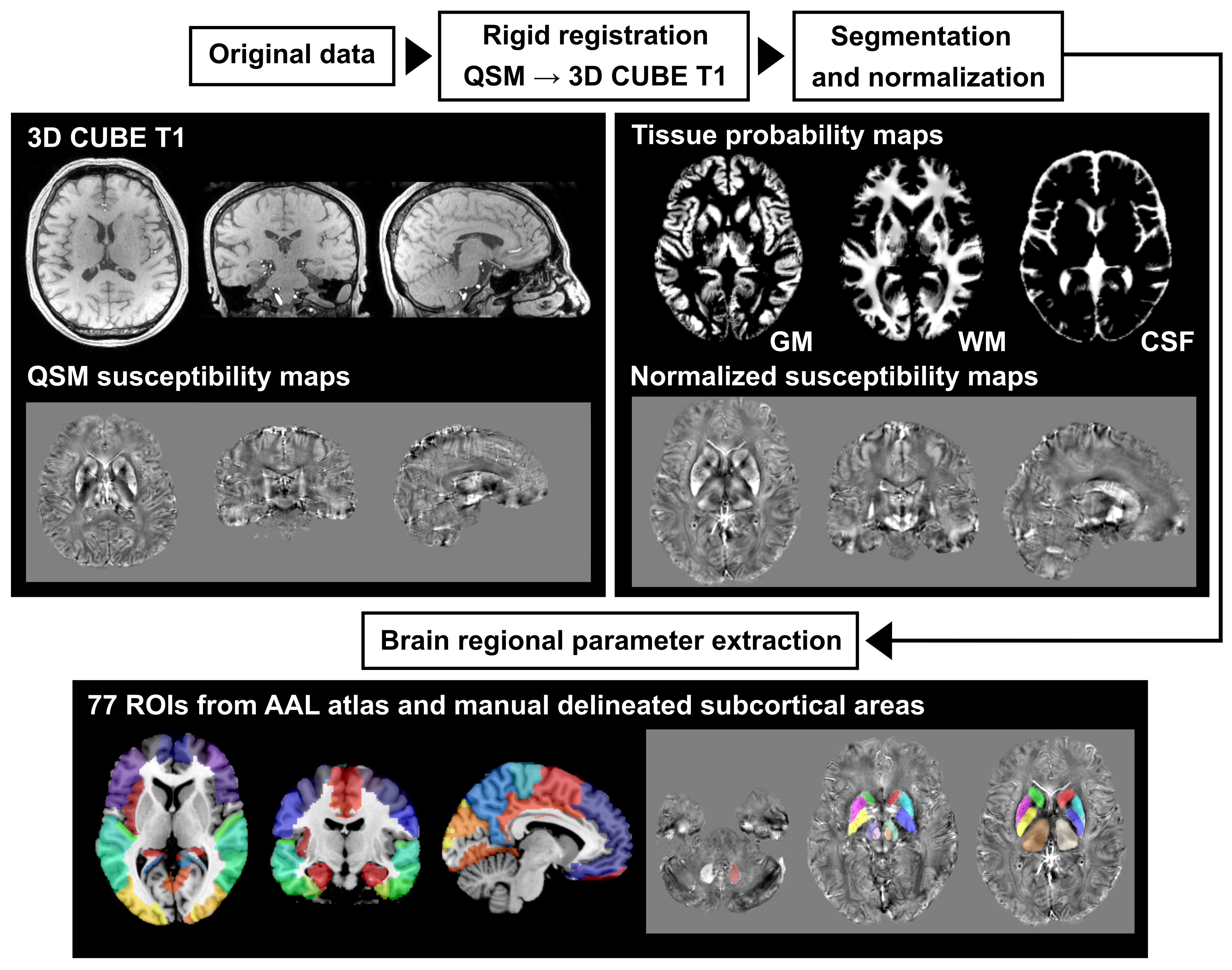
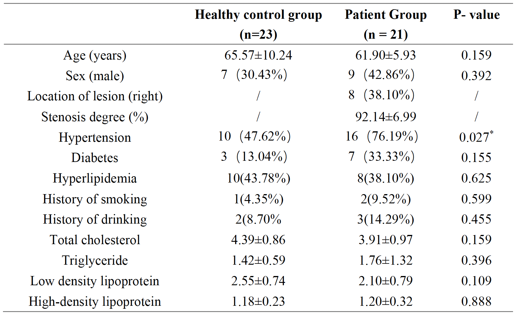
The project was approved by the Ethics Committee of our hospital. A total of 21 unilateral MCA severe stenosis patients and 23 age-matched healthy controls (HCs) were included in this study. The inclusion criteria for patients with unilateral MCA stenosis were as follows: 1) unilateral MCA severe stenosis (> 70%), with other large vessels (both in neck and brain) showing no or only mild stenosis; 2) long- term ischemic attack symptoms associated with unilateral MCA stenosis (≥ 4 weeks); 3) ages > 50 years; 4) no previous history of cerebral hemorrhage, brain tumor, brain injury, or dementia. MRI examinations were performed on a 3-Tesla MRI scanner (Discovery MR750; GE Healthcare, Milwaukee, WI, USA) equipped with a 32-channel head coil. QSM and T1WI images were obtained with following parameters: 1) QSM was acquired by a 3D FSPGR sequence, TR = 41.5 ms, TE = 3:3:27 ms, FOV = 240 x 240 mm2, matrix size = 240 x 192, slice thickness = 1 mm, flip angle = 20°; (3) T1WI was acquired by a 3D CUBE sequence, TR/TE = 800/25 ms, FOV = 240 × 240 mm2, matrix size = 288 × 288, slice thickness = 1 mm. For the brain regional analysis (Figure 1), we first conducted a rigid registration between QSM and CUBE T1WI using SPM12 software. Subsequently, we employed CAT12 toolbox to perform segmentation and normalization, aligning data to the MNI space. Considering the suboptimal accuracy of auto-segmentation on deep GM nucleus, we further manually delineated bilateral caudate nucleus (CN), putamen (PU), globus pallidus (GP), thalamus, substantia nigra (SN), red nucleus, and dentate nucleus (DN). Finally, brain regional iron-related susceptibility values were extracted from 77 ROIs. Clinical variables were evaluated. Independent sample t-test (or Mann-Whitney U test) and paired sample t-test (or Wilcoxon signed-rank test) were performed to assess susceptibility differences between HCs and patients, and between ipsilateral and contralateral sides of patients, respectively. ROC curve analysis was used to evaluate the efficacy of susceptibility value in distinguishing patients with unilateral MCA stenosis from HCs.Results
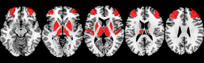
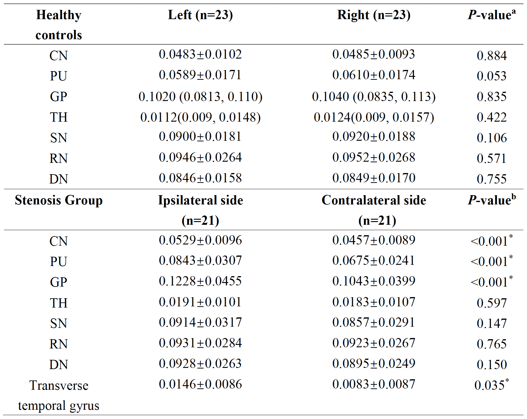
No significant differences were found in clinical factors between two groups except for hypertension (P = 0.027) (Table 1). Regarding the comparison between bilateral brain regions, we found that susceptibility values were comparable in all brain regions in HCs, whereas for patients with unilateral MCA stenosis had significantly higher susceptibility values in ipsilateral CN, PU, GP, and transverse temporal gyrus than the contralateral side (all P < 0.05) (Table 2). Regarding the comparison between groups, we found that patients had significantly higher susceptibility values in ipsilateral PU, GP, TH, middle frontal gyrus, and transvers temporal gyrus than HCs (all P < 0.05), while no differences were observed in contralateral sides (Table 3 and Figure 2). ROC analysis demonstrated that susceptibility values in ipsilateral PU, GP, TH, middle frontal gyrus, and transvers temporal gyrus had AUCs of 0.735, 0.662, 0.640, 0.721, and 0.721, respectively in distinguishing patients with unilateral MCA severe stenosis from HCs.Discussion
By brain regional analysis of susceptibility values, this study demonstrated abnormal iron accumulation in the deep GM nuclei, middle frontal gyrus, and transvers temporal gyrus after chronic MCA severe stenosis. Therefore, iron deposition, as measured by QSM, may be a potential biomarker for evaluating long-term cerebral ischemia.Acknowledgements
No acknowledgement found.References
1. Kuriakose, D., & Xiao, Z. (2020). The current state and future prospects of stroke pathophysiology and treatment. International Journal of Molecular Sciences, 21, 7609.
2. Ran, Y., Wang, Y., Zhu, M., Wu, X., Malhotra, A., Lei, X., Zhang, F., Wang, X., Xie, S., Zhou, J., Zhu, J., Cheng, J., Zhu, C. (2020). Increased plaque burden in the middle cerebral artery correlates with a higher risk of recurrent ischemic stroke: Evidence from a quantitative magnetic resonance imaging investigation. Stroke, 51(2), 659–662.
3. Sekerdag, E., Solaroglu, I., Gursoy-Ozdemir, Y. (2018). Stroke-related mechanisms of cell death and emerging molecular and cellular therapeutic strategies. Current Neuropharmacology, 16, 1396-1415.
4. Stankiewicz, J., Panter, S.S., Neema, M., Arora, A., Batt, C.E., Bakshi, R. (2007). Iron in chronic brain disorders: insights from imaging and potential therapeutic implications. In Neurotherapeutics, 4, 371-386.
5. Hayflick, S.J., Kurian, M.A., Hogarth, P. (2018). Neurodegeneration with brain iron accumulation: An overview. In Handbook of Clinical Neurology, 147, 293-305.
6. Tuo, Q.-z., Lei, P., Jackman, K.A., Li, X.-l., Xiong, H., Li, X.-l., Liuyang, Z.-y., Roisman, L., Zhang, S.-t., Ayton, S., Wang, Q., Crouch, P.J., Ganio, K., Wang, X.-c., Pei, L., Adlard, P.A., Lu, Y.-m., Cappai, R., Wang, J.-z., Liu, R., Bush, A.I. (2017). Tau-mediated iron export as a protective mechanism against ferroptosis in ischemic stroke. In Molecular Psychiatry, 22 (11), 1520-1530.
7. Vinayagamani S, Sheelakumari R, Sabarish S, et al. Quantitative Susceptibility Mapping: Technical Considerations and Clinical Applications in Neuroimaging. Journal of magnetic resonance imaging: JMRI. 2021;53(1):23-37.
8. Mao H, Dou W, Chen K, et al. Evaluating iron deposition in gray matter nuclei of patients with unilateral middle cerebral artery stenosis using quantitative susceptibility mapping. NeuroImage Clinical. 2022; 34:103021.
Figures