2300
Stem Cell-Derived Extracellular Vesicles Restore Sodium & Energetic Homeostasis in Ischemic Stroke as Quantified by Longitudinal MRI/S at 21.1 T1Center for Interdisciplinary Magnetic Resonance, National High Magnetic Field Laboratory, Tallahassee, FL, United States, 2Chemical & Biomedical Engineering, Florida State University, Tallahassee, FL, United States
Synopsis
Keywords: Stroke, High-Field MRI, Spectroscopy, Metabolites, Stem Cell Therapy, Neuroinflammation, Preclinical
Goal(s): This work administers extracellular vesicles (EV) derived from human mesenchymal cells (hMSC) to salvage tissue while monitoring recovery and metabolic changes longitudinally using ultra-high field MRI/S.
Approach: T2-W MRI,23Na-CSI, and 1H-MRS were used to quantify lesion reduction, sodium homeostasis and energetic remodeling.
Results: Upon EV treatment, sodium (and to a lesser extent proton) lesions were reduced by day 3, while lactate, creatine and NAA were stabilized compared to control.
Impact: Combined sodium MRI and proton MRS provide a more sensitive and early quantitative metric to evaluate the efficacy of stem cell-derived therapy following ischemia and longitudinal metabolic, ionic and functional recovery. Such methods can evaluate other treatments against different pathologies.
Introduction
Stroke is a leading cause of death and disability worldwide. Of the limited treatment options, tissue plasminogen activator (tPA) is the only FDA-approved drug for ischemic stroke treatment, which works by dissolving blood clots to restore blood flow to the brain. However, it has a therapeutic window of only 4.5 h1 while surgical thrombectomy extends this window to approximately 24 h. Beyond clearance of the initial occlusion, human mesenchymal stem cells (hMSC) have shown promise as a biotherapeutic for ischemic stroke due to reparative mechanisms that induce an anti-inflammatory response, angiogenesis and neurogenesis via endogenous recruitment2.As an alternative to direct hMSC injection, extracellular vesicles (EV) derived from hMSC have been shown to have therapeutic potential. EV are membrane-enclosed, cell-derived vesicles that cannot independently replicate. EV contain protein and genetic cargo secreted by hMSC, but are more readily able to cross the blood-brain barrier than injected cells. To increase their therapeutic potential further, EV were derived from hMSC that have been aggregated as 3D spheroids3. Resulting EV are smaller (~100-150 nm) with higher miRNA expression and upregulation of cytokines and anti-inflammatory factors3.
Stem cell-based therapies must work to correct an excitotoxic environment created following a stroke, preferably restoring metabolic and energetic homeostasis4,5. Ultra-high field MRI at 21.1 T is able to track ultra-small iron oxide nanoparticle (USPIO) labeled EV using gradient recalled echo imaging, monitor metabolic and ionic alterations resulting from stroke and treatment, and provide increased sensitivity to assess lesion recovery using 1H T2-weighted. Previous studies have shown evidence of the restoration of sodium homeostasis and reduced sodium lesion volume over a period of 7 d6.
The goal of this study is to use combined 1H and 23Na MRI/S techniques, specifically, 1H T2W MRI and spectroscopy and 23Na CSI to monitor recovery longitudinally following EV treatment in a preclinical stroke model.
Methods
EV Isolation: hMSC were aggregated at passage 4 in ultra-low attachment using a WAVE Bioreactor4. Over a three-day period, EV were harvested from aggregates, labeled with 0.5-1 mg/mL of USPIO and purified for injection using ultracentrifugation.Animal Model: A transient middle cerebral artery occlusion model7 was instituted in female Sprague-Dawley rats for 1 h. Immediately following the occlusion, the animals received the treatment (EV or saline control) via intra-arterial injection.
Imaging: Data were acquired using the 21.1-T (900-MHz) vertical bore magnet at the NHMFL. In vivo assessment utilized a linear birdcage double-tuned 23Na/1H radio frequency coil on 0, 1, 3, 7 and 21-day post-ischemia to assess tissue recovery and treatment efficacy. EV administration was confirmed with gradient recalled echo (GRE) images (50x50-µm in-plane resolution). Lesion volume was evaluated using T2W RARE at a 100x100-µm resolution. 3D 23Na CSI was acquired at 1-mm isotropic resolution. Relaxation-enhanced (RE) MRS using semi-LASER evaluated metabolites in both ischemic and contralateral hemispheres. T2W images enabled anatomical reference to the ischemic lesion and contralateral alignment. Metabolite phantom studies were acquired using semi-LASER to quantify absolute metabolite concentrations.
Analysis: 3D 23Na CSI data were reconstructed in MATLAB to a zero-filled 0.5-mm isotropic resolution. Volumetric and signal analyses for the 3D 23Na CSI and T2W were performed in Amira 3D Visualization Software. A signal threshold generated from the contralateral hemisphere was used to define the ischemic lesion. MRS data was processed in TOPSPIN 4.1.4 to monitor metabolites (total choline (Cho), total creatine (Cre), N-acetyl aspartate (NAA) and lactate) longitudinally according to previous literature8.
Results and Discussion
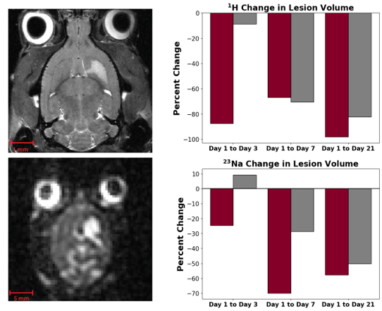
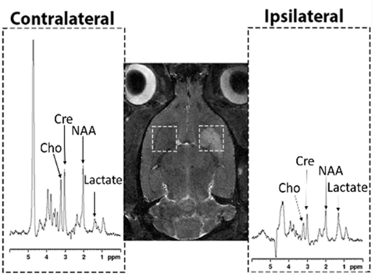
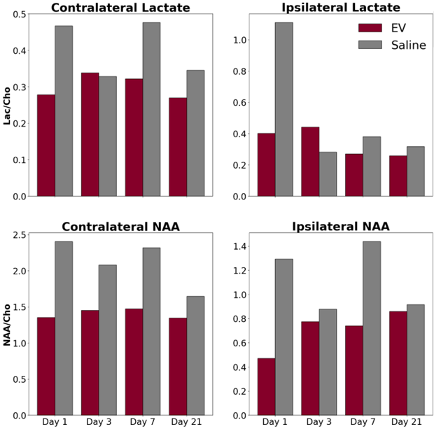
3D 23Na CSI data indicates that the lesion volume in the EV-treated animals decreased from days 1 to 3, whereas the animals receiving the saline control treatment had an increase in lesion volume over this period. By day 7, both groups began to exhibit a decrease in the lesion volume (Figure 1). MR spectroscopy data shows elevated levels of lactate in the penumbral region (Figure 2&3) and decreased NAA (Figure 3) and total creatine. These metabolites begin to recover by day 7 in the EV-treated animals and by day 21 in the saline control. These results suggest that there is a faster recovery in sodium and energetic homeostasis of the EV-treated animals.Conclusion
Results indicate that the EV treatment could offer significant improvement for ischemic stroke recovery. The re-establishment of sodium homeostasis was observed as early as day 3 following the occlusion in the EV-treated animals. MRS data indicates that the transplantation of the 3D-EV resulted in improved metabolite recovery compared to the control PBS group.Acknowledgements
All work has been conducted in accordance with FSU Animal Care and Use Committee. Funding support has been provided by the NSF (DMR-1644779) and NIH (RO1-NS102395 to SCG and R01-NS125016 to YL)References
1. K. M. Rexrode, T. E. Madsen, A. Y. X. Yu, C. Carcel, J. H. Lichtman, and E. C. Miller, “The Impact of Sex and Gender on Stroke,” Circ. Res., vol. 130, no. 4, pp. 512–528, Feb. 2022, doi: 10.1161/CIRCRESAHA.121.319915.
2. Y. Zhang, N. Dong, H. Hong, J. Qi, S. Zhang, and J. Wang, “Mesenchymal Stem Cells: Therapeutic Mechanisms for Stroke,” Int. J. Mol. Sci., vol. 23, no. 5, p. 2550, Feb. 2022, doi: 10.3390/ijms23052550.
3. X. Yuan et al., “Engineering extracellular vesicles by three-dimensional dynamic culture of human mesenchymal stem cells,” J. Extracell. Vesicles, vol. 11, no. 6, p. e12235, Jun. 2022, doi: 10.1002/jev2.12235.
4. C. Davis, S. I. Savitz, and N. Satani, “Mesenchymal Stem Cell Derived Extracellular Vesicles for Repairing the Neurovascular Unit after Ischemic Stroke,” Cells, vol. 10, no. 4, Art. no. 4, Apr. 2021, doi: 10.3390/cells10040767.
5. S. Sarvari, F. Moakedi, E. Hone, J. W. Simpkins, and X. Ren, “Mechanisms in blood-brain barrier opening and metabolism-challenged cerebrovascular ischemia with emphasis on ischemic stroke,” Metab. Brain Dis., vol. 35, no. 6, pp. 851–868, Aug. 2020, doi: 10.1007/s11011-020-00573-8.
6. X. Yuan, J. T. Rosenberg, Y. Liu, S. C. Grant, and T. Ma, “Aggregation of human mesenchymal stem cells enhances survival and efficacy in stroke treatment,” Cytotherapy, vol. 21, no. 10, pp. 1033–1048, Oct. 2019, doi: 10.1016/j.jcyt.2019.04.055.
7. E. Z. Longa, P. R. Weinstein, S. Carlson, and R. Cummins, “Reversible middle cerebral artery occlusion without craniectomy in rats,” Stroke, vol. 20, no. 1, pp. 84–91, Jan. 1989, doi: 10.1161/01.str.20.1.84.
8. S. Helsper et al., “Multinuclear MRI Reveals Early Efficacy of Stem Cell Therapy in Stroke,” Transl. Stroke Res., Jul. 2022, doi: 10.1007/s12975-022-01057-w.
Figures