2299
A Multi-task Deep learning Model for Simultaneous Segmentations of Penumbra and Infarct in Patients with Acute Ischemic Stroke1Institute of Research and Clinical Innovations, Neusoft Medical Systems Co., Ltd, Shanghai, China, 2Department of Radiology, The First Affiliated Hospital of Jinan University, Guangzhou, Guangdong, China, 3Institute of Research and Clinical Innovations, Neusoft Medical Systems Co., Ltd, Guangzhou, China, 4Institute of Research and Clinical Innovations, Neusoft Medical Systems Co., Ltd, Beijing, China, 5Neusoft Medical Systems Co. Ltd,, Shenyang, China
Synopsis
Keywords: Stroke, Segmentation
Motivation: Arterial spin labeling (ASL) has shown comparable results with dynamic susceptibility contrast magnetic resonance imaging in evaluating hypoperfused lesions in patients with acute ischemic stroke (AIS). However, the precise delineation of penumbra in ASL is still challenging.
Goal(s): To develop a deep learning (DL) model based on ASL to identify eligible candidates for endovascular treatment in AIS patients.
Approach: A multi-task DL model was proposed for simultaneous segmentations of penumbra and infarct by combining cerebral blood flow and DWI images.
Results: The multi-task segmentation performed well, which is comparable to the results achieved by radiologists.
Impact: The proposed approach performed well for the segmentation of penumbra and infarct, which could provide a promising approach for assisting decision-making for endovascular treatment in patients with acute ischemic stroke.
Introduction
The ischemic penumbra refers to the area where blood flow is reduced but irreversible damage has not yet occurred1, and therefore the identification of the location and size of the ischemic penumbra is crucial for treatment decision making in patients with acute ischemic stroke (AIS)2. The ischemic penumbra delineation typically depends on dynamic susceptibility contrast magnetic resonance imaging (DSC-MRI) or computed tomography perfusion (CTP) 3-4. However, DSC-MRI requires intravenous administration of contrast agent, whereas CTP has the extra disadvantage of high radiation exposure. Arterial spin labeling (ASL) is a non-invasive imaging technique without either radiation exposure or contrast agent injection5, which has shown potential for the assessment of penumbral tissue6-7. This study aimed to develop and validate a deep learning (DL) model that can simultaneously delineate ischemic penumbra and infarct based on ASL and DWI sequences, which would provide robust inputs for the subsequent identification of the suitable patients with AIS for endovascular treatment.Methods
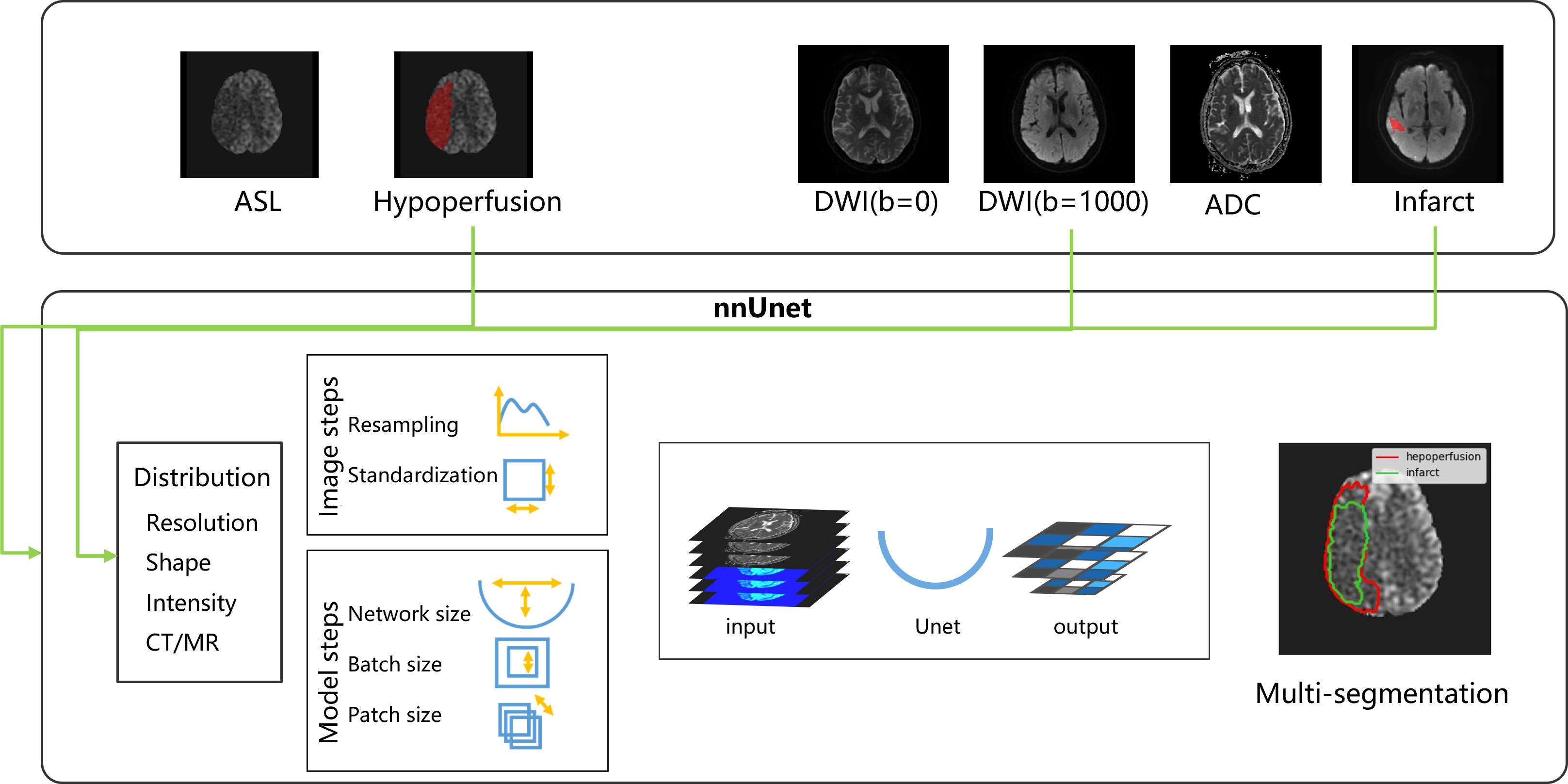
Data acquisition: We retrospectively collected the patients with AIS from January 2018 to December 2022. A total of 151 patients with AIS were enrolled in this retrospective study (96 males and 54 females, mean age: 62.3±12.7 years old). All diffusion-weighted imaging (DWI) and ASL images were collected. The ground truth segmentation: The cerebral blood flow (CBF) maps were generated from ASL images. The ischemic core and hypoperfused areas were segmented semi-automatically on the apparent diffusion coefficient (ADC) and CBF maps by applying certain thresholds, which were then manually corrected and segmented by a neuroradiologist (6 years of experience in neuroimaging) who were blinded to the patients’ clinical information. A senior neuroradiologist with 10 years of experience in neuroimaging confirmed the segmentation accuracy. Any disagreement between the two observers was reconciled until a final consensus was reached. The multi-task DL segmentation model: The dataset was randomly divided into a training cohort (n=106) and a test cohort (n=45) at a ratio of 7:3. No new U-Net (nnU-Net) is a specialized DL framework for medical image segmentation, which has demonstrated state-of-the-art performance in many medical image segmentation challenges8. The penumbra and infarct were segmented simultaneously by using the DL framework (see Figure 1) with the combinations of CBF, DWI, and ADC map as inputs. The Dice score was used to evaluate the accuracy for segmentation.Results
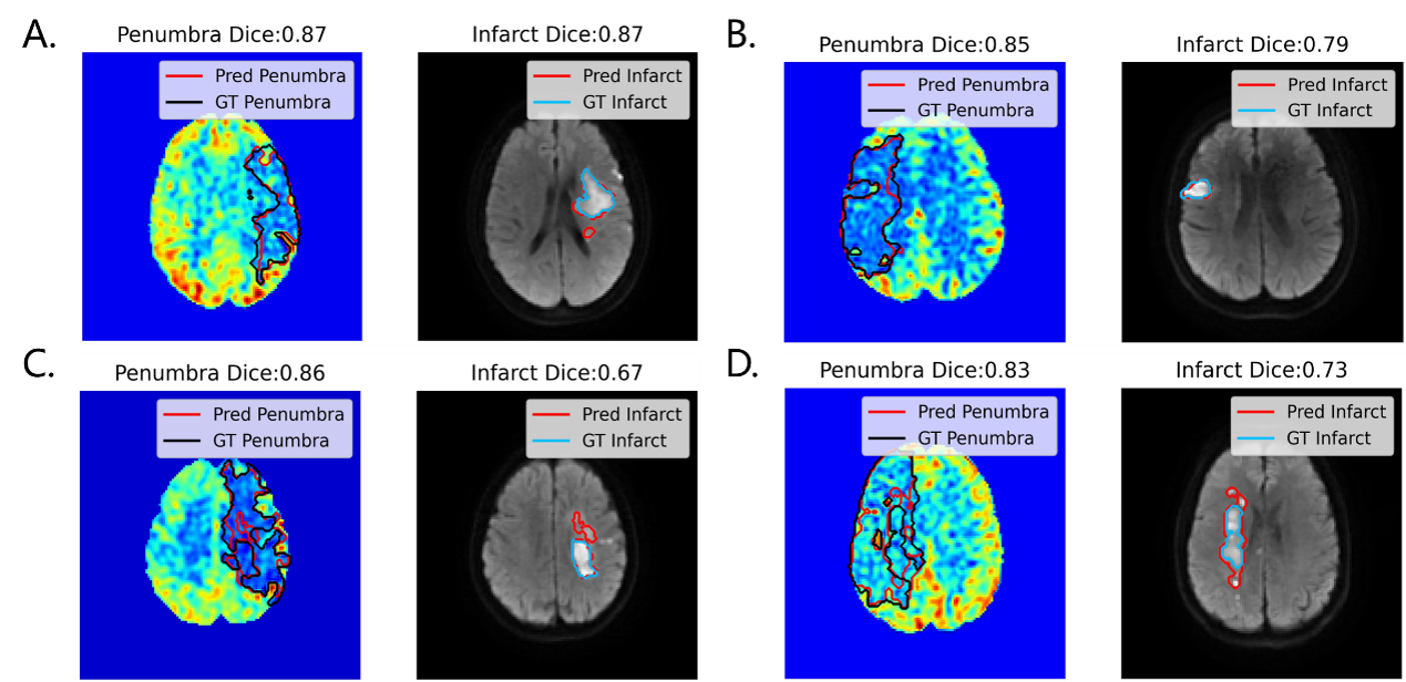
For voxel-level evaluation of hypoperfusion and infarct segmentation, the group average Dice coefficient of best candidate model was 0.582±0.22 and 0.755±0.13 respectively. Notably, for either the segmentation of the ischemic penumbra or the infarct core, deep learning-based methods yield smoother boundaries in comparison to threshold-based segmentation results, aligning well with clinical evidence. The multi-sequence fusion model achieves the highest segmentation performance by combining CBF, ADC and DWI, Figure 2 shows that the segmentation results of the model are comparable to the results yielded by radiologists.Discussion
The aim of ischemic stroke therapy is to salvage the penumbra and achieve the reperfusion of ischemic brain tissue. Therefore, the accurate identification and evaluation of ischemic penumbra is vital for AIS patients to make correct clinical treatment decision to improve clinical outcomes9-10. In this study, the segmentation models achieved satisfactory results for both penumbra and infarct segmentations. Compared with a previous study6, our segmentation models performed better for infarct segmentation, which achieved a higher Dice coefficient. Importantly the whole pipeline has been fully automated, which effectively reduces the workload for radiologists. DSC-MRI requires intravenous administration of contrast agent which is not suitable for some patients with advanced stage of chronic kidney disease11. As an alternative non-invasive perfusion imaging technique, ASL can be an important complement in these circumstances. On the other hand, DWI is essential to extract infarct cores. Furthermore, the ADC map derived from DWI can better describe the diffusion characteristics of the ischemic lesions than DWI. In fact, Table 1 shows that the Dice coefficient has been improved by combining ADC, likely suggesting that the ADC map and DWI can provide complementary information for improving segmentation performance.Conclusion
The proposed DL-based algorithm that integrated ASL and DWI images provided a promising approach for delineating ischemic penumbra in patients with AIS robustly, which could assist clinicians in formulating corresponding personalized treatment plans at the early stage of stroke onset, thus improving the ultimate outcomes for patients with AIS.Acknowledgements
We would like to acknowledge the equal contributions of Jing Zhang and Xiaoling Wu to this work. Both authors contributed equally to the experimental design, data analysis, and manuscript preparation.References
1. Ermine CM, Bivard A, Parsons MW, Baron JC. The ischemic penumbra: From concept to reality. Int J Stroke. 2021;16(5):497-509.
2. Powers WJ, Rabinstein AA, Ackerson T, et al. 2018 guidelines for the early Management of Patients with Acute Ischemic Stroke: A guideline for healthcare professionals from the American Heart Association/American Stroke Association. Stroke 2018;49:e46-e110.
3. Nogueira RG, Jadhav AP, Haussen DC, et al. Thrombectomy 6 to 24 hours after stroke with a mismatch between deficit and infarct. N Engl J Med 2018;378:11-21.
4. Campbell BCV, Majoie CBLM, Albers GW, et al. Penumbral imaging and functional outcome in patients with anterior circulation ischaemic stroke treated with endovascular thrombectomy versus medical therapy: A meta-analysis of individual patient-level data. Lancet Neurol 2019;18:46-55.
5. Hernandez-Garcia L, Aramendía-Vidaurreta V, Bolar DS, et al. Recent Technical Developments in ASL: A Review of the State of the Art. Magn Reson Med 2022;88(5):2021-2042.
6. Wang K, Shou Q, Ma SJ, et al. Deep Learning Detection of Penumbral Tissue on Arterial Spin Labeling in Stroke. Stroke 2020;51(2):489-497.
7. Lyu J, Duan Q, Xiao S, et al. Arterial Spin Labeling-Based MRI Estimation of Penumbral Tissue in Acute Ischemic Stroke. J Magn Reson Imaging 2023;57(4):1241-1247.
8. Isensee, F., Jaeger, P.F., Kohl, S.A.A. et al. nnU-Net: a self-configuring method for deep learning-based biomedical image segmentation. Nat Methods 18, 203–211 (2021).
9. Albers GW, Marks MP, Kemp S, et al. Thrombectomy for stroke at 6 to 16 hours with selection by perfusion imaging. N Engl J Med 2018;378:708-718.
10. Tang TY, Jiao Y, Cui Y, et al. Penumbra-based radiomics signature as prognostic biomarkers for thrombolysis of acute ischemic stroke patients: a multicenter cohort study. J Neurol. 2020;267(5):1454-1463.
11. Mehdi A, Taliercio JJ, Nakhoul G. Contrast media in patients with kidney disease: An update. Cleve Clin J Med 2020;87:683-694.
Figures