2297
Identification of culprit plaques in stroke patients using radiomics based on three-dimensional high-resolution vascular wall imaging1Tongji Hospital of Tongji Medical college of Huazhong University of science and technology, Wuhan, China
Synopsis
Keywords: Stroke, Vessels
Motivation: Identifying the culprit plaque among the plaques in stroke patients is important. Previous studies were based on 2D sequences, 3D HRMR-VWI is a novel imaging examination to evaluate vessel wall.
Goal(s): To establish a high performance model to identify the culprit plaques in stroke patients.
Approach: We used traditional method and five different radiomics methods to identify the culprit plaques in stroke patients based on 3D HRMR-VWI.
Results: In traditional information, intraplaque hemorrhage is an independent predictor for culprit plaques, the efficacy of radiomics is much higher than traditional model, the extreme gradient boosting method showed the best performance in radiomics models.
Impact: Our study established an accurate method to identify the culprit plaques in stroke patients, to help clinicians make a more precise treatment plan, it will improve the prognosis and prevent the recurrence in stroke patients.
Introduction
Atherosclerosis is the most common cause of stroke, accounting for approximately 18% to 25% of all strokes(1). The culprit plaque refers to the plaque causes cerebrovascular events among multiple plaques in stroke patients. Finding the culprit plaque and treated with active intervention will improve the prognosis and prevent the recurrence in patient with cerebrovascular events. Previous studies were based on 2D sequences and the typical slice(2-5), it means only one slice of radiomics features were extracted, the other characters such as the shape and area were not considered. Three-dimensional High-resolution magnetic resonance vascular wall imaging (3D HRMR-VWI) is a novel imaging examination to evaluate blood vessel wall(6,7). We aim to identify the culprit and nonculprit plaques in cerebrovascular events patients with middle cerebral artery plaque using traditional and radiomic method based on three-dimensional high-resolution magnetic resonance vascular wall imaging (3D HRMR-VWI),and compare the efficacy of traditional method five different radiomics methods in identifying culprit plaques.Methods
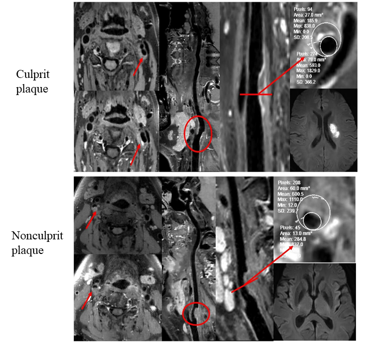
A total of 117 patients with 139 plaques in the middle cerebral arteries were enrolled and divided into training and validation sets in a ratio of 7:3. Magnetic resonance examinations included 3D HRMR-VWI (before and after enhancement), magnetic resonance angiography(MRA) and diffusion weighted imaging(DWI), Each identified plaque was classified as 69 culprit plaques and 70 nonculprit plaques according to the imaging of 3D HRMR-VWI, MRA, DWI and clinical symptoms. The plaque is confirmed based on observing the stenosis of the middle cerebral artery on MRA and eccentric thickening in vessel wall on reconstructing HRMR-VWI. The plaque is identified as culprit when conformed to one of the following conditions(4) (Fig1): 2).DWI lesion is observed in the middle cerebral artery blood supply area, and this is the only one plaque on the blood supply vessel; 2) DWI lesion is observed in the middle cerebral artery blood supply area, cause the most severe stenosis among the multiple plaques on the blood supply vessel. The radiomics model was constructed using 3D HRMR-VWI before and after enhancement. Traditional information included plaque characteristics (plaque diameter, minimum lumen area, intraplaque hemorrhage, minimum lumen diameter, stenosis rate, plaque burden, enhancement rate and remodeling index) and clinical risk factors (sex, age, hypertension, hyperlipidemia, diabetes, smoking, drinking, history of coronary heart disease and stroke history). Mann-Whitney U test and chi-square test were used for traditional factors, and factors with predictive value in univariate analysis were further analyzed by multivariate logistic regression. The radiomics models were modeled by the least absolute shrinkage selection operator (LASSO) method, the random forest (RF) method, the extreme learning machine (ELM) method, the linear discriminant analysis (LDA) method and the extreme gradient boosting (XGB) method. The Delong test was used to compare the differences of the modles’ efficacy in identifying culprit plaques.Results
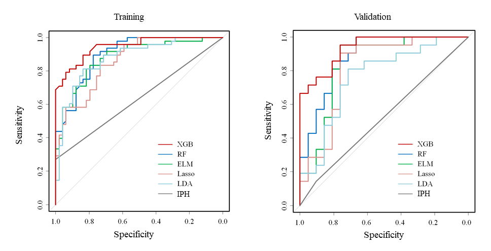
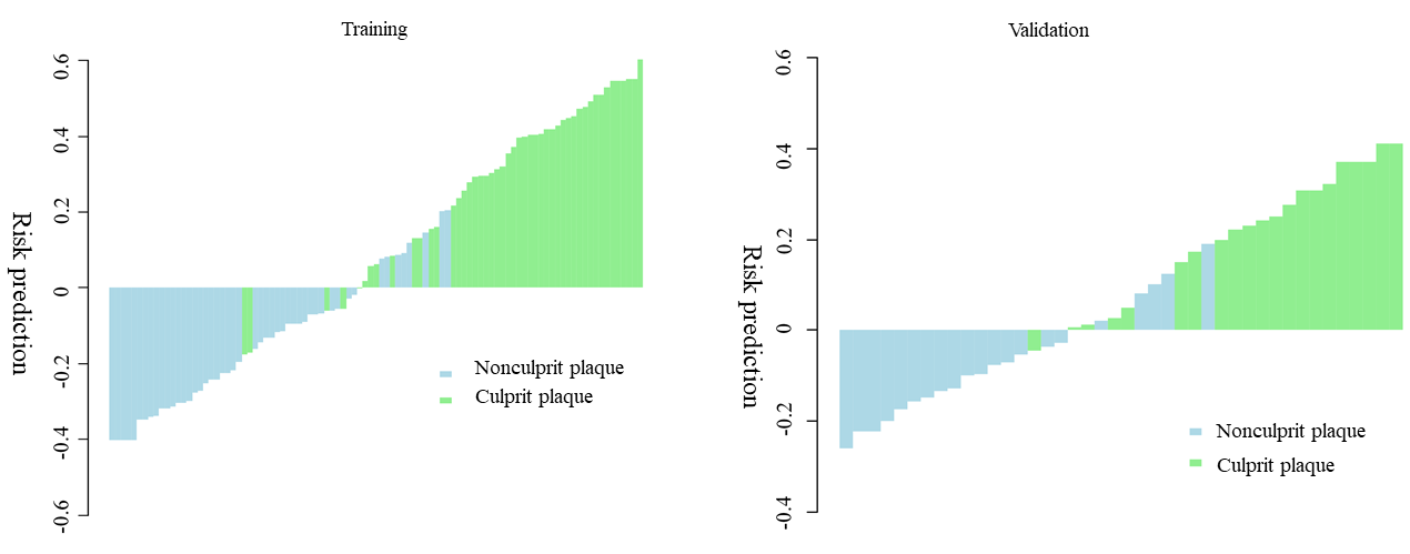
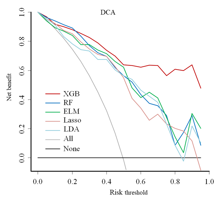
In traditional information, only intraplaque hemorrhage was an independent predictor for culprit plaques; Radiomics played an important role in identifying culprit plaques, and its efficacy was much higher than traditional information; Enhanced 3D HRMR-VWI showed better efficiency than before enhanced 3D HRMR-VWI, and the performance of combing the two sequences is the best; Among different radiomics models, the XGB method shows the best performance, the final fusion prediction model was established by the XGB method based on intra-plaque hemorrhage and 3D HRMR-VWI radiomics, the area under curve (AUC) in the training set is 0.949, and in the validation set is 0.939(Fig 2-4).Conclusion
In this study, the use of radiomics in 3D HRMR-VWI can accurately identify culprit plaques in symptomatic middle cerebral artery plaques, the extreme gradient boosting method showed the best performance in radiomics models, it can help clinicians find the culprit plaque and improve the prognosis and prevent the recurrence in patient with cerebrovascular events.Acknowledgements
No acknowledgement found.References
1. WONG L K.Global burden of intracranial atherosclerosis[J]. Int J Stroke, 2006, 1 (3): 158-9.
2. ZHANG R Y, ZHANG Q W, JI A H, et al.Identification of high-risk carotid plaque with MRI-based radiomics and machine learning[J]. European Radiology, 2020.
3. SHI Z, ZHU C C, DEGNAN A J, et al.Identification of high-risk plaque features in intracranial atherosclerosis: initial experience using a radiomic approach[J]. European Radiology, 2018, 28 (9): 3912-3921.
4. QIAO Y, ZEILER S R, MIRBAGHERI S, et al.Intracranial plaque enhancement in patients with cerebrovascular events on high-spatial-resolution MR images[J]. Radiology, 2014, 271 (2): 534-542.
5. SHI Z, LI J, ZHAO M, et al.Quantitative Histogram Analysis on Intracranial Atherosclerotic Plaques: A High-Resolution Magnetic Resonance Imaging Study[J]. Stroke, 2020, 51 (7): 2161-2169.
6. YARNYKH V L, YUAN C.Multislice double inversion-recovery black-blood imaging with simultaneous slice reinversion[J]. J Magn Reson Imaging, 2003, 17 (4): 478-483.
7. CHOI Y J, JUNG S C, LEE D H.Vessel Wall Imaging of the Intracranial and Cervical Carotid Arteries[J]. J Stroke, 2015, 17 (3): 238-255.
Figures