2295
Correlation between cartilage degeneration in IR-ZTE MR imaging and histological findings in knee osteoarthritis1Department of Radiology, Tri-Service General Hospital, National Defense Medical Center, Taipei, Taiwan, 2Department and Graduate Institute of Biology and Anatomy, National Defense Medical Center, Taipei, Taiwan
Synopsis
Keywords: Cartilage, MSK, cartilage, osteoarthritis, IR-ZTE MR imaging
Motivation: The recently developed IR-ZTE MR technique is being proposed to have osteochondral junction (OCJ) region detection capability. The signal source in IR-ZTE imaging of the OCJ remains to be investigated. Validation by histology would provide strong corroboration of the signal sources.
Goal(s): Our goal was to evaluate the correlation between the knee cartilage signal using IR-ZTE and degree of histological degeneration.
Approach: Three patients were imaged using IR-ZTE before total knee replacement. Histological analysis was performed using the OARSI osteoarthritis cartilage histopathology assessment system.
Results: Results demonstrated that the signal changes of IR-ZTE was correlated strongly degree of histological degeneration in knee cartilage.
Impact: IR-ZTE signal seems to serve as an in vivo indicator of the cartilage degeneration in knee OA progression.
Introduction
The recently developed MR technique with the advantage of zero echo time (ZTE) acquisition has bone structure detection capability1. With appropriate inversion recovery (IR) magnetization preparation prior to ZTE acquisition, the signal contrast of cartilage tissue, bone, and muscle would be raised. Recent study showed that IR-ZTE allows direct imaging of the osteochondral junction (OCJ) region of the human knee and provided more direct information on lesions2. The signal source in IR-ZTE imaging of the OCJ remains to be investigated. It is likely that the deepest radial layer of articular cartilage, the calcified cartilage, and the subchondral bone plate all contribute to the IR-ZTE signal. In vivo T2 mapping, a physiological cartilage imaging technique, was correlated strongly with histological degeneration, suggesting that T2 mapping enables the detection and quantification of early compositional changes of the meniscus in knee OA3. However, the interrelationship of the IR-ZTE and degree of histological degeneration in knee cartilage and meniscus has not been cleared. Validation by histology would provide strong corroboration of the signal sources. The purposes of this study were to optimize IR-ZTE parameters in enhancing knee cartilage structure and then to evaluate the correlation between the knee cartilage signal using IR-ZTE and degree of histological degeneration.Methods
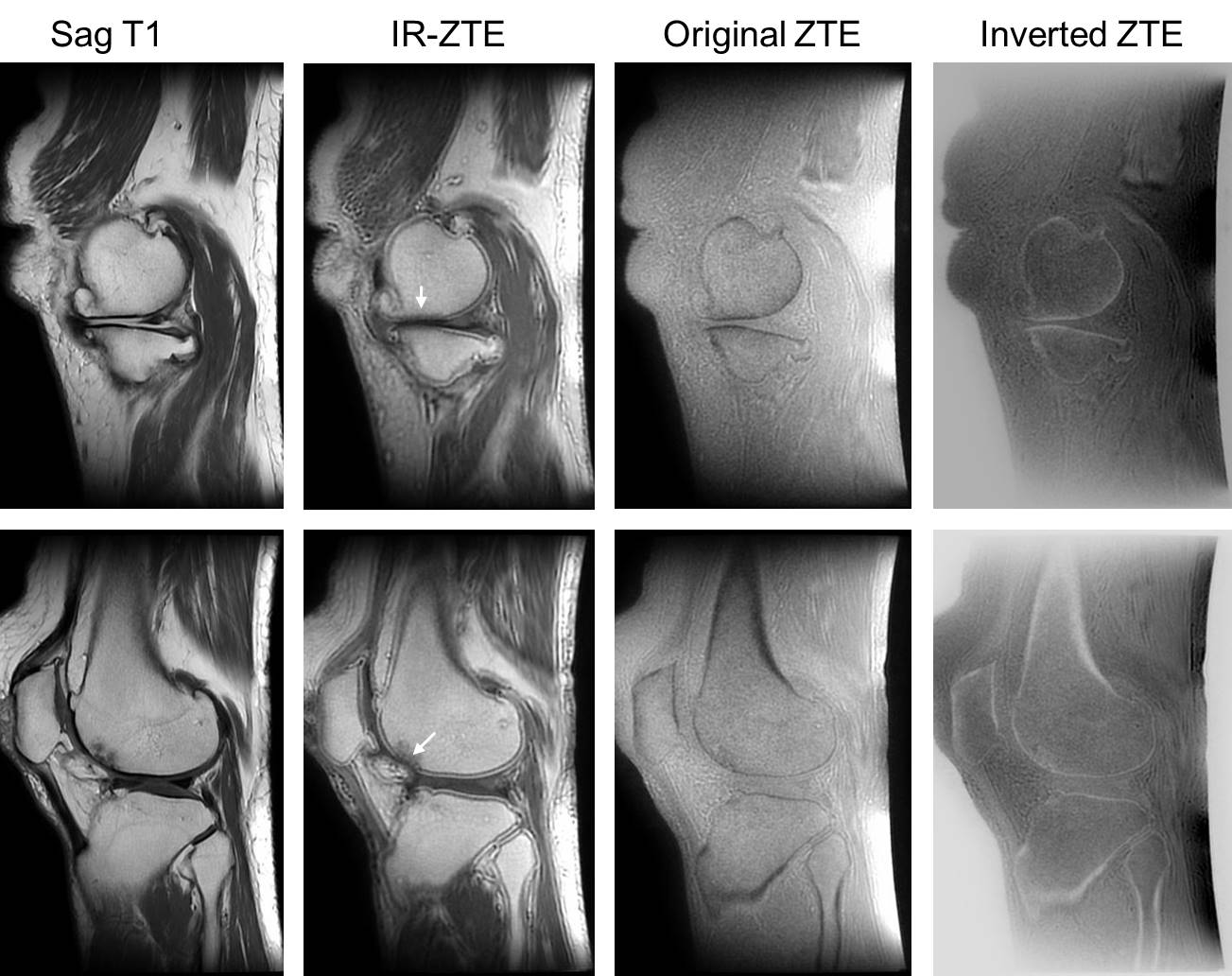
We prospectively enrolled three patients with primary end-stage knee OA undergoing elective total knee replacement surgery for this study (74F, 73F, and 63M). Upon approval of the Institutional Review Board, all subjects underwent 3.0T MR examinations (Discovery MR750, GE Healthcare, Milwaukee, USA) before total knee replacement surgery and then histopathology assessment.ZTE sequence was carried out using a non-selective hard pulse excitation followed immediately by 3D center-out radial sampling to achieve a nominal TE of zero. To enhance the contrast of cartilage tissue, an inversion recovery (IR) magnetization-prepared image was acquired prior to ZTE readout (IR-ZTE). The Figure 1 showed the IR-ZTE image quality in the knee cartilage with primary end-stage knee OA. IR-ZTE data was acquired with the scanning parameters as follows: TR=1019 ms, nominal TE=0 ms, TI=800 ms, flip angle=5 degree, receiver bandwidth= 62 kHz, field-of-view= 22 cm, resolution interpolated to 0.7 × 0.7 × 1 mm, scan time= 5 min 52 s. For IR-ZTE signal analysis, we used the contrast-to-noise ratios (CNRs) between the femoral condyle and tibial condyle tissue and its neighboring low signal region were measured in IR-ZTE images. CNR was calculated by taking the absolute difference of average signal intensities in two regions normalized by standard deviation of background noise.
The cartilage specimens from OA patients who have undergone total knee replacement surgery were examined for histopathological assessment. The cartilage specimens were fixed in 10% neutral buffered formalin for 12 h and decalcified in a rapid decalcifier with ethylene-diamine-tetra-acetic acid (EDTA) for 24 h at room temperature (Nihon Shiyaku Industries, Osaka, Japan). All slides were scanned using the AT Turbo method (Aperio®, Leica Microsystems, Nanterre, France), and images will be captured using the dedicated software Image Scope (Aperio®, Leica Microsystems, Vista, CA, USA). The Osteoarthritis Research Society International (OARSI) system was used to evaluate differences in articular cartilage histopathology [4].
The statistical analyses were performed with SPSS software (version 20, SPSS, Inc, Chicago, IL, USA). Spearman rank correlation analysis of degree of histological degeneration, CNRs, condyle tissue signal intensity, neighboring region signal intensity and SNR in the femoral condyle tissue.
Results
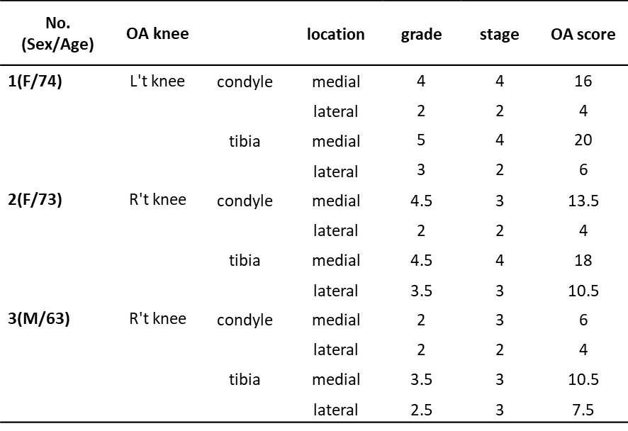
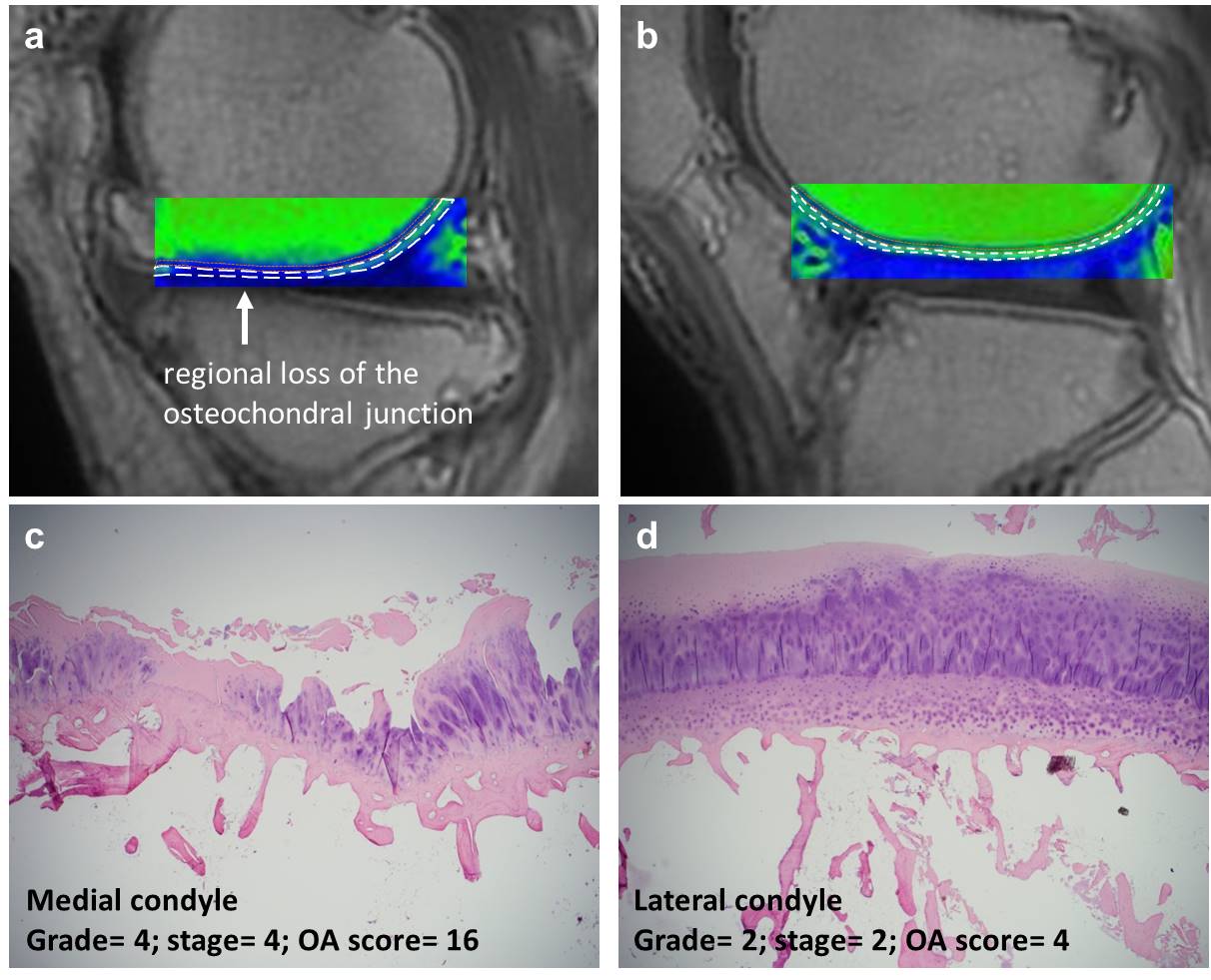
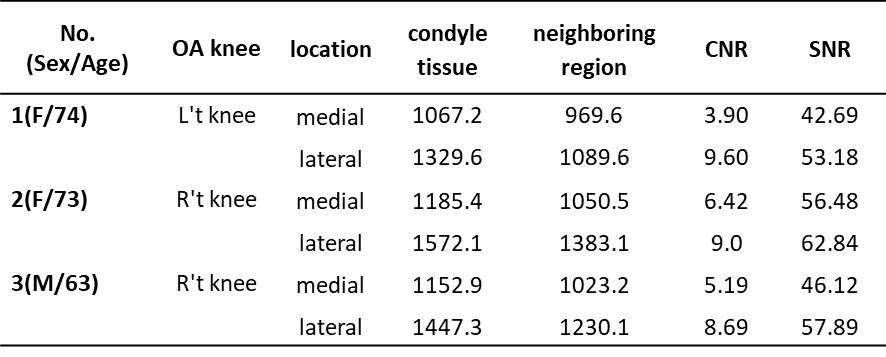
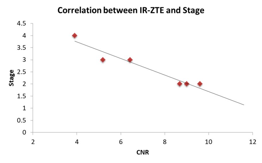
In total, 12 cartilage regions were used for histological analysis (medial condyle: n = 3, lateral condyle: n = 3, medial tibia: n = 3, lateral tibia: n = 3). The histological scores per cartilage are shown in Table 1. Figure 2 shows the CNRs measured using regions of interest (ROIs) on the femoral condyle in IR-ZTE images from a patient with OA before total knee replacement surgery and corresponding histological findings. The CNRs per femoral condyle are shown in Table 2. Correlation analysis between CNRs values and stage was performed. The two parameters were negatively correlated with a coefficient of -0.939 (p=0.005), indicating the consistency between IR-ZTE signal and the histopathological scores (n=6). It appears that IR-ZTE signal can be used as an in vivo indicator of the state of osteoarthritis (Figure 3).Discussion and Conclusion
The preliminary result showed that the signal changes of IR-ZTE MR imaging was correlated strongly degree of histological degeneration in knee cartilage, suggesting that IR-ZTE MR imaging enables direct visualization of the compositional changes of the articular cartilage in knee OA. Further assessment of more patients with different degrees of cartilage degeneration to understand the efficacy of IR-ZTE MR imaging in cartilage physiology is currently underway.Acknowledgements
No acknowledgement found.References
1. Fujisaki A, et al. Zero Echo Time Magnetic Resonance Imaging; Techniques and Clinical Utility in Musculoskeletal System. J Magn Reson Imaging. 2023.
2. Jang H, et al. Feasibility of an Inversion Recovery-Prepared Fat-Saturated Zero Echo Time Sequence for High Contrast Imaging of the Osteochondral Junction. Front Endocrinol (Lausanne). 2021.
3. Eijgenraam SM, et al. T2 mapping of the meniscus is a biomarker for early osteoarthritis. Eur Radiol. 2019.
4. Waldstein W, et al. OARSI osteoarthritis cartilage histopathology assessment system: A biomechanical evaluation in the human knee. J Orthop Res. 2016.
Figures