2292
A fully automated implant mode MRI scan workflow for asymptomatic patients.1GE HealthCare, Waukesha, WI, United States, 2GE HealthCare, Niskayuna, NY, United States, 3GE HealthCare, New York, NY, United States, 4GE HealthCare, Bangalore, India, 5Hospital for Special Surgery, New York, NY, United States
Synopsis
Keywords: Other Musculoskeletal, MSK, Implant mode scan, Prescan, Calibration, Mavric
Motivation: Patients are often unaware of the presence and location of metal implants, which may be confirmed only after localizer scan, thus potentially disrupting workflow.
Goal(s): To detect the presence of metal implant within the first few seconds of prescan and further characterize the location and shape of the metal from the calibration.
Approach: Prescan data of phantom with metal screws and human volunteer with metallic implants were used.
Results: The skewness of the center frequency signal is from 1.37 to 4.47 for implants, while for normal subjects, the signal is very symmetric and the calculated skewness is <0.3.
Impact: Automated MR scanning workflow for the patients with implants to improve efficiency of the MR technologists.
Introduction
MRI has been shown to be a valuable measure of assessing the soft tissue envelope around metal implants. Patients are often unaware of the presence and location of metal implants, which may be confirmed only after localizer scan, thus potentially disrupting workflow. Our proposal aims to address this problem and provides a more efficient and automated manner to detect metal and trigger metal scanning workflows (HyperMavric SL [1] calibration scans) automatically. Depending on the composition and extent, the patient can be shifted to 1.5T MRI (or lower field strength scanner) to reduce susceptibility and maximize image quality with the use of 3D MSI techniques or appropriate parameter modification. In this work, we demonstrate the presence of metal is identified within the first few seconds of prescan based on the B0 map and can further characterize the location and shape of the metal from the calibration volume [2], or even trigger specific spectral scans like MAVRIC to determine the location of implant.Goal
We demonstrate a methodology to detect and localize the metallic implant using the prescan data of the localizer scan from MRI in asymptomatic subjects, in whom the presence and location of the implant is unknown. This information can be used to optimize the pulse sequence and scan prescription parameters for metal scanning sequences.Methods
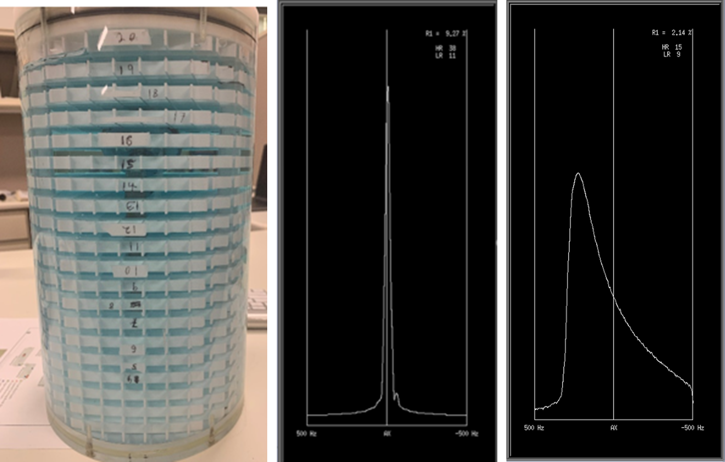
Phantom Data:A hip phantom with metal screws and a DQA phantom which are available in-house were scanned in a 1.5T Voyager GEHC scanner. Regular three-plane localizer (protocol from GE library) and prescan data were acquired. 3D Coil sensitivity maps and center frequency data from the prescan were collected from the scanner. Phase images of coil sensitivity maps and center frequency raw data are analyzed and compared for these phantoms.
Human Subject Data:
Human subject data (N=11) with variety of metal implants (size and material) were collected from clinical site (1.5T 450w GE Scanner) for retrospective analysis. Normal subjects without metal implant data from 1.5T (GEHC Artist) and 3T (GEHC Architect) scanner were collected within our institution. All data was collected with multi-channel coils. The center frequency plot was collected for each scan. Prescan raw data was read and separated for each channel and center frequency high and the pattern of center frequency (in terms of peak width, spectrum skewness) was analyzed. We also studied the impact of coil proximity to implant and its impact on frequency spectrum and if that can provide more specificity to metal detection. The phase and magnitude images of coil sensitivity maps were reconstructed.
Results and Discussion:
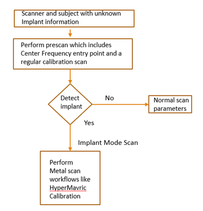
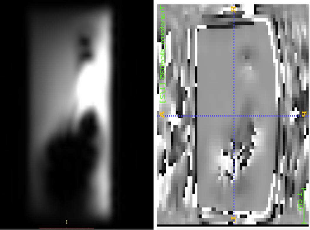
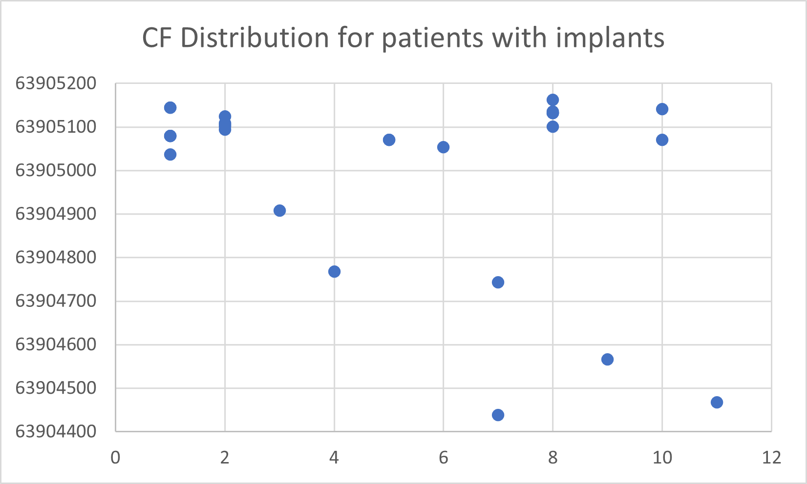
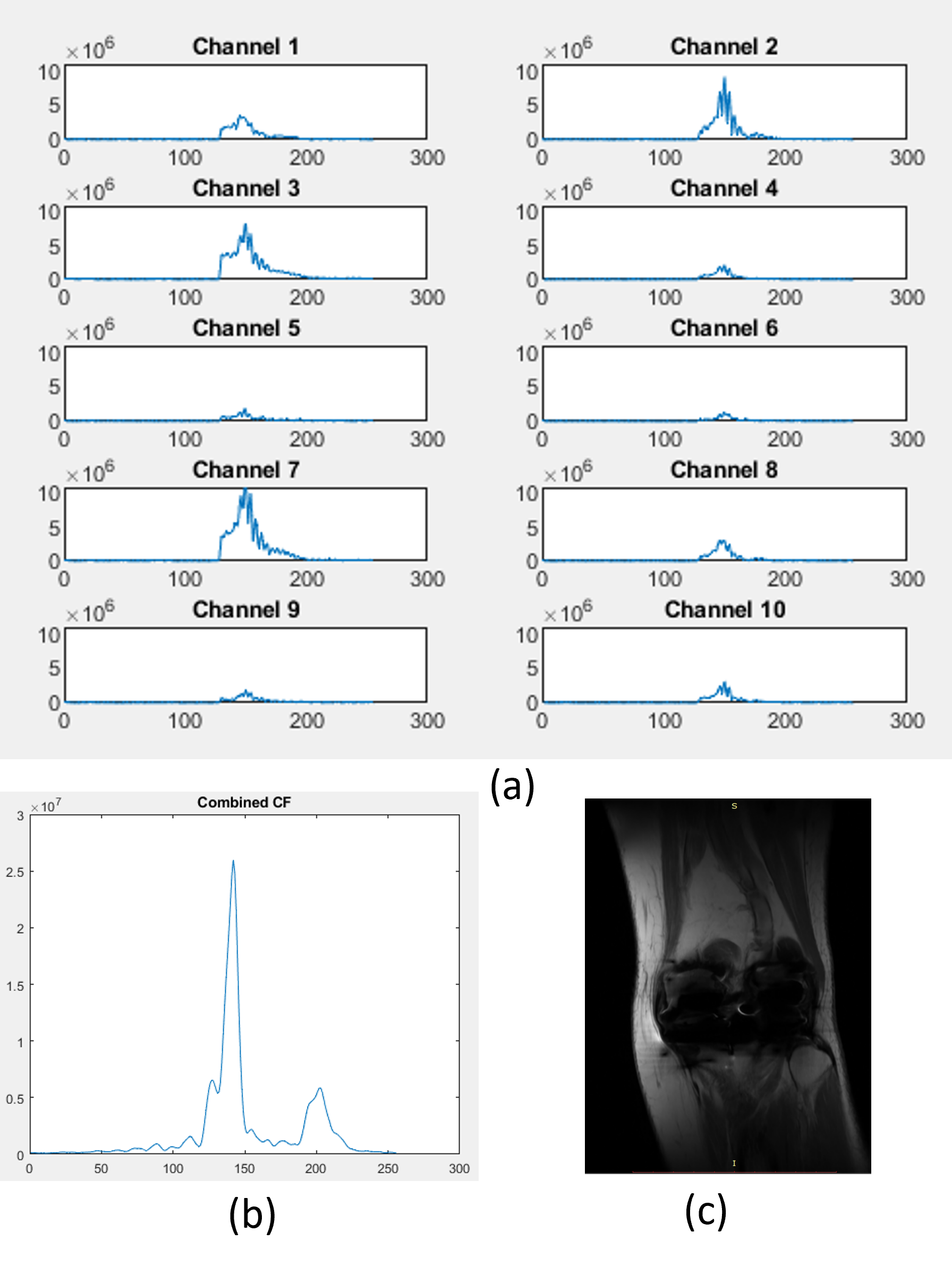
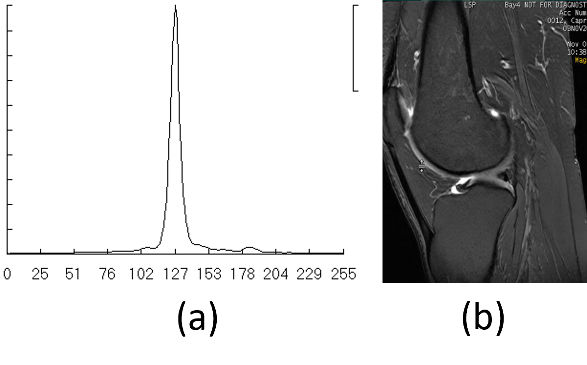
Figure (1) presents the flowchart of the automated workflow for metal scans. For an asymptomatic patient where the metal information is not known, the metal detection algorithm is run on the prescan entry point. If the metal is detected, scanner will be switched to implant mode where the technologist can follow metal scanning workflow with Hyper MAVRIC or adjust the scanner prescription and pulse sequence parameters or triage patients to a more appropriate scanner. In this way, the scanner automatically performs the scanning without any interruption. Figure (2) compared the center frequency pattern for metal implant phantom compared with DQA phantom. Figure (3) shows the phase images of calibration with metal implant where the location and rough shape can be detected by segmenting the area which has high phase disturbance [2]. Figure (4) shows the distribution of CF spectrum for various metal implants. Figure (5) presents the center frequency plots of the individual channel for one series. The signal is sampled with 256 points. The multi-channel combined data were shown with their corresponding high-resolution images in Figure (5) b and c. Similar plots are presented from a subject without implant in Figure(6). For the cases with implants there are multiple peaks, and the peak and spectral width are broader. The skewness of the multi-channel combined signal is calculated, and it is within 1.37 to 4.47, while for normal subjects, the signal is very symmetric and the calculated skewness is <0.3.Conclusion
We introduced a novel method to enable a fully automated MR scanning workflow for the patients with implants. The generalized fully automated workflow can help to increase the efficiency of the MR technologists, as the scanner automatically adjusts the scan parameters based on the presence and burden of metallic implants.Acknowledgements
No acknowledgement found.References
1. Kevin M. Koch et al. A Multispectral Three-Dimensional Acquisition Technique for Imaging Near Metal Implant, Magnetic Resonance in Medicine 61:381–390 (2009)
2. Deepa Anand, Dattesh Shanbhag, Chitresh Bhushan, Kavitha Manickam and Radhika Madhavan, "Automatic localization of metal artifacts regions on MRI scout images", ISMRM 2023
Figures