2291
FRACTURE compared to CT for detecting structural lesions of the sacroiliac joint in axial spondyloarthritis1the first affiliated hospital of nanjing medical university, nanjing, China, 2Philips healthcare,shanghai, China, shanghai, China, 3nanjing medical university, nanjing, China
Synopsis
Keywords: Other Musculoskeletal, MSK
Motivation: The detection of structural damage of the sacroiliac joint is essential for diagnosing axial spondyloarthritis.
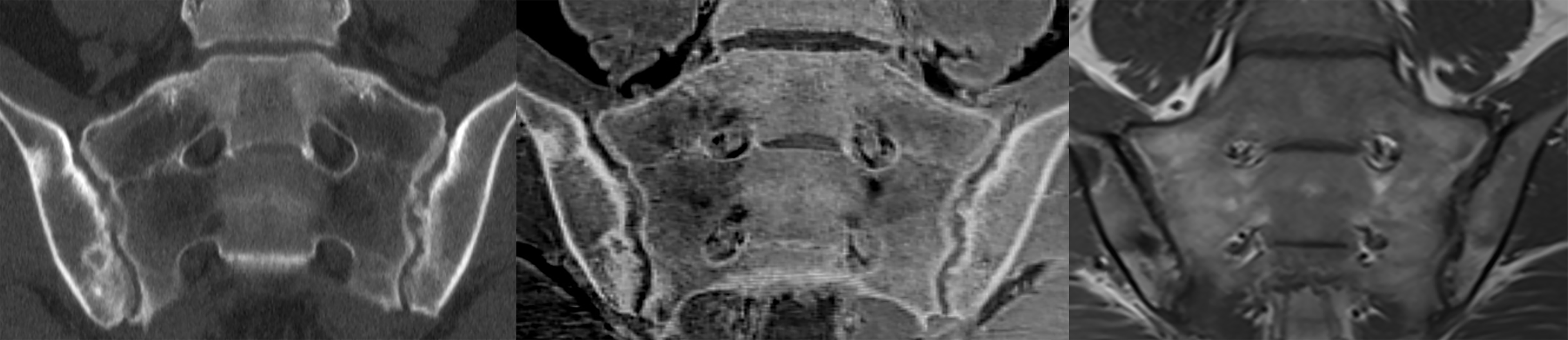
Goal(s): To assess the diagnostic performance of FRACTURE sequence in detecting structure lesions compared to CT.
Approach: A retrospective study was conducted on 83 patients with definite axial SpA who underwent MRI and CT scans of the sacroiliac joint between 2021 and 2022. An overall impression of sacroiliitis grade was also provided according to the scoring system of the modified New York criteria.
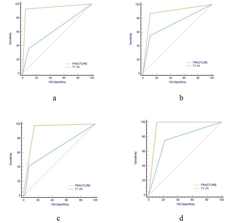
Results: When compared to CT as the standard, FRACTURE demonstrated high agreement with AUC values of 0.908, 0.943, 0.918 and 0.944 for sacroiliitis grade.
Impact: FRACTURE can evaluate structural lesions and demonstrate good diagnostic performance in patients with SpA.
Background
The detection of structural damage, such as erosions, sclerosis, and ankyloses, of the sacroiliac joint (SIJ) is essential for diagnosing axial spondyloarthritis (axSpA). However, CT is considered more effective than conventional MRI in assessing structural lesions.Purpose
To assess the diagnostic performance of FRACTURE (fast field echo resembling a CT using restricted echo-spacing) sequence in detecting structure lesions in axial spondyloarthritis (SpA) compared to T1W imaging and CT.Materials and Methods
A retrospective study was conducted on 83 patients with definite axial SpA who underwent MRI and CT scans of the sacroiliac (SI) joint between 2021 and 2022. The FRACTURE and TIWI sequence of MRI and CT scan of the SIJ were available at the same time. The image of SIJ were scored for structural lesions, including joint space changes (score 0–5), subchondral erosions (score 0–3) and subchondral sclerosis (score 0–2). An overall impression of sacroiliitis grade was also provided according to the scoring system of the modified New York criteria.Results
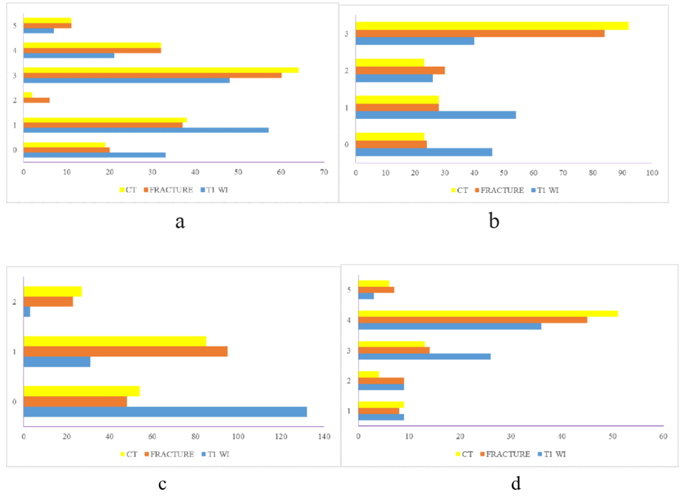
A total of 166 SIJ (83 participants) were available for analysis. When compared to CT as the standard, FRACTURE demonstrated high agreement with AUC values of 0.908, 0.943, 0.918 and 0.944 for joint space changes, subchondral erosions, subchondral sclerosis, and overall impression of sacroiliitis grade. FRACTURE had a higher diagnostic accuracy than T1WI for joint space changes (91.8% vs 70.5%), subchondral erosion (93.3% vs 56.4%), subchondral sclerosis (94% vs 77.6%), and overall impression of sacroiliitis grade (98.9% vs 75.3%).Conclusion
FRACTURE imaging can evaluate structural lesions and demonstrate good diagnostic performance in patients with SpA.Acknowledgements
I would like to give my heartfelt thanks to all the people who have ever helped me in this paper.References
References
1. Sieper J, Poddubnyy D. Axial spondyloarthritis. Lancet. 2017;390:73-84.
2. Taurog JD, Chhabra A, Colbert RA. Ankylosing Spondylitis and Axial Spondyloarthritis. N Engl J Med. 2016;374(26):2563-2574.
3. Rudwaleit M, van der Heijde D, Landewé R, et al. The development of Assessment of SpondyloArthritis international Society classification criteria for axial spondyloarthritis (part II): validation and final selection. Ann Rheum Dis 2009;68:777–783.
4. Mullenders L, Atkinson M, Paretzke H, et al. Assessing cancer risks of low-dose radiation. Nature Rev Cancer. 2009;9:596-604.
5. Johnson B, Alizai H, Dempsey M. Fast field echo resembling a CT using restricted echo-spacing (FRACTURE): a novel MRI technique with superior bone contrast. Skeletal Radiol. 2021;50(8):1705-1713. 6. Poddubnyy D, Gaydukova I, Hermann KG, et al. Magnetic resonance imaging compared to conventional radiographs for detection of chronic structural changes in sacroiliac joints in axial spondyloarthritis. J Rheumatol. 2013;40:1557–1565.
7. Van der Linden S, Valkenburg HA, Cats A. Evaluation of diagnostic criteria for ankylosing spondylitis. A proposal for modification of the New York criteria. Arthritis Rheum. 1984;27:361-368.
8. Baraliakos X, Ghadir A, Fruth M et al. Which magnetic resonance imaging lesions in the sacroiliac joints are most relevant for diagnosing axial spondyloarthritis? A prospective study comparing rheumatologists’evaluations with radiologists’ findings. Arthritis Rheumatol. 2021;73:800–805.
9. Poddubnyy D, Vahldiek J, Spiller I, et al. Evaluation of 2 screening strategies for early identification of patients with axial spondyloarthritis in primary care. J Rheumatol. 2011;38:2452–2460.
10. Hermann KG, Baraliakos X, van der Heijde D, et al. Descriptions of spinal MRI lesions and definition of a positive MRI of the spine in axial spondyloarthritis: a consensual approach by the ASAS/OMERACT MRI study group. Ann Rheum Dis. 2012;71:1278–1288.
11. Heuft-Dorenbosch L, Landewé R, Weijers R, et al. Combining information obtained from magnetic resonance imaging and conventional radiographs to detect sacroiliitis in patients with recent onset inflammatory back pain. Ann Rheum Dis. 2006;65:804–808.
12. Weber U, Lambert RG, Pedersen SJ, et al. Assessment of structural lesions in sacroiliac joints enhances diagnostic utility of magnetic resonance imaging in early spondylarthritis. Arthritis Care Res. 2010;62:1763-1771.
13. Rudwaleit M, Jurik AG, Hermann KG, et al. Defining active sacroiliitis on magnetic resonance imaging (MRI) for classification of axial spondyloarthritis: A consensual approach by the ASAS/OMERACT MRI group. Ann Rheum Dis. 2009;68(10):1520-1527.
14. Jans LBO, Chen M, Elewaut D, et al. MRI-based synthetic CT in the detection of structural lesions in patients with suspected Sacroiliitis: Comparison with MRI. Radiology. 2021;298 (2):343-349.
15. Diekhoff T, Hermann KG, Greese J, et al. Comparison of MRI with radiography for detecting structural lesions of the sacroiliac joint using CT as standard of reference: Results from the SIMACT study. Ann Rheum.Dis 2017;76(9):1502-1508
16. Van Gaalen FA, Bakker PA, de Hooge M, et al. Assessment of sacroiliitis by radiographs and MRI: where are we now? Curr Opin Rheumatol. 2014;26:384–388.
17. Puhakka KB, Jurik AG, Schiøttz-Christensen B, et al. MRI abnormalities of sacroiliac joints in early spondylarthropathy: a 1-year follow-up study. Scand J Rheumatol. 2004;33(5):332-338.
18. Geijer M, Gadeholt Göthlin G, Göthlin JH. Diagnosis and progression of sacroiliitis in repeated sacroiliac joint computed tomography. Arthritis. 2013;2013:659487.
19. Hahn S, Song JS, Choi EJ, et al. Can Bone Erosion in Axial Spondyloarthropathy be Detected by Ultrashort Echo Time Imaging? A Comparison With Computed Tomography in the Sacroiliac Joint. J Magn Reson Imaging. 2022;56(5):1580-1590.
20. Chang EY, Du J, Chung CB. UTE imaging in the musculoskeletal system. J Magn Reson Imaging. 2015;41(4):870-883.
21. Bharadwaj UU, Coy A, Motamedi D, et al. CT-like MRI: a qualitative assessment of ZTE sequences for knee osseous abnormalities. Skeletal Radiol. 2022;51(8):1585-1594.
22. Hou B, Liu C, Li Y, et al. Evaluation of the degenerative lumbar osseous morphology using zero echo time magnetic resonance imaging (ZTE-MRI). Eur Spine J. 2022;31(3):792-800.
Figures