2290
Texture Analysis of the Infrapatellar Fat Pad Within and Between Collegiate Basketball and Swimming Athletes1Department of Radiology and Imaging, Hospital for Special Surgery, New York, NY, United States, 2School of Medicine, Radiology, University of California San Francisco, San Francisco, CA, United States, 3Stanford Medicine Department of Radiology and Biomedical Imaging, Stanford University, Stanford, CA, United States
Synopsis
Keywords: Other Musculoskeletal, MSK
Motivation: MRI-based texture features have been used to identify abnormalities within the infrapatellar fat pad (IPFP) of middle-aged to elderly adults, yet no existing investigations have evaluated IPFP quality in young athletes.
Goal(s): To quantify and compare texture features of the IPFP in collegiate athletes comprised of men and women basketball players and swimmers.
Approach: Image texture features of the IPFP were calculated from bilateral MRIs acquired at 2 time points. (baseline and 1 yr. follow-up) for collegiate men and women basketball and swimming athletes.
Results: The IPFP of collegiate basketball players significantly differed from the swimmers, with greater global and local image homogeneity.
Impact: While the texture features of swimmer IPFPs remained consistent across sexes and timepoints, swimmers had significantly greater texture heterogeneity than basketball players at baseline. Observed variability indicates the need for further longitudinal investigation of IPFP structure within low impact sports.
Introduction
As a vascularized, intracapsular, extra-synovial structure, the infrapatellar fat pad (IPFP) provides cushioning and regulates inflammation throughout the anterior knee compartment1-3. A pathological inflammatory response within the IPFP can arise from prior damage within the knee4,5, injuries of which commonly occur in high impact sports (e.g. basketball, volleyball, and high jump)6 and certain swim strokes (butterfly and breast stroke)7. Magnetic resonance imaging (MRI) is commonly used to evaluate the inflammatory response to injury as shown by a concomitant increase in fat-pad signal intensity8. Efforts have been made to remove reader-dependence of this evaluation by quantitatively analyzing gray-level signal intensities to obtain texture, or ‘radiomic’, features within a region of interest (ROI)1. Texture analyses quantify variation in voxel intensity within an ROI9 and texture features from MRI of the IPFP have shown greater discrimination for IPFP synovitis than clinical scoring alone1. As prior work focused on an aged cohort (mean age > 60), this study aimed to: 1) quantify texture features of the IPFP in collegiate basketball and swimming athletes, and 2) compare texture features between sports.Methods
Following IRB approval with informed consent, bilateral morphologic MRIs, consisting of water and fat images derived from a 3D CUBE FLEX acquisition, were acquired at baseline (TP0) from collegiate men and women basketball and swimming athletes from Division 1 schools. A second dataset was obtained approximately 1 year after the initial visit (TP1). Two left knees were excluded from the analysis due to poor image quality. The entire IPFP volume in the athletes’ knees in the fat images was automatically segmented (Mathworks, Natick MA) and manually edited, as needed, in ITK-Snap10. Image texture analysis software, maZda11 (V18.07), was utilized to evaluate voxel intensity data within each IPFP to calculate the gray level co-occurrence matrix (GLCM). Intensity data was normalized to a range defined by the 1st and 99th percentiles of voxel intensity within the ROI. Texture features derived from the GLCM included: angular second moment (ASM, image homogeneity), contrast (image variability), correlation (interdependence of intensities between neighboring voxels), inverse difference moment (IDM, local homogeneity), and entropy (image disorder)9. Mann-Whitney U tests were performed to detect differences of texture features and IPFP volume between the basketball players and swimmers at TP0. Paired t-tests were performed to compare differences in texture features between swimmers at TP0 and TP1.Results
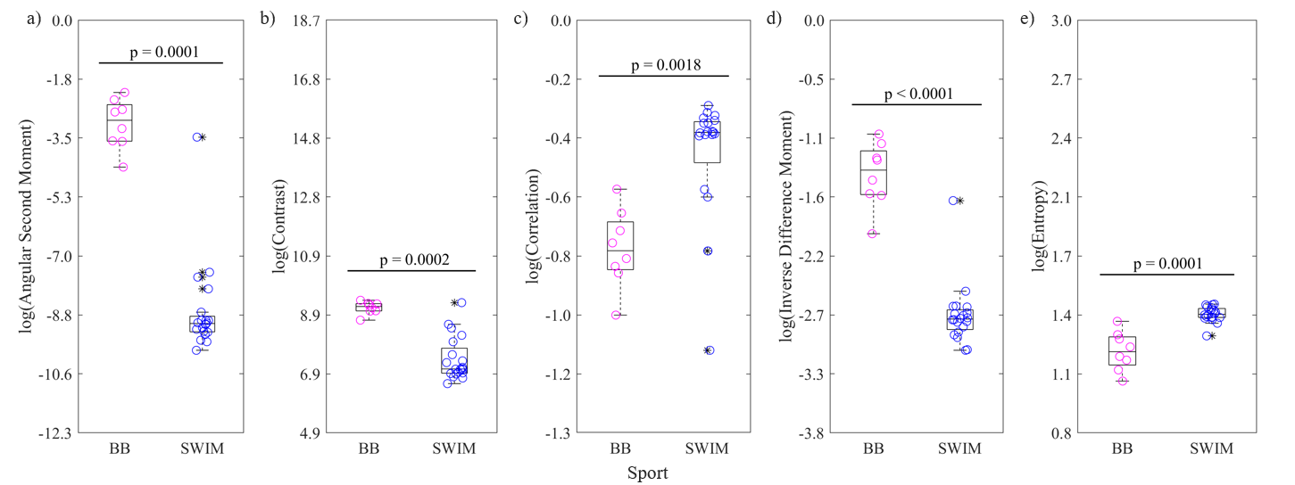
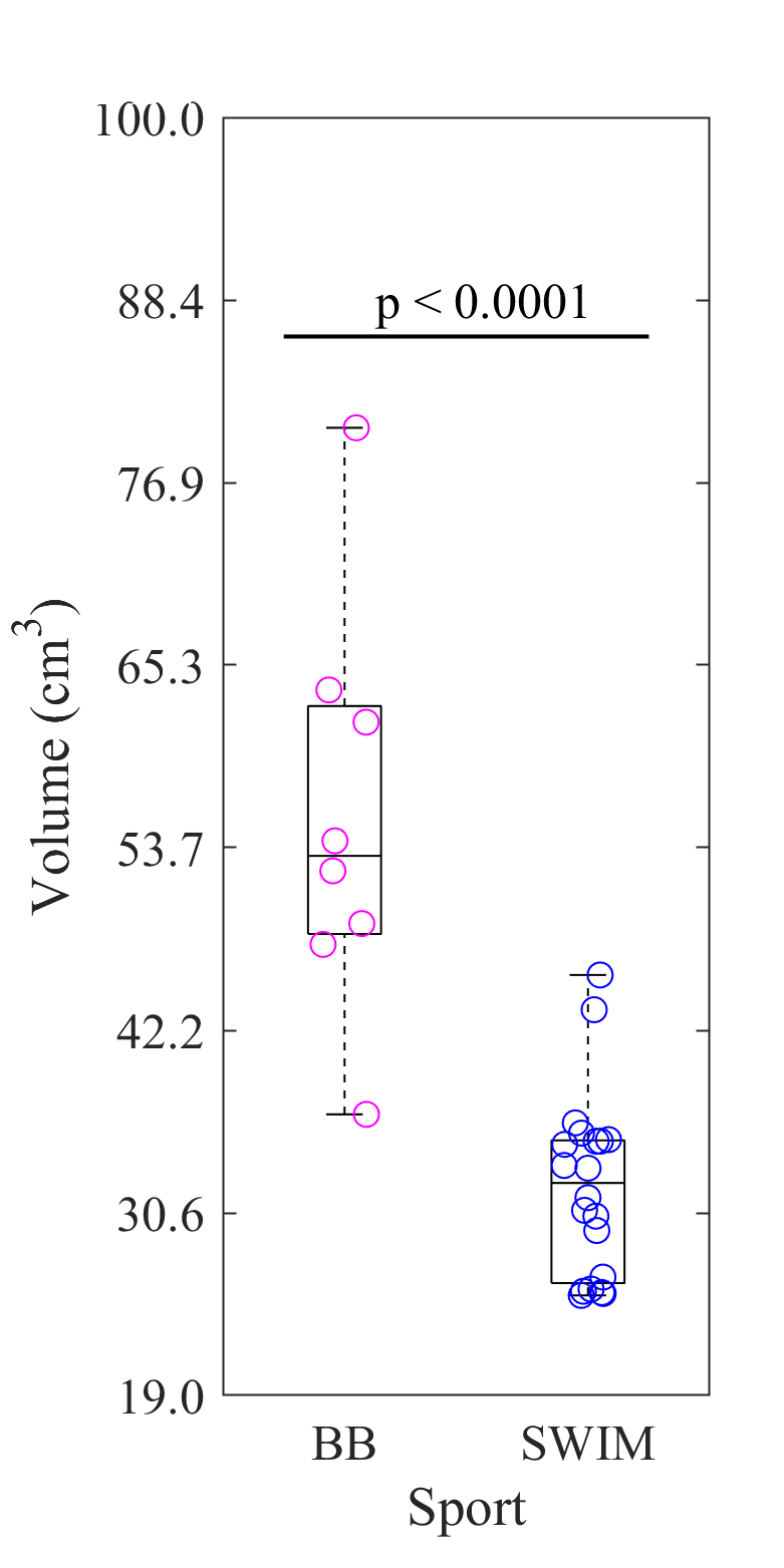
The data from 5 basketball athletes (5 male; Age: 18.8 ± 0.45 y.o.; BMI: 22.9 ± 2.5) and 10 swimming athletes (5 female, 5 male; Age: 18.2 ± 0.4 y.o.; BMI: 23.9 ± 2.2) have been evaluated to date. Basketball vs Swimming: At TP0, all image texture features were significantly different between the sports. The basketball players had higher values of ASM (p=0.0001), contrast (p=0.0002), IDM (p<0.0001), and lower values for correlation (p=0.0018) and entropy (p=0.0001) (Figure 1). The IPFP volume of the basketball players was also significantly larger than the swimmers (p<0.0001) (Figure 2). Swimming - TP0 vs TP1: No differences were detected for any texture feature between TP0 and TP1.Discussion
Higher ASM and IDM with lower entropy indicate that the IPFP of the basketball players are significantly more homogeneous than those of collegiate swimmers. Contrast differences suggest increased heterogeneity in the IPFP of basketball players, however, contrast has been previously shown to be influenced by the extent of the defined ROI12. Smaller ROIs often have higher correlation values9, consistent with the IPFPs of the swimmers which were smaller in volume as compared to the basketball players. We anticipated greater variability in basketball player IPFP texture because of the high number of knee injuries in the sport6; however, our results indicate that constant low impact exercise of swimmers may contribute to increased IPFP texture heterogeneity. Chronic overuse knee injuries are common among butterfly and breaststroke swimmers7 and recurring knee pain has been positively associated with number of years of swimming13. These findings, and the current lacking report of knee injuries in swimmers7,14, highlight the need for a more extensive look into swimmer knee anatomy over longer periods of time (> 1 yrs). Finally, while greater heterogeneity in the IPFP of swimmers was observed, when compared to basketball players, future studies would benefit by also evaluating a control group age-matched non-athletes.Conclusion
The texture features of the IPFP of basketball players were significantly different from the IPFP of swimmers at baseline. Non-significant longitudinal changes in texture parameters suggest that texture heterogeneity may be activity specific, with basketball players having more homogeneous fat pads, while swimmers have greater texture variability.Acknowledgements
The authors would like to acknowledge funding from the GE/NBA consortium who provided funding support for this study and the support of the staff of the HSS MRI Department for their assistance with scanning.References
- Li J, Fu S, Gong Z et al. MRI-base texture analysis of infrapatellar fat pad to predict knee osteoarthritis incidence. Radiology. 2022; 304:611-621.
- Dragoo JL, Johnson C, McConnell J. Evaluation and treatment of disorders of the infrapatellar fat pad. Sports Medicine. 2012; 42(1):51-67.
- Atukorala I, Kwoh CK, Guermazi A et al. Synovitis in knee osteoarthritis: a precursor of disease? Annals of Rheumatic Diseases. 2016; 75(2):390-395.
- Myers SL, Brandt KD, Ehlich JW et al. Synovial Inflammation in patients with early osteoarthritis of the knee. J. Rheumatol. 1990; 17(12):1662-1669.
- Fuerst M, Lammers L, Schafer F. Investigation of calcium crystals in OA knees. Rheumatology International. 2010; 30:623-631.
- Majewski M, Habelt S, Steinbruck K. Epidemiology of athletic knee injuries: A 10-year study. The Knee. 2006; 13(3):184-188.
- Trinidad A, Gonzalez-Garcia H, Lopez-Valenciano A. An updated review of the epidemiology of swimming injuries. Physical Medicine and Rehabilitation. 2020; 13(9):1005-1020.
- Hunter DJ, Zhang W, Conaghan PG et al. Systematic review of the concurrent and predictive validity of MRI biomarkers in OA. Osteoarthritis and Cartilage. 2011; 19(5): 557-588.
- Hall-Beyer, Mryka. GLCM texture: a tutorial. 2023.
- Yushkevich PA, Piven J, Hazlett HC et al. User-guided 3D active contour segmentation of anatomical structures: Significantly improved efficiency and reliability. Neuroimage. 2006; 31(3):1116-1128.
- Szczypinski PM, Strzelecki M,
Materka A et al. MaZda—a software package for image texture analysis. Computer
Methods and Programs in Biomedicine. 2009; 94(1):66-76.
- Zaworski C, Cheah J, Koff MF et al. MRI-based texture analysis of trabecular bone for opportunistic screening of skeletal fragility. 2021; 106(8);2233-2241.
- Hahn T, Foldspang A. Prevalent knee pain and sport. Scandinavian Journal of Social Medicine. 1998; 26(1): 44-52.
- Hill L, Mountjoy M, Miller J. Non-shoulder injuries in swimming: A systemic review. Clinical Journal of Sport Medicine. 2022; 32(3):256-264.
Figures