2289
Reactive oxygen species responsive theranotics nanoprobe for chemical exchange saturation transfer imaging in osteoarthritis1Department of Radiology, Zhujiang Hospital, Southern Medical University, Guangzhou, China, 2Department of Rehabilitation Medicine, Zhujiang Hospital, Southern Medical University, Guangzhou, China, 3Department of Joint and Orthopedics, Orthopedic Center, Zhujiang Hospital, Southern Medical University, Guangzhou, China, 4Guangdong Key Lab of Orthopedic Technology and Implant, Guangzhou, China
Synopsis
Keywords: Osteoarthritis, Contrast Agent
Motivation: There is still a lack of noninvasive technique in quantifying the acidic extracellular pH of damaged cartilage in early osteoarthritis (OA).
Goal(s): We aimed to fabricate an advance reactive oxygen species (ROS) responsive CEST-MRI contrast agent for damaged cartilaged imaging and treatment in OA.
Approach: We use CEST-MRI to investigate whether our sythesized PLGE-TK-PEG@PDA-SA nanoparticles repond to ROS accumulating in damaged cartilage could result in the exsistance of CEST signal, while no CEST signal would be tested in normal cartilage without excessive ROS.
Results: The PLGA-TK-PEG@PDA-SA nanoparticles could effectively respond to ROS and then produce significant CEST effect for precisely pH measurement.
Impact: The novel PLGA-TK-PEG@PDA-SA CEST- MRI contrast could be used to noninvasively monitor the extracellular pH of cartilage in osteoarthritis, and eliminate excessive ROS to defend against its damage to cartilage. This smart, sensitive ROS-responsive nanoprobe is promising for OA theranostics.
INTRODUCTION
The acidic extracellular pH of cartilage in osteoarthritis is often associated with deregulated cartilage cell metabolism[1]. Chemical exchange saturation transfer (CEST) magnetic resonance imaging (MRI) has emerged as a noninvasive technique for absolute pH measurement[2, 3]; therefore, it has potential utility for disease diagnoses and evaluating therapeutic responses. Herein, we aimed to fabricate an advance ROS responsive and damaged cartilage targeting CEST-MRI contrast for imaging and treatment of OA, which may provide reference for clinical application.METHODS
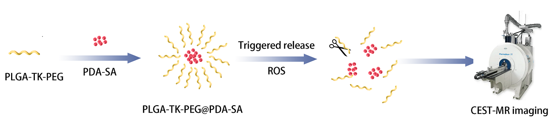
Materials: We conjugated the PDA with SA, and then modified with a ROS-sensitive PLGE-TK-PEG nanoparticles (NPs) (Fig.1). We prepared two phantoms as follows. One phantom was prepared by dissolving PLGA-TK-PEG@PDA-SA NPs in PBS. A second phantom was prepared using PLGA-TK-PEG@PDA-SA NPs in PBS and adding H2O2. The second phantom was then titrated to pH values of 6.0, 6.5, 7.0 and 7.5 using hydrogen chloride and sodium hydroxide solutions, respectively.CEST-MRI: CEST-MRI experiments of phantoms were performed on a Bruker 9.7T(Bruker BioSpin MRI GmbH, Ettlingen, Germany). All experiments were carried out at 37℃. CEST MR images were acquired with follow parameters: Repetition time/echo time/saturation time (TR/TE/TS) = 6000/15/3000 ms, saturation power =2 and 6 µT, number of averages = 2, RARE factor = 24, matrix size = 128×128, FOV = 30×30 mm2. The saturation frequency was from -16 to 16 ppm with 0.25 interval. The total acquisition time of CEST phantom experiment was 48 min 48 sec.
Data analysis: All images were processed in MATLAB using CEST codes downloaded from www.cest-sources.org with modifications.
RESULTS
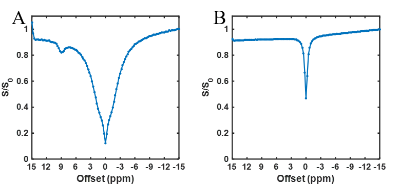
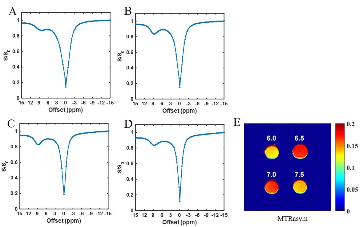
The CEST signal of PLGA-TK-PEG@PDA-SA NPs in PBS was disappeared (Fig.2B), whereas the CEST signal appeared in H2O2 (Fig.2A). The PLGA-TK-PEG@PDA-SA NPs in H2O2 had significant CEST effects at 9 ppm. The Z-spectra of PLGA-TK-PEG@PDA-SA NPs in H2O2 were obtained at different pH values (Fig.3A-D). A log10 ratio based on CEST signals at 5µT and 6µT was calculated and linearly correlated with the measured pH (Fig.4).DISCUSSION
At present, there is still a lack of noninvasive techniques for precisely detecting damaged cartilage in early OA. We designed the ROS-responsive theranostic nanoprobe for effective CEST-MR imaging and therapy in OA. Results showed that the CEST-MRI signal of PLGA-TK-PEG@PDA-SA NPs can smartly “turn on” in response to excessive ROS and “turn off” in the normal environment without ROS, which resulted in specific imaging of damaged cartilage without perturbations associated with normal cartilage. PLGE-TK-PEG@PDA-SA could effectively respond to ROS and then remarkably produce significant CEST effect for precisely pH measurement of damaged cartilage where accumulated excessive ROS. What’s more, it could eliminate abnormal over-production of endogenous ROS within cartilage tissue for treatment of OA.CONCLUSION
The novel sythesized PLGA-TK-PEG@PDA-SA nanoparticle produce excellent characteristics as CEST-MRI agent, which presented a promising newnanoplatform in OA theranostic applications.Acknowledgements
The authors have no acknowledgements to note.References
1. Lombardi AF, Ma Y, Jang H, et al. AcidoCEST-UTE MRI Reveals an Acidic Microenvironment in Knee Osteoarthritis. Int J Mol Sci[J]. 2022;23(8):4466.
2. Ma YJ, High RA, Tang Q, et al. AcidoCEST-UTE MRI for the Assessment of Extracellular pH of Joint Tissues at 3 T. Invest Radiol[J]. 2019;54(9):565-571.
3. Tao Q, Yi P, Cai Z, et al. Ratiometric chemical exchange saturation transfer pH mapping using two iodinated agents with nonequivalent amide protons and a single low saturation power. Quant Imaging Med Surg[J]. 2022;12(7):3889-3902.
Figures