2288
Accelerated 3D IR-FS-UTE Knee Imaging with High Short-T2 Contrast in Less Than 5 minutes1UIH America, Inc, Houston, TX, United States, 2Bioengineering, Stanford University, Stanford, CA, United States, 3United Imaging Research Institute of Innovative Medical Equipment, Shenzhen, China
Synopsis
Keywords: Other Musculoskeletal, MSK
Motivation: 3D DIR-UTE can provide high short T2 contrast that is useful in knee imaging, however with a prohibited long acquisition time.
Goal(s): The goal is to maintain high short-T2 contrast while significantly reducing the acquisition time.
Approach: The approach involves employing an accelerated IR-prepared fat-saturation UTE sequence along with advanced compressive sensing reconstruction.
Results: This approach achieved a threefold acceleration without sacrificing image quality and produced high short-T2 contrast, making structures like the meniscus and ligament clearly visible.
Impact: This technique holds potential for various musculoskeletal applications, such as in the early detection of conditions such as osteochondral junction alterations, osteoarthritis and meniscal tears.
Introduction
For MR knee imaging, structures with short T2 relaxation times, such as the articular cartilage's deep radial and calcified layers, menisci, ligaments, tendons, and both cortical and trabecular bones, hold considerable interest for both research and clinical practice [1][2]. While UTE sequences can detect short-T2 species, the contrast can be compromised by the high signal intensity of surrounding long-T2 species. To enhance short-T2 contrast, the long-T2 signals can be suppressed through subtraction between UTE echo and normal TE echo [3]. Recently, a method called dual adiabatic inversion recovery ultrashort echo time (DIR UTE) imaging was introduced to effectively suppress water and fat signals, thus enhancing the visibility of short-T2 species [2][4]. However, one drawback was the extensive acquisition time due to the use of DIR pulses. In this study, we demonstrate that high short-T2 contrast can be achieved using a single inversion recovery (IR) along with a fat-saturation pulse in a UTE sequence. Compressed Sensing (CS) MRI were employed to achieve a 3-fold acceleration without compromising image quality, resulting in an acquisition time of <5 minutes.Methods
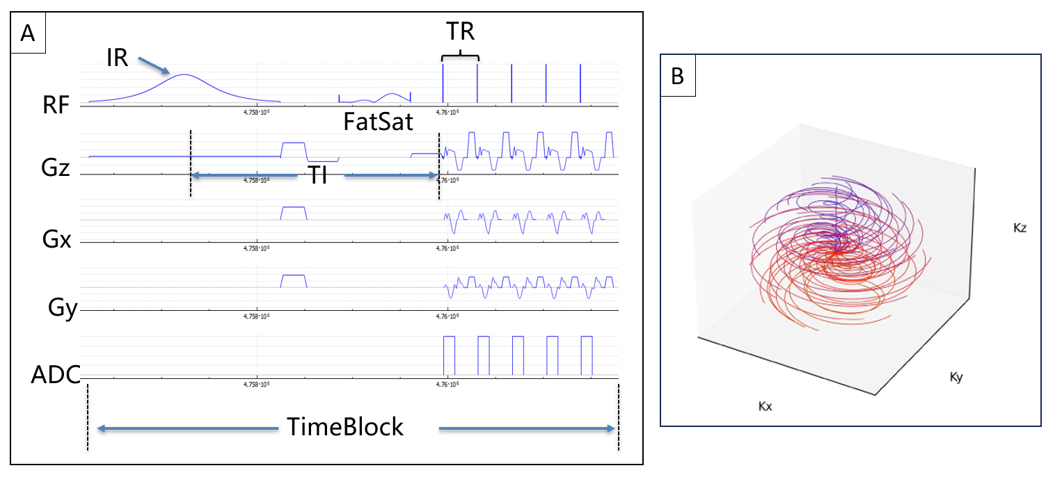
IR-FS-UTE with 3D WHIRL-Cones Trajectory:As depicted in Figure 1A, an IR and FS pulse were incorporated before the gradient-spoiled sequence. To expedite acquisition, multiple lines (TRs) were acquired after each IR and FS pulse, forming a time-block. To achieve UTE, a selective hard pulse was employed as the excitation pulse. 3D WHIRL-cones trajectory was employed to provide higher data acquisition efficiency [5] (Figure 1B).
Data Acquisition:
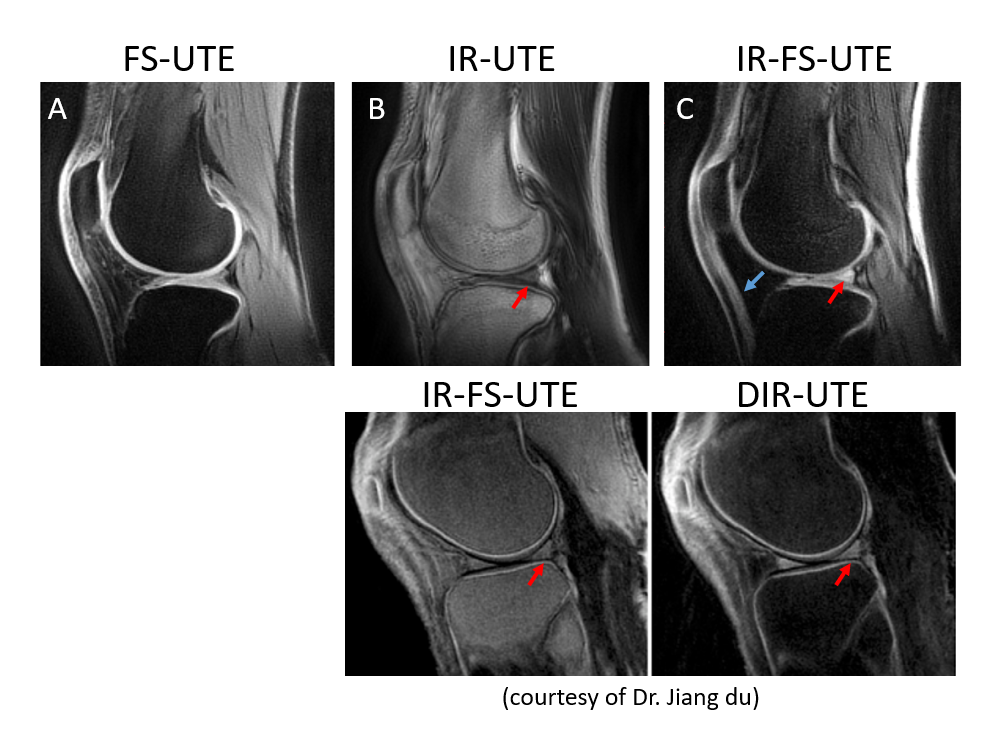
The sequence from Figure 1 was implemented on a 3T system (uMR 790, United Imaging Healthcare, Shanghai, China). With IRB approval, images were acquired on both phantom and human knees. Key sequence parameters were: TR=3.7ms, TE=0.05ms, TI=500ms, time-block=600ms, slice thickness=2mm, FOV=18cm×18cm, matrix=240×240, slices=48. The acquisition was repeated three times with different magnetic preparation pulses: FS-UTE, IR-UTE and IR-FS-UTE, respectively. The acquisition time was 3:29, 12:19 and 12:38, respectively.
Image Reconstruction:
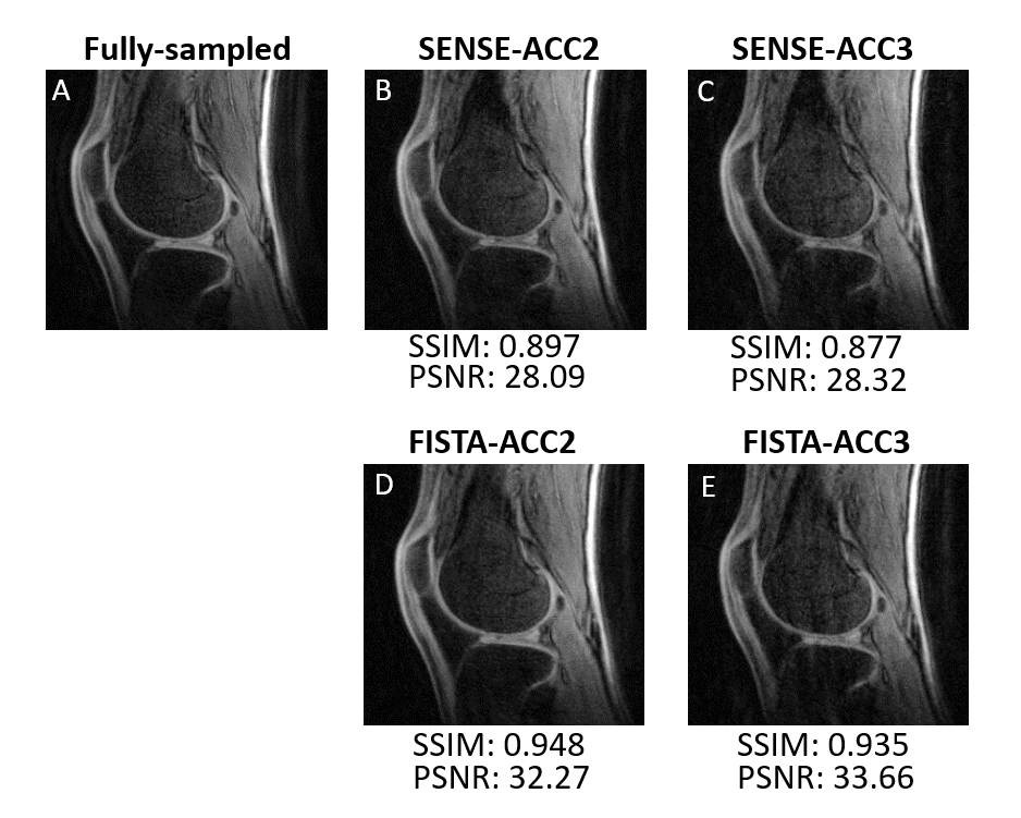
Fully-sampled data were reconstructed using NUFFT based on sigpy [6]. To expedite acquisition, the data was retrospectively undersampled with acceleration factors of 2 and 3, then reconstructed iteratively with two compressed sensing (CS) reconstruction techniques: SENSitivity Encoding (SENSE) and the Fast Iterative Shrinkage-Thresholding Algorithm (FISTA).
For SENSE reconstruction, consider the problem: $$\min_x \frac{1}{2} \| \sqrt {(D)} (F S x) - y \|_2^2 + \frac{\lambda}{2} \| x \|_2^2$$ For FISTA reconstruction, also known as L1-wavelet regularized reconstruction, consider the problem: $$\min_x \frac{1}{2} \| P F S x - y \|_2^2 + \lambda \| W x \|_1$$ where D is the density compensation function, F is the Fourier transform operator, S is the SENSE operator, P is the sampling operator, W is the wavelet operator, x is the image, and y is the k-space measurements. The reconstruction was performed based on sigpy [6] on a desktop PC with NVIDIA RTX-3090 24GB graphics card. SSIM and PSNR were calculated for quantitative comparison.
Results
Representative images acquired using different magnetic preparation pulses are displayed in Figure 2 (A-C), showcasing different contrasts. IR-FS-UTE offers excellent contrast for short T2 tissues, such as ligaments (blue arrow) and meniscus (red arrow), similar to DIR-UTE images.The quality of the under-sampled images closely matched that of the fully-sampled data (SSIM > 0.93), irrespective of the reconstruction method and acceleration rate, as shown in Figure 3. While SENSE-recon exhibits lower SSIM and PSNR values compared to FISTA, it also displays fewer noticeable image artifacts. With an acceleration factor of 3, the acquisition time can be reduced to less than 5 minutes, aligning well with clinical expectations.
Discussion and Conclusion
In summary, IR-FS-UTE demonstrates its ability to provide high contrast for short-T2 species. With this technique, short-T2 tissues such as the meniscus and ligament can be clearly visible on the image, which are not typically distinguishable with routine clinical imaging. The contrast of IR-FS-UTE from this study is quite different from the literature, and is closer to DIR-UTE [4]. This may be due to the fat-saturation pulse used in this study providing a better suppression of the bone marrow. A significant advancement is the threefold faster data acquisition facilitated by CS reconstruction, trimming scan times to under five minutes without quality compromise. Further acceleration is conceivable with deep learning techniques that have proven successful in various imaging applications [7]–[9]. To conclude, the accelerated IR-FS-UTE sequence is a promising tool for musculoskeletal imaging, enabling the detection of short-T2 tissue changes such as osteochondral junction alterations, osteoarthritis and meniscal tears.Acknowledgements
No acknowledgement found.References
[1] G. E. Gold, J. M. Pauly, A. Macovski, and R. J. Herfkens, “MR Spectroscopic imaging of collagen: Tendons and knee menisci,” Magn. Reson. Med., vol. 34, no. 5, pp. 647–654, Nov. 1995, doi: 10.1002/mrm.1910340502.
[2] J. Du, A. M. Takahashi, W. C. Bae, C. B. Chung, and G. M. Bydder, “Dual inversion recovery, ultrashort echo time (DIR UTE) imaging: Creating high contrast for short‐ T 2 species,” Magn. Reson. Med., vol. 63, no. 2, pp. 447–455, Feb. 2010, doi: 10.1002/mrm.22257.
[3] P. E. Z. Larson, S. M. Conolly, J. M. Pauly, and D. G. Nishimura, “Using adiabatic inversion pulses for long‐ T 2 suppression in ultrashort echo time (UTE) imaging,” Magn. Reson. Med., vol. 58, no. 5, pp. 952–961, Nov. 2007, doi: 10.1002/mrm.21341.
[4] A. F. Lombardi et al., “High‐contrast osteochondral junction imaging using a 3D dual adiabatic inversion recovery‐prepared ultrashort echo time cones sequence,” NMR Biomed., vol. 34, no. 8, p. e4559, Aug. 2021, doi: 10.1002/nbm.4559.
[5] J. G. Pipe, “An optimized center-outk-space trajectory for multishot MRI: Comparison with spiral and projection reconstruction,” Magn. Reson. Med., vol. 42, no. 4, pp. 714–720, Oct. 1999, doi: 10.1002/(SICI)1522-2594(199910)42:4<714::AID-MRM13>3.0.CO;2-G.
[6] F. Ong and Lustig, Michael, “SigPy: a python package for high performance iterative reconstruction,” in Proceedings of the International Society of Magnetic Resonance in Medicine, Montréal, QC, 4819 2019.
[7] Y. Han, L. Sunwoo, and J. C. Ye, “k-Space Deep Learning for Accelerated MRI,” ArXiv180503779 Cs Stat, Jul. 2019, Accessed: May 23, 2020. [Online]. Available: http://arxiv.org/abs/1805.03779
[8] D. Lee, J. Yoo, S. Tak, and J. C. Ye, “Deep Residual Learning for Accelerated MRI using Magnitude and Phase Networks,” ArXiv180400432 Cs Stat, Apr. 2018, Accessed: Oct. 25, 2021. [Online]. Available: http://arxiv.org/abs/1804.00432
[9] C. M. Sandino, J. Y. Cheng, F. Chen, M. Mardani, J. M. Pauly, and S. S. Vasanawala, “Compressed Sensing: From Research to Clinical Practice With Deep Neural Networks: Shortening Scan Times for Magnetic Resonance Imaging,” IEEE Signal Process. Mag., vol. 37, no. 1, pp. 117–127, Jan. 2020, doi: 10.1109/MSP.2019.2950433.
Figures