2287
Validating accelerated T1rho dispersion measurement with samples from spontaneous human osteoarthritis1Department of Technical Physics, University of Eastern Finland, Kuopio, Finland
Synopsis
Keywords: Cartilage, Quantitative Imaging
Motivation: Validation of compressed sensing accelerated T1rho dispersion measurement of spontaneously degenerated human cartilage tissue samples.
Goal(s): To study the effect the data reduction has on the ability to detect differences between the intact and degenerated articular cartilage specimens at different spin-lock amplitudes.
Approach: T1rho dispersion measurement with compressed sensing technique for reconstruction.
Results: Four-fold acceleration of T1rho dispersion measurement by compressed sensing approach was feasible without loss in the sensitivity to osteoarthritic changes within the articular cartilage. Differences were significant between intact and OA groups in the superficial and transitional zones, and T1rho correlated moderately with the reference methods.
Impact: Compressed sensing allowed reducing the scan-time required for measurement of T1rho dispersion, while still retaining the ability to detect degenerative changes in articular cartilage. Thus, the study brings T1rho dispersion measurements closer to clinical viability.
Introduction
T1rho dispersion is sensitive in detecting degenerative changes, that can be related to histological and biomechanical properties of cartilage1–3. Compressed sensing (CS) based acceleration has previously been applied to T1rho-mapping4–6. For good performance, leveraging the correlations over the spin-lock time (TSL) dimension in the reconstruction is essential. The T1rho dispersion measurements allows for similar approach over the spin-lock amplitude (SLA) dimension. The aim of the study was to validate the usability of accelerated T1rho dispersion measurement with CS-based reconstruction by studying the effect that the acceleration factor has on the ability to detect differences between the intact and degenerated articular cartilage specimens, at different SLAs. Biomechanical properties and OARSI grades of the samples were used as reference data.Materials and Methods
Osteochondral plugs (n = 27, 4 mm diameter) from the femur (n=14) and tibia (n=13) of human cadaver knee joints were obtained from a commercial biobank (Science Care, USA) under Ethical permission 134/2015 of North Savo hospital district ethical committee. Samples ranged from visually intact to severely degenerated. Prior to the MRI, equilibrium and dynamic Young’s moduli were measured using indentation testing7. After the MRI, the samples were processed to histological sections. The sections were stained with safranin-O to perform OARSI grading. OARSI grade was assigned to each sample as a consensus of three reviewers8. The samples were retrospectively divided into intact and OA groups by the OARSI grades. The intact group was defined as OARSI grade≤1 and the OA group as OARSI grade>1.MRI was performed with 9.4T pre-clinical scanner using a 19-mm quadrature RF volume transceiver (RAPID Biomedical GmbH, Rimpar, Germany) and VnmrJ 3.1 Varian/Agilent DirectDrive console (Varian Associates Inc., Palo Alto, CA, USA). The samples were mounted on custom-made two-sample holder, immersed in a test tube filled with perfluoropolyether (Galden HS-240, Solvay Solexis, Italy). Imaging was performed with CW-T1rho magnetization prepared radial bSSFP sequence (Table 1). In total, 2048 k-space lines were acquired per contrast with 64 lines after each preparation, followed by a relaxation delay of 6 seconds, using tiny Golden angle increments for the line orientations9. The acquisition time was approximately 80 minutes. The acquired data was retrospectively down sampled to study the potential acceleration factor achievable with compressed sensing. We defined the acceleration factor (AF) as the amount of acceleration compared to theoretical radial full sampling; the original acquisition had roughly 8x oversampling.
The Mann-Whitney U-test was used for evaluating differences in the T1rho relaxation times between intact and OA groups in superficial, transitional, and radial zone ROIs as well as in a full thickness ROI (bulk value). Spearman’s correlation coefficients were calculated for pooled data between bulk relaxation times and equilibrium and dynamic modulus, and OARSI grades. Bland-Altmann plots of the bulk T1rho-values were used to evaluate the agreement between the compressed and full data over the SLA-dimension. All data analyses were performed using in-house software and Aedes (http://aedes.uef.fi) in Matlab (Matlab R2021b, MathWorks, Natick, MA, USA) and CS reconstructions10 were done in Python.
Results
T1rho relaxation times were elevated in the OA group samples at all AFs, especially in the superficial zone, indicating tissue disruption (Fig. 1). No meaningful bias was observed with respect to reference reconstruction in the T1rho-values over different SLAs (Fig. 2). Zonal analysis revealed statistically significant differences between the intact and OA groups at all SLAs and AFs for superficial and transitional zones (Fig. 3). In the full thickness ROIs, statistically significant differences were noted between the groups at all SLAs for AF1, while for AF4, the differences were significant towards the higher SLAs (500Hz and above) (Fig. 3). The correlations between the T1rho and equilibrium modulus or OARSI grades were moderate, for all SLAs and AFs, while for the dynamic modulus, similar correlations were noted at the higher SLAs (Table 2).Discussion
The CW-T1rho data showed significant dispersion in the current study, in agreement with previous reports in human cartilage1,11. The results indicated that T1rho were sensitive to the degenerative changes, especially in the superficial and transitional zones of the articular cartilage. In the radial zone, differences between the intact and OA group were minimum, suggesting either no degenerative changes, or not detectable with T1rho measurements described. Furthermore, T1rho relaxation times agreed with the biomechanical properties and OARSI grading, especially in differentiating between the healthy and degenerated cartilage, as demonstrated earlier1,2.Conclusion
The findings indicate that there is feasibility for T1rho dispersion measurements, and for reducing the data acquisition time via compressed sensing, while still maintaining the ability to detect degenerative changes in cartilage.Acknowledgements
This study was made possible by the Academy of Finland projects (#315820, #324529, #324994, #348410, #352666, #357787, #325146 and #354693), Sigrid Jusélius Foundation, Olvi-Foundation, Päivikki and Sakari Sohlberg and funding from Horizon 2020 framework programme (780598, H2020-ICT-2017-1). Funding sources had no role in the design of the study, analysis, and interpretation of the results, or writing and submission of the abstract. This work was carried out with the support of Kuopio Biomedical Imaging Unit, University of Eastern Finland, Kuopio, Finland (part of Biocenter Kuopio, Finnish Biomedical Imaging Node,and EuroBioImaging).References
1. Rautiainen J, Nissi MJ, Salo EN, et al. Multiparametric MRI assessment of human articular cartilage degeneration: Correlation with quantitative histology and mechanical properties. Magn Reson Med. 2015;74(1):249-259. doi:10.1002/mrm.254012. Kajabi AW, Casula V, Ojanen S, et al. Multiparametric MR imaging reveals early cartilage degeneration at 2 and 8 weeks after ACL transection in a rabbit model. J Orthop Res. 2020;38(9):1974-1986. doi:10.1002/jor.24644
3. Elsayed H, Karjalainen J, Nissi MJ, et al. Assessing post-traumatic changes in cartilage using T1rho dispersion parameters. Magn Reson Imaging. 2023;97(December 2022):91-101. doi:10.1016/j.mri.2022.12.012
4. Pandit P, Rivoire J, King K, Li X. Accelerated T1rho acquisition for knee cartilage quantification using compressed sensing and data-driven parallel imaging: A feasibility study. Magn Reson Med. 2016;75(3):1256-1261. doi:10.1002/mrm.25702
5. Zibetti MVW, Sharafi A, Otazo R, Regatte RR. Accelerating 3D-T1rho mapping of cartilage using compressed sensing with different sparse and low rank models. Magn Reson Med. 2018;80(4):1475-1491. doi:10.1002/mrm.27138
6. Zhou Y, PandiT P, Pedoia V, et al. Accelerating t1ρ cartilage imaging using compressed sensing with iterative locally adapted support detection and JSENSE. Magn Reson Med. 2016;75(4):1617-1629. doi:10.1002/mrm.25773
7. Ebrahimi M, Ojanen S, Mohammadi A, et al. Elastic, Viscoelastic and Fibril-Reinforced Poroelastic Material Properties of Healthy and Osteoarthritic Human Tibial Cartilage. Ann Biomed Eng. 2019;47(4):953-966. doi:10.1007/s10439-019-02213-4
8. Hänninen NE, Nykänen O, Prakash M, Hanni M, Nieminen MT, Nissi MJ. Orientation anisotropy of quantitative MRI parameters in degenerated human articular cartilage. J Orthop Res. 2021;39(4):861-870. doi:10.1002/jor.24778
9. Wundrak S, Paul J, Ulrici J, et al. Golden ratio sparse MRI using tiny golden angles. Magn Reson Med. 2016;75(6):2372-2378. doi:10.1002/mrm.25831
10. Paajanen A, Hanhela M, Hänninen N, Nykänen O, Kolehmainen V, Nissi MJ. Fast Compressed Sensing of 3D Radial T1 Mapping with Different Sparse and Low-Rank Models. J Imaging. 2023;9(8):1-15. doi:10.3390/jimaging9080151
11. Keenan KE, Besier TF, Pauly JM, et al. T1rho Dispersion in Articular Cartilage: Relationship to Material Properties and Macromolecular Content. Cartilage. 2015;6(2):113-122. doi:10.1177/1947603515569529
Figures
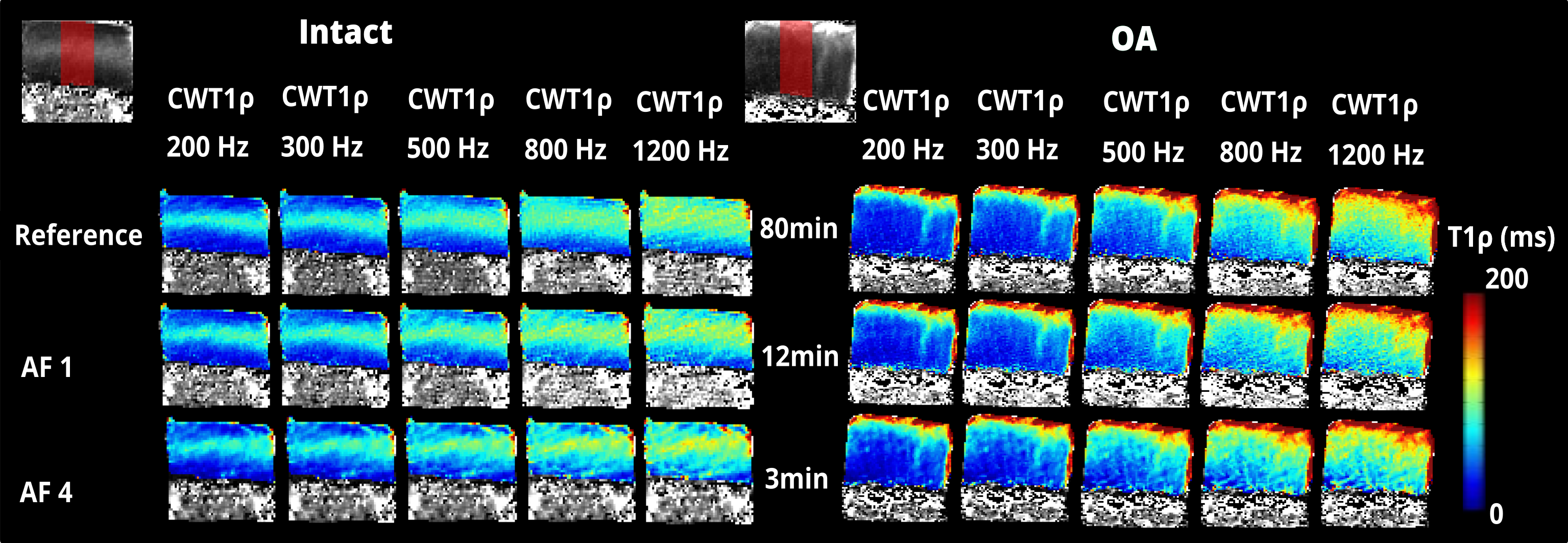
Figure 1. CW-T1rho relaxation time maps of the articular cartilage in intact and OA samples, along with anatomical images and the ROIs used for the depth-wise and bulk analysis. CW-T1rho dispersion is shown with different spin-locking frequencies (200, 300, 500, 800, and 1200 Hz). In comparison to intact, the OA sample shows elevated relaxation times in the superficial cartilage in the T1rho maps, indicating superficial tissue disruption. The reference reconstruction is from full data (8x oversampling).
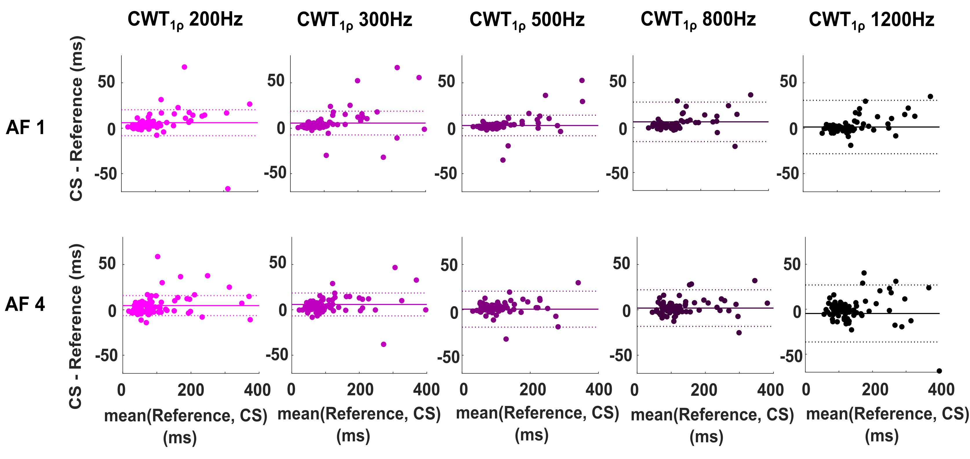
Figure 2. Bland-Altman plots illustrating the agreement between full data (reference) and two different acceleration factors with optimized regularization. The plots show the ROI-wise difference (superficial, transitional, and radial ROIs) between the full data and reduced data versus the average of the two. The solid line is the mean of differences, and the dotted lines represent the standard deviation of differences from three ROIs.
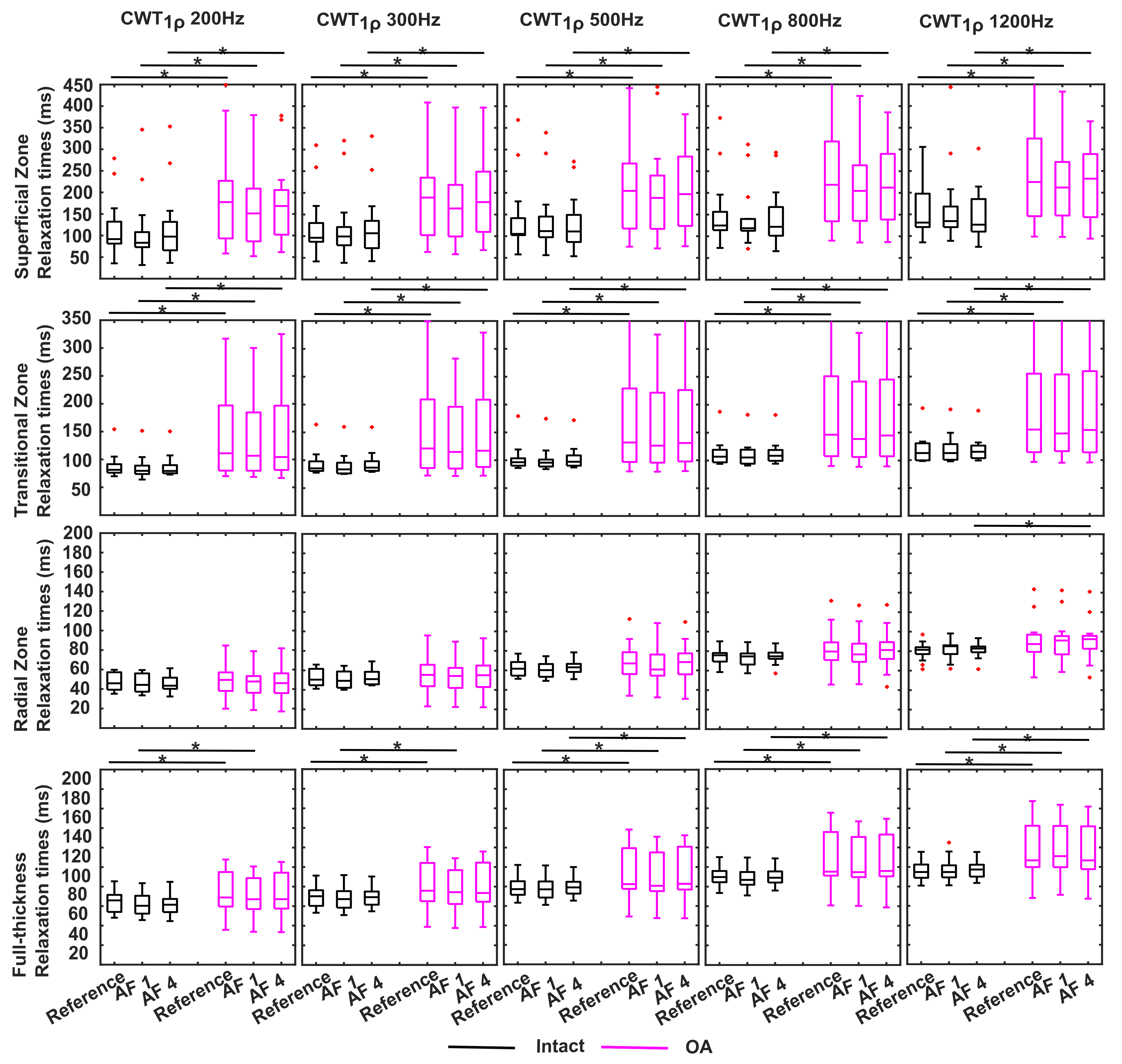
Figure 3. CW-T1rho relaxation times from superficial, transitional, radial zones and full-thickness ROI for each SLA. The lines inside the boxes are the median values of the measurements and the red crosses indicate the outliers within the data limits. Black indicates intact group and pink indicates the OA group. Reference is the full data. Significances between intact and OA groups are indicated with black asterisks.
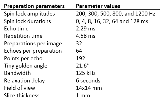
Table 1. Measurement parameters for magnetization prepared bSSFP radial readout sequence for CW-T1rho.
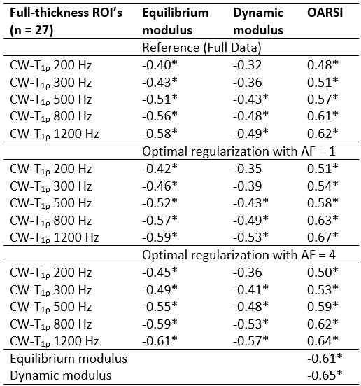
Table 2. Spearman’s correlation coefficients between CW-T1rho (Full thickness ROI) and equilibrium modulus, dynamic modulus and OARSI grade. Asterisks represent the statistically significant correlations (p<0.05).