2284
The application of T2 mapping, DWI and SPARCC in monitoring the treatment of sacroiliac bone marrow edema in patients with ankylosing spondylitis1Guiyang City, Guizhou Province, Guiyang City, Guizhou Province, China
Synopsis
Keywords: Bone, fMRI
Motivation: Evaluation of sacroiliac subarticular bone marrow edema for active inflammation of ankylosing spondylitis(AS)before and after biologic treatment.
Goal(s): MRI T2 mapping, DWI and SPARCC scores were used to compare the changes of BME in patients with AS before and after biologic treatment.
Approach: DWI, SPARCC scores and T2mapping were compared to evaluate the changes of bone marrow edema in patients with sacroiliac arthritis alone or in combination.
Results: The integrated diagnostic efficacy of DWI and T2 mapping demonstrates an optimal performance.
Impact: The combined application of magnetic resonance diffusion-weighted imaging (DWI), T2 mapping, and STIR-T2WI SPARCC scores provides an effective quantitative method for the clinical evaluation of the therapeutic efficacy of biologics in AS.
Introduction
AS is a chronic irreversible inflammatory disease with unknown etiology[1], characterized by axial bone inflammation and rigidity, which mainly affects young and middle-aged men[2]. Monitoring the alterations in BME of the sacroiliac joint is vital for evaluating the progression of AS patients' condition and the efficacy of medications [3]. Accurate and non-invasive imaging can enable AS patients to receive precise diagnosis and early treatment [3]. Therefore, this study mainly discussed the application value of MRI T2 mapping, DWI and SPARCC scores in evaluating the bone marrow edema in sacroiliarthritis of patients with AS in the active inflammatory stages[4]. Furthermore, provide a better imaging program for clinicians to aid in the early diagnosis of AS and in evaluating the efficacy of treatments.Methods
Thirty patients (23 males, aged 30.40±6.00 years)with AS who received biologics from September 2021 to January2023 were included as the patient group, and 32 healthy individuals (15 males , aged 27.53±7.28 years)were included as the control group. Routine MRI, T2 mapping and DWI sequences of Sacroiliac joint were performed in AS patients before treatment, 6 months after treatment with biological agents, and so as to control group. The paired sample t-test was employed to compare the mean T2 values, ADC values and SPARCC scores before and 6 months following AS treatment, as well as between the treatment and control groups, respectively. A Correlation ROC curve was constructed to evaluate the diagnostic efficacy of SPARCC score, DWI and T2mapping before and after AS treatment. Binary Logistic regression was used to analyze the correlation diagnostic efficacy of pairwise combination and combination of the three quantitative values.Results
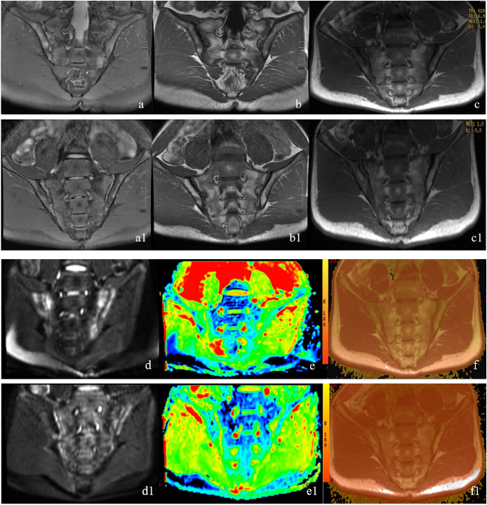
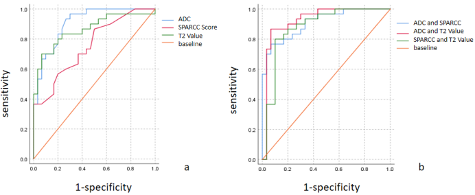
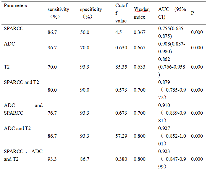
The SPARCC score, ADC value and T2 value following AS treatment exhibited significant decrement compared to their pre-treatment counterparts, were significantly lower than those before AS treatment, and these differences were statistically significant (P <0.001)(Figure1). The sensitivity of ADC before and after treatment was the highest (96.7%), the specificity was 70.0%, and the area under the curve was about 0.908 (95% confidence interval: 0.837-0.980), P < 0.001. The diagnostic sensitivity of the combined ADC and T2 values before and after AS treatment was 86.7%, the specificity was the highest (93.3%), and the area under the curve was the largest (about 0.927 (95% confidence interval: 0.852-1.001), P < 0.001(Table、figure2).Discussion
In this study, the ADC and T2 values decreased after treatment, indicating that DWI and T2 mapping can effectively evaluate water molecule changes of BME [5,6]. The combined use of DWI and T2 mapping can more accurately assess the diffusion motion and content changes of water molecules in BME under the sacroiliac joint in AS [7], thus providing high value for evaluating the therapeutic effect of AS[8]. The effectiveness of SPARCC score is slightly lower when used alone to evaluate the therapeutic effect of AS or when combined with other quantitative MRI parameters to evaluate its therapeutic effect.Conclusions
The combination of DWI and T2 mapping demonstrated superior efficacy in assessing drug efficacy in AS sacroiliac arthritis. The diagnostic efficiency of DWI alone is better than that of SPARCC score combined with T2 mapping.Acknowledgements
Associate Professor Song Lingling of Guizhou Medical University, thanks to the experts of the Imaging Technology Group of the Affiliated Hospital of Guizhou Medical University, thanks to all the members of the skeletal muscle system group for their help and support.References
1.Khan MA, Yong SB, Wei JC. Ankylosing spondylitis: History, epidemiology, and HLA-B27. Int J Rheum Dis. 2023 Mar;26(3):413-414. 2.Xiong Y, Cai M, Xu Y, Dong P, Chen H, He W, Zhang J. Joint together: The etiology and pathogenesis of ankylosing spondylitis. Front Immunol. 2022 Oct 17;13:996103. 3.Du MS, Xiong XQ, Liu H, Qin X, Hu XF, Chen W. The evaluation of bone marrow edema in sacroiliac joint in patients with ankylosing spondylitis using magnetic resonance imaging Dixon sequence. BMC Musculoskelet Disord. 2021 Nov 1;22(1):919. 4.Ning Q, Fan T, Ren H, Ye H, Wang W. Differentiating active from Inactive Sacroiliitis in ankylosing spondylitis by combination of DWI and Magnetization Transfer Imaging. Pak J Med Sci. 2023 Mar-Apr;39(2):417-422. 5.Wang D, Yin H, Liu W, Li Z, Ren J, Wang K, Han D. Comparative analysis of the diagnostic values of T2 mapping and diffusion-weighted imaging for sacroiliitis in ankylosing spondylitis. Skeletal Radiol. 2020 Oct;49(10):1597-1606. 6.Kucybała I, Ciuk S, Urbanik A, Wojciechowski W. The usefulness of diffusion-weighted imaging (DWI) and dynamic contrast-enhanced (DCE) sequences visual assessment in the early diagnosis of axial spondyloarthritis. Rheumatol Int. 2019 Sep;39(9):1559-1565. 7.Lin M, Chen X, Yu S, Gao F, Ma M. Monitoring the efficacy of tumor necrosis factor alpha antagonists in the treatment of Ankylosing spondylarthritis: a pilot study based on MR relaxometry technique. BMC Med Imaging. 2021 Jul 30;21(1):117. 8.Hededal P, Østergaard M, Sørensen IJ, Loft AG, Hindrup JS, Thamsborg G, Asmussen K, Hendricks O, Nørregaard J, Møller JM, Jurik AG, Morsel L, Balding L, Pedersen SJ. Development and Validation of MRI Sacroiliac Joint Scoring Methods for the Semiaxial Scan Plane Corresponding to the Berlin and SPARCC MRI Scoring Methods, and of a New Global MRI Sacroiliac Joint Method. J Rheumatol. 2018 Jan;45(1):70-77.Figures