2283
Quantitative Assessment of Meniscal Degeneration Using Three-dimensional Ultrashort Echo Time Adiabatic T1ρ (3D UTE-AdiabT1ρ) Imaging1Department of Radiology, Guangzhou First People's Hospital, School of Medicine, South China University of Technology, Guangzhou, China, 2Department of Radiology, University of California San Diego, San Diego, CA, United States, 3Department of Radiology, the third affiliated hospital of Southern Medical University, Guangzhou, China, 4Tianhe District Maternal and Child Hospital of Guangzhou, Guangzhou, China, 5Department of Orthopaedic Surgery, University of California San Diego, San Diego, CA, United States, 6Radiology Service, Veterans Affairs San Diego Healthcare System, San Diego, CA, United States
Synopsis
Keywords: Osteoarthritis, Osteoarthritis
Motivation: The three-dimensional ultrashort echo time adiabatic T1ρ (3D UTE-AdiabT1ρ) sequence can provide magic angle-insensitive evaluation of proteoglycan depletion in both short- and long-T2 tissues in the knee joint.
Goal(s): Our goal was to assess its clinical performance.
Approach: We applied this sequence to healthy volunteers and patients with different degrees of osteoarthritis.
Results: The sequence could be used for quantitative evaluation of meniscus degeneration. The UTE-AdiabT1ρ values in different subregions showed a significant positive relationship with WORMS, Stoller grades and KL groups. The 3D UTE-AdiabT1ρ biomarker can diagnose meniscal degeneration at an early stage.
Impact: The 3D UTE-AdiabT1ρ sequence allows quantitative imaging of the menisci. The UTE-AdiabT1ρ biomarker can diagnose meniscal degeneration at an early stage. Early detection of compositional changes in the menisci is of critical importance in avoiding or slowing down OA progression.
Introduction
The pathogenesis and progression of osteoarthritis (OA) result from degeneration in articular cartilage and other joint tissues, including the menisci, ligaments, tendons, and subchondral bone. The menisci play a vital protective role in knee joint health by facilitating shock absorption and load distribution. Meniscal degeneration has shown a strong association with the development and progression of OA (1, 2). Early detection of compositional changes in the menisci is of critical importance in avoiding or slowing down OA progression. Unfortunately, the menisci have short T2s and show little or no signal with clinical sequences (3). The three-dimensional ultrashort echo time (3D UTE) Cones sequence allows fast volumetric imaging of both short- and long-T2 tissues in the knee joint (4, 5). While T1ρ is a promising biomarker for the detection of early OA, a major confounding factor is the magic angle effect (6-9). Recent studies have shown AdiabaticT1ρ is much less sensitive to the magic angle effect than conventional T1ρ and T2 (10). The combination of 3D UTE sequence with AdiabT1ρ preparation (UTE-AdiabT1ρ) allows for magic angle-insensitive imaging of various knee joint tissues and can provide effective non-invasive assessment of proteoglycan (PG) depletion in the meniscus. In this study we aim to further evaluate its efficacy in assessing meniscus degeneration in healthy volunteers and OA patients.Method
A total of 63 human subjects (aged 23-80 years, mean age 50±15 years; 32 males, 31 females) were recruited for this study. Written informed consent was obtained from all subjects. The whole knee joint (26 left knees, 37 right knees) was scanned on a 3T MR750 scanner (GE Healthcare Technologies, Milwaukee, WI). The 3D UTE-AdiabT1ρ imaging parameters were: repetition time (TR)=500 ms; flip angle (FA)=10°; acquisition matrix=256×256×36, spin-locking times (TSLs)=0, 12, 24, 36, 48, 72, and 96 ms. The whole knee menisci were segmented as follows: medial meniscus anterior horn (MMAH), medial meniscus posterior horn (MMPH), medial meniscus body (MMB), lateral meniscus anterior horn (LMAH), lateral meniscus posterior horn (LMPH) and lateral meniscus body (LMB). Each meniscus region was independently scored slice-by-slice by two experienced musculoskeletal radiologists (M.W. and Y.W.) with 26 and 10 years of experience, respectively, according to Kellgren-Lawrence (KL) grade (11), Stoller grade and WORMS. Stoller grade was classified as follows: Stoller 1 = shapeless or spherical high-signal shadows. Stoller 2 = linear high signal, neither of which extends to the articular surface. Stoller 3 = linear or diffuse hypersignal extending to the articular surface, i.e., meniscus tear (12, 13). WORMS was graded as follows: WORMS 1 = intrasubstance abnormalities. WORMS 2 = non-displaced tear. WORMS 3 = displaced or complex tear. WORMS 4 = complete destruction or maceration (14-16). Then, all subjects were classified into three groups according to KL grade: normal controls (KL= 0), doubtful-minimal OA (KL≤2), and moderate-severe OA (KL≥3) (17). Two radiologists individually drew the regions of interest (ROIs) onto images of each segmentation slice by slice. The DICOM images were analyzed using MATLAB 2017b and respective correlations between 3D UTE-AdiabT1ρ values and KL group, Stoller grade and WORMS were analyzed.Results
Figure 1 shows representative UTE-AdiabT1ρ fitting of MMPH, where the normal meniscus showed a UTE-AdiabT1ρ value of 23.8±2.3 ms and the abnormal menisci (WROMS=2, 3, and 4) showed UTE-AdiabT1ρ values of 36.6±3.8 ms, 42.1±4.0 ms, and 53.3±7.5 ms, respectively. Increased UTE-AdiabT1ρ values were observed in abnormal menisci.Table 1 shows UTE-AdiabT1ρ values in different WORMS, Stoller grades and KL groups. Figure 2 shows the corresponding boxplots. The Spearman correlation coefficient showed a positive relationship between UTE-AdiabT1ρ values in different subregions and the corresponding WORMS, Stoller grades and KL groups (P<0.05). Differences in UTE-AdiabT1ρ values among different groups were all statistically significant (P<0.05).
Figure 3 shows the ROC curves, which suggest that UTE-AdiabT1ρ could distinguish mild meniscus degeneration (WORMS=1 and Stoller=1) from normal controls (WORMS 0 and Stoller=0), and doubtful-minimal OA (KL=1-2) from normal controls (KL=0). The AUC values of UTE-AdiabT1ρ were 0.90 and 0.87 for mild meniscus degeneration (WORMS=1 and Stoller=1), and 0.77 for doubtful-minimal OA (KL=1-2). The diagnostic threshold value of UTE-AdiabT1ρ was 26.1ms and 25.9 ms with a sensitivity of 78.5% and 73.0% and a specificity of 87.1% and 85.8% for mild meniscus degeneration with WORMS=1 and Stoller=1, and 26.8 ms with a sensitivity of 62.7% and a specificity of 76.1% for doubtful-minimal OA.
Conclusion
The 3D UTE-AdiabT1ρ sequence allows quantitative imaging of the menisci. UTE-AdiabT1ρ values in different subregions showed a significant positive relationship with WORMS, Stoller grades and KL groups. The 3D UTE-AdiabT1ρ biomarker can diagnose meniscal degeneration at an early stage.Acknowledgements
The authors are thankful for support from NIH (R01AR075825, R01AR062581, and R01AR068987), Veterans Affairs (I01RX002604 and I01CX001388), and GE Healthcare.
References
1. Antony B, Driban JB, Price LL, Lo GH, Ward RJ, Nevitt M, et al. The relationship between meniscal pathology and osteoarthritis depends on the type of meniscal damage visible on magnetic resonance images: data from the Osteoarthritis Initiative. Osteoarthritis Cartilage 2017; 25(1): 76-84.
2. Link TM, Steinbach LS, Ghosh S, Ries M, Lu Y, Lane N, et al. Osteoarthritis: MR imaging findings in different stages of disease and correlation with clinical findings. Radiology 2003; 226(2): 373-381.
3. Chang EY, Du J, Chung CB. UTE imaging in the musculoskeletal system. J Magn Reson Imaging. 2015; 41(4):870-883.
4. Ma YJ, Carl M, Shao H, Tadros AS, Chang EY, Du J. Three-dimensional ultrashort echo time cones T1ρ (3D UTE-cones-T1ρ) imaging. NMR Biomed. 2017; 30:3709.
5. Wu M, Zhao W, Wan L, et al. Quantitative three-dimensional ultrashort echo time cones imaging of the knee joint with motion correction. NMR in Biomedicine. 2020; 33(1): e4214.
6. Wang N, Xia Y. Dependencies of multi-component T2 and T1ρ relaxation on the anisotropy of collagen fbrils in bovine nasal cartilage. J Magn Reson. 2011; 212:124–132.
7. Du J, Statum S, Znamirowski R, Bydder GM, Chung CB. Ultrashort TE T1rho magic angle imaging. Magn Reson Med. 2013; 69:682-687.
8. Shao H, Pauli C, Li S, et al. Magic angle effect plays a major role in both T1rho and T2 relaxation in articular cartilage. Osteoarthritis Cartilage. 2017; 25:2022-2030.
9. Roemer FW, Kijowski R, Guermazi A. Editorial: from theory to practice – the challenges of compositional MRI in osteoarthritis research. Osteoarthritis Cartilage. 2017; 25:1923-1925.
10. Hänninen N, Rautiainen J, Rieppo L, Saarakkala S, Nissi MJ. Orientation anisotropy of quantitative MRI relaxation parameters in ordered tissue. Sci Rep. 2017; 7:9606.
11. Zhang X, Ma YJ, Wei Z, Wu M, Ashir A, Jerban S, Li S, Chang EY, Du J. Macromolecular fraction (MMF) from 3D ultrashort echo time cones magnetization transfer (3D UTE-Cones-MT) imaging predicts meniscal degeneration and knee osteoarthritis. Osteoarthritis Cartilage. 2021; 29(8): 1173-1180.
12. Stoller DW. Magnetic Resonance Imaging in Orthopaedics and Sports Medicine. 3rd ed. Philadelphia, PA: Lippincott Williams & Wilkins; 2006.
13. Balik MS, Turan A, Celik FB. Is There A Relationship Between Three-Dimensionally Measured Baker's Cyst Volume and Knee Pathologies? Eurasian J Med. 2019; 51(1): 64-69.
14. Stehling C, Liebl H, Krug R, Lane NE, Nevitt MC, Lynch J, McCulloch CE, Link TM. Patellar cartilage: T2 values and morphologic abnormalities at 3.0-T MR imaging in relation to physical activity in asymptomatic subjects from the osteoarthritis initiative. Radiology. 2010 Feb;254(2):509-520.
15. Laberge MA, Baum T, Virayavanich W, Nardo L, Nevitt MC, Lynch J, McCulloch CE, Link TM. Obesity increases the prevalence and severity of focal knee abnormalities diagnosed using 3T MRI in middleaged subjects ddata from the Osteoarthritis Initiative. Skeletal Radiol 2012; 41(6): 633-641.
16. Stehling C, Lane NE, Nevitt MC, Lynch J, McCulloch CE, Link TM. Subjects with higher physical activity levels have more severe focal knee lesions diagnosed with 3T MRI: analysis of a non-symptomatic cohort of the osteoarthritis initiative. Osteoarthritis Cartilage. 2010; 18(6): 776-786.
17. Wang L, Chang G, Xu J, Vieira RL, Krasnokutsky S, Abramson S, Regatte RR. T1rho MRI of menisci and cartilage in patients with osteoarthritis at 3T. Eur J Radiol. 2012; 81(9): 2329-2336.
Figures
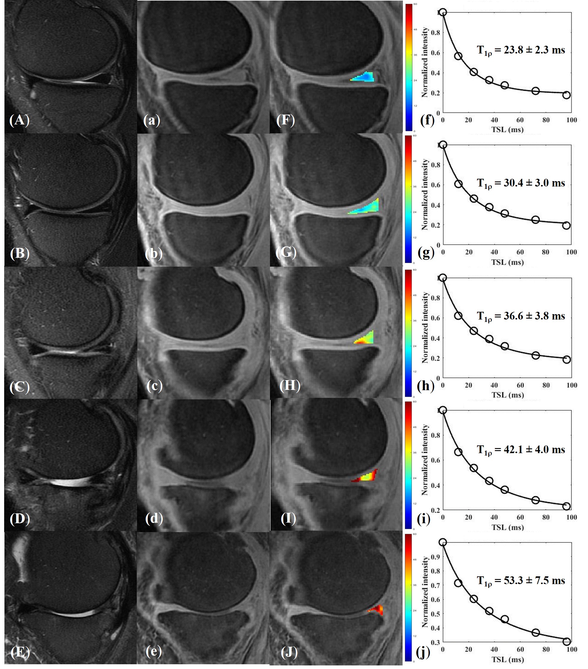
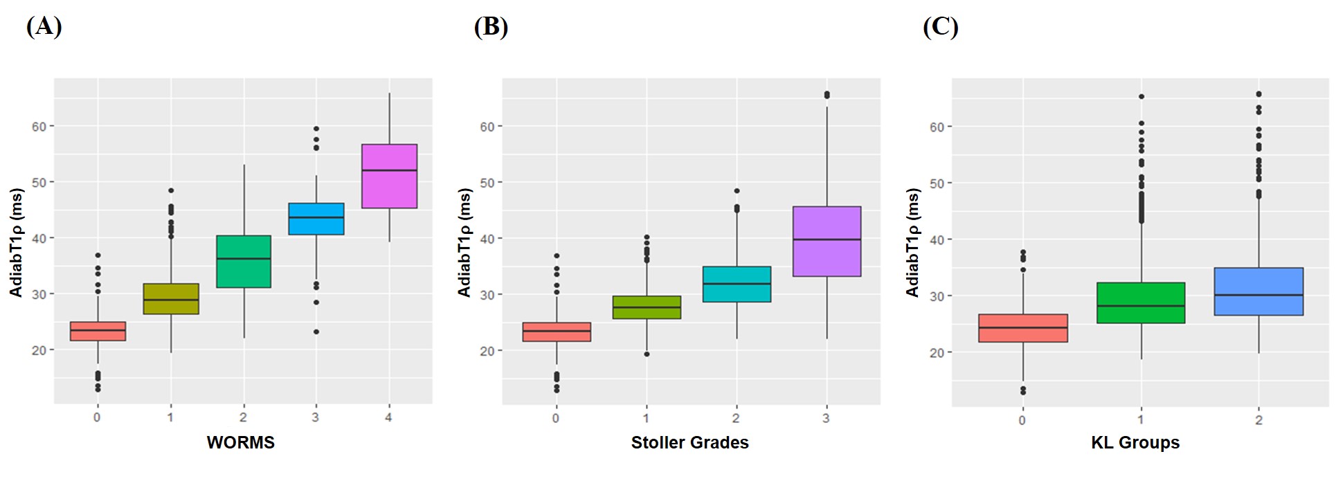
Figure 2. Boxplot of UTE-AdiabT1ρ values in different groups based on WORMS (A), Stoller grades (B) and KL groups (C).
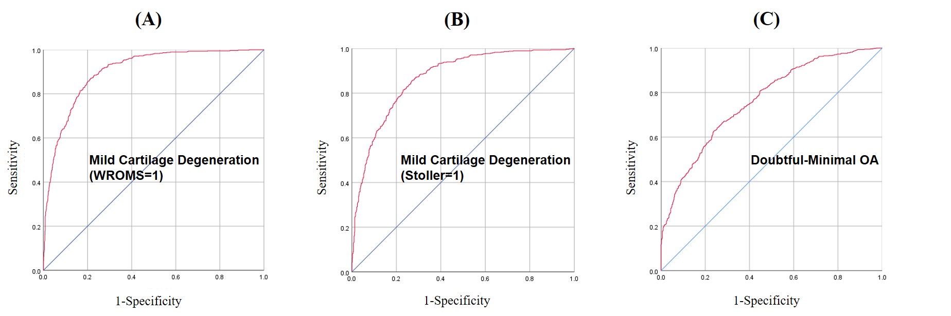
Figure 3. ROC curves of UTE-AdiabT1ρ for the diagnosis of mild meniscal degeneration with WORMS=1 (A) or Stoller=1 (B), and doubtful-minimal OA (KL=1-2), with the corresponding AUCs of 0.90, 0.87 and 0.77, and UTE-AdiabT1ρ cutoff values of 26.1 ms, 25.9 ms, and 26.8 ms, respectively.
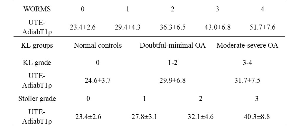
Table 1. Mean UTE-AdiabT1ρ (ms) in different WORMS groups (WORMS=0, 1, 2, 3, 4), KL groups (KL=0,1-2,3-4), and Stoller groups (Sroller grade = 0, 1, 2, 3).