2281
Prediction of Knee Osteoarthritis using MRI-based Radiomic Features of the Subchondral Bone and Infrapatellar Fat Pad1Department of Radiology & Nuclear Medicine, Erasmus MC University Medical Center, Rotterdam, Netherlands, 2Faculdade de Engenharia, Universidade do Porto, Porto, Portugal, 3Instituto de Ciências Biomédicas Abel Salazar, Universidade do Porto, Porto, Portugal, 4Department of Radiology, University of Wisconsin-Madison, Madison, WI, United States, 5Instituto de Engenharia de Sistemas e Computadores, Tecnologia e Ciência (INESC TEC), Porto, Portugal, 6Department of Biomechanical Engineering, Delft University of Technology, Delft, Netherlands
Synopsis
Keywords: Osteoarthritis, MSK
Motivation: While radiomics has been applied to various MRI data to predict knee osteoarthritis (KOA) incidence, there is a lack of knowledge on the combination of radiomics features from different knee structures.
Goal(s): To assess the ability of MRI-based radiomic features extracted from automatically segmented femur, tibia, patella, and infrapatellar fat pad to predict KOA incidence.
Approach: 710 DESS MRIs were segmented using deep learning, trained with 30 manually delineated images. KOA incidence was predicted using Elastic Net, based on radiomic features from the four knee structures.
Results: The model combining features from the four knee structures resulted in a ROC AUC of 0.65.
Impact: While further research should be conducted to improve the accuracy of the developed radiomics pipeline in order to improve its applicability in clinical practice, radiomic features gathered from different knee structures are promising imaging biomarkers for early KOA prediction.
Introduction
The infrapatellar fat pad (IPFP) has been proposed as a source of knee pain in patients suffering from knee osteoarthritis (KOA) or its supposed precursor, patellofemoral pain (PFP)1. It has been shown that magnetic resonance imaging (MRI)-based radiomic features extracted from the femoral and tibial subchondral bones differ between knees without and with KOA2-4 and that texture features extracted from the IPFP are associated with future development of KOA and PFP5-7. However, the combination of radiomic features extracted from the subchondral bone and the IPFP for KOA assessment has yet to be studied. This study aims to automatically extract radiomic features from the femoral, tibial, and patellar bones as well as the IPFP to assess their ability to predict KOA development over a 48-month period.Methods
The 3D double echo steady state water excitation MRI scans of the Pivotal Osteoarthritis Initiative (OAI) MRI Analyses (POMA) dataset were used in this study8. The POMA study includes 355 patients that developed incident radiographic KOA (KL>1) during the 48-month follow-up, and 355 controls that did not develop the disease during the same period. Automatic segmentation of the femur, tibia, patella, and IPFP was achieved using nnU-Net9 (3D U-Net configuration). The 3D nnU-Net was trained with 30 manually segmented knees from the OAI-ZIB dataset10 and 5-fold cross-validation. The accuracy of the automatic segmentation was assessed using the Dice similarity coefficient (DSC).The Workflow for Optimal Radiomics Classification toolbox (v. 3.6.0)11 was used for feature extraction (n=452). Radiomic features were related to orientation (n = 3), histogram (n = 13), shape (n = 21), original phase (n = 39), and texture (n = 376). Elastic-Net was selected for dimensionality reduction and prediction, computed using the Stochastic Gradient Descent Classifier (SGDClassifier). The hyperparameters were optimized using 10-fold stratified cross-validation with a grid search and 10 repetitions. Three main models were evaluated based on: 1) patients’ information (age, sex, and BMI), 2) radiomic features gathered from the four knee structures, and 3) combination of models 1 and 2. Moreover, radiomic features were split into subgroups for evaluation, according to their type (orientation, histogram, shape, phase and texture). In order to assess the influence of each knee structure on the development of KOA, radiomic features extracted from the femoral, tibial and patellar bone as well as the IPFP were also analyzed individually. Models’ performance for predicting KOA incidence was evaluated using the area under the receiver operating characteristic curve (ROC AUC).
Results
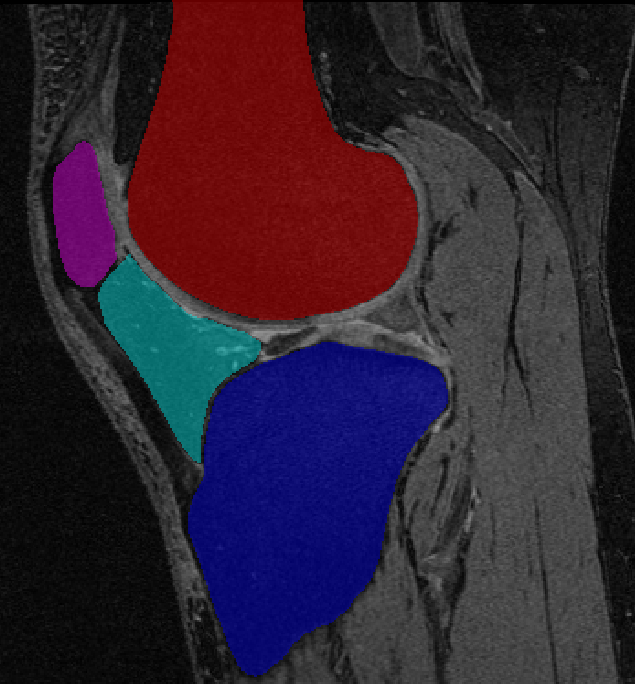
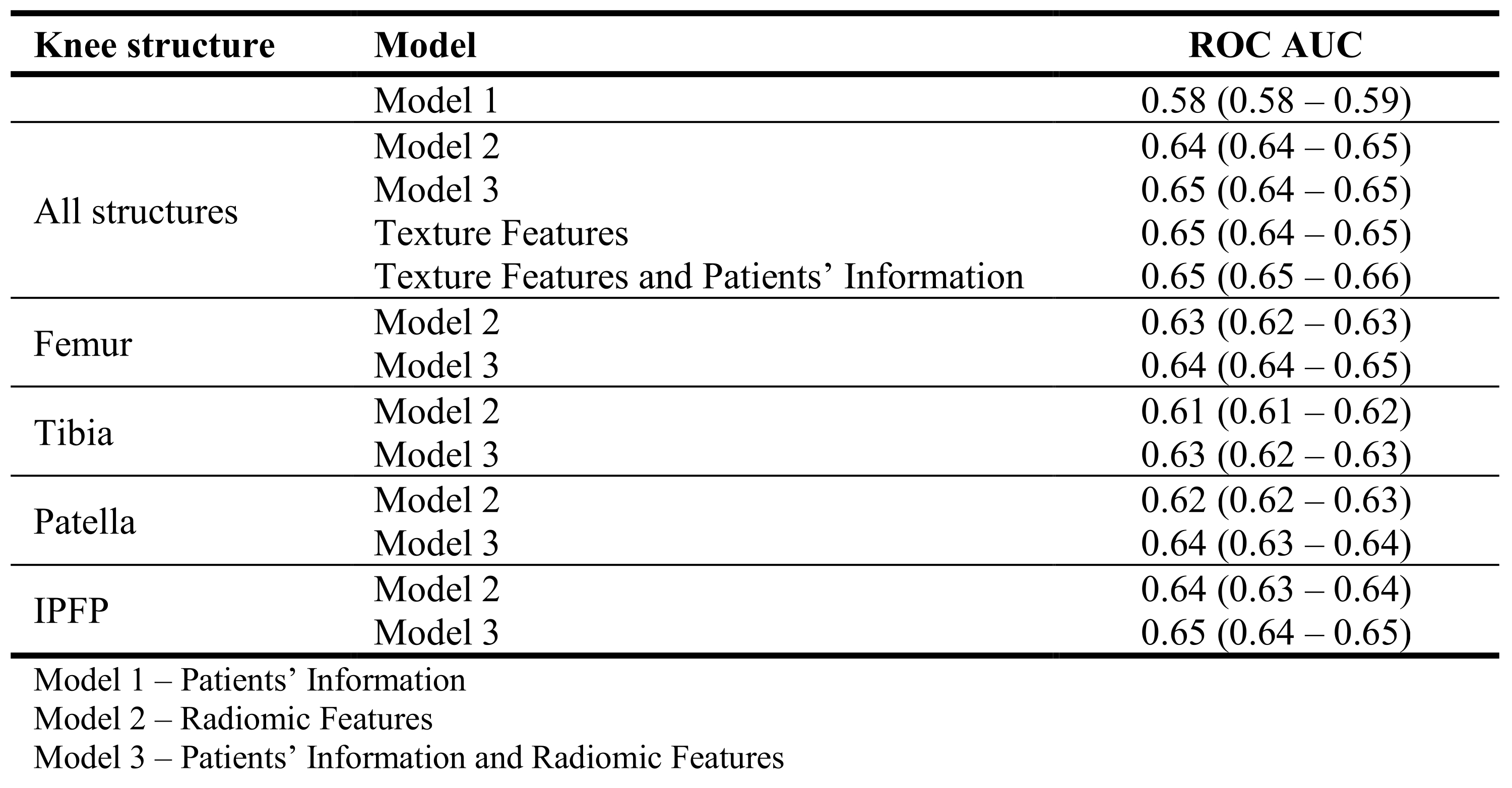
Automatic segmentation resulted in a mean (standard deviation) DSC of 0.99 (0.01) for the femur and tibia, 0.96 (0.01) for the patella, and 0.94 (0.01) for the IPFP. An example of the automatically segmented tissues is shown in Figure 1.Model 3 applied on all knee structures resulted in the highest performance, with a ROC AUC (95% confidence interval) of 0.65 (0.64-0.65). Similar performance was obtained for model 2 (ROC AUC of 0.64 (0.64-0.65)), while model 1 yielded lower metrics (ROC AUC of 0.58 (0.58-0.59)). The model based on all texture features extracted from the four knee structures, resulted in a ROC AUC of 0.65 (0.64-0.65) and, when combined with the patients’ information, a ROC AUC of 0.65 (0.65-0.66) was achieved. When assessing the models with features from each structure individually, the model including features extracted from the IPFP resulted in the highest performance when combined with the patients’ information (0.65 (0.64-0.65)). The performance metrics obtained for the studied models are presented in Table 1.
Discussion
The accuracy of the automatic segmentation of the IPFP was higher than in previous literature (DSC of 0.90)6, while the accuracy of femur, tibia and patella segmentation was equal to state-of-the-art performance10.To predict KOA incidence, the model based on the patients’ information and radiomic features extracted from the femur, tibia, patella as well as the IPFP, resulted in the highest performance metrics. The models including all texture features, extracted from the four tissues, resulted in analogous performance indicating the importance of texture features in the models. When assessing the models with features from each of the four structures individually, models based on IPFP features had the highest performance metrics, emphasizing the importance of whole-joint assessment for KOA evaluation.
Further research should be conducted to improve the robustness of the models and radiomics pipeline.
Conclusion
Radiomic features gathered from different knee structures are promising imaging biomarkers for early KOA detection.Acknowledgements
No acknowledgement found.References
- Ioan-Facsinay A, Kloppenburg M. An emerging player in knee osteoarthritis: the infrapatellar fat pad. Arthritis Res Ther. 2013;15(6):225.
- Hirvasniemi J, Klein S, Bierma-Zeinstra S, Vernooij MW, Schiphof D, Oei EHG. A machine learning approach to distinguish between knees without and with osteoarthritis using MRI-based radiomic features from tibial bone. Eur Radiol. 2021;31(11):8513-8521.
- MacKay JW, Kapoor G, Driban JB, et al. Association of subchondral bone texture on magnetic resonance imaging with radiographic knee osteoarthritis progression: data from the Osteoarthritis Initiative Bone Ancillary Study. Eur Radiol. 2018;28(11):4687-4695.
- Xue Z, Wang L, Sun Q, et al. Radiomics analysis using MR imaging of subchondral bone for identification of knee osteoarthritis. J Orthop Surg Res. 2022;17:414.
- Li J, Fu S, Gong Z, et al. MRI-based Texture Analysis of Infrapatellar Fat Pad to Predict Knee Osteoarthritis Incidence. Radiology. 2022;304(3):611-621.
- Yu K, Ying J, Zhao T, et al. Prediction model for knee osteoarthritis using magnetic resonance-based radiomic features from the infrapatellar fat pad: data from the osteoarthritis initiative. Quant Imaging Med Surg. 2023;13(1):352-369.
- Campos IR, van der Heijden RA, Oei EHG, et al. MRI-based radiomics for assessment of the infrapatellar fat pad’s influence on patellofemoral pain. Osteoarthritis Imaging 2023;3:100134.
- Roemer FW, Kwoh CK, Hannon MJ, et al. What comes first? Multitissue involvement leading to radiographic osteoarthritis: magnetic resonance imaging-based trajectory analysis over four years in the osteoarthritis initiative. Arthritis Rheumatol. 2015;67(8):2085-96.
- Isensee F, Jaeger PF, Kohl SAA, Petersen J, Maier-Hein KH. nnU-Net: a self-configuring method for deep learning-based biomedical image segmentation. Nat Methods. 2021;18(2):203-211.
- Ambellan F, Tack A, Ehlke M, Zachow S. Automated segmentation of knee bone and cartilage combining statistical shape knowledge and convolutional neural networks: Data from the Osteoarthritis Initiative. Med Image Anal. 2019;52:109-118.
- Starmans M, van der Voort SR, Phil T, et al. Reproducible radiomics through automated machine learning validated on twelve clinical applications. 2021. arXiv preprint arXiv:2108.08618.
Figures