2276
Deep Learning Reconstruction for Turbo Spin Echo Imaging to Accelerate Ankle MRI: A Multi-Reader Study1Huashan hospital, Shanghai, China, 2Siemens Shenzhen Magnetic Resonance Ltd, Shenzhen, China, 3Siemens Healthineers, Shanghai, China, 4Siemens Healthineers, Erlangen, Germany
Synopsis
Keywords: Whole Joint, MSK
Motivation: Ankle MRI usually requires a long examination time. A faster scan with adequate image qualities is desired in clinical practice.
Goal(s): To evaluate a deep-learning reconstruction accelerated turbo spin echo (DLR-TSE) sequence in ankle application.
Approach: Four radiologists independently assessed the image quality and reviewed structural abnormalities of 56 consecutive patients on DLR-TSE and compared these results with those of conventional TSE.
Results: Overall, DLR-TSE achieved superior image qualities with a 57.4% reduction in total acquisition time compared with conventional TSE images. There were no differences in the differentiation of anatomic details, diagnostic confidence, or assessments of structural abnormalities between the two techniques.
Impact: Deep learning reconstruction can accelerate the turbo spin echo imaging without compromising the image quality or lesion detectability in ankle MRI.
Introduction
Deep-learning reconstruction (DLR) accelerated MRI may reduce the scan time while maintaining optimal image quality[4,5]. Studies exploring the clinical use of DLR-TSE for the musculoskeletal system have shown improvements in overall image quality, signal-to-noise ratio, contrast-to-noise ratio, and sharpness without decreasing diagnostic performance in the spine[1], knee[2,6], and shoulder[3]. However, few studies have focused on the ankle. The anatomic complexity and relatively small size of the ankle may hinder DLR’s ability to accelerate ankle MRI in a manner similar to the spine and knee.The present study was conducted to evaluate a DLR-TSE sequence of the ankle MRI in terms of acquisition time, image quality, and lesion detectability in comparison with conventional TSE.Methods
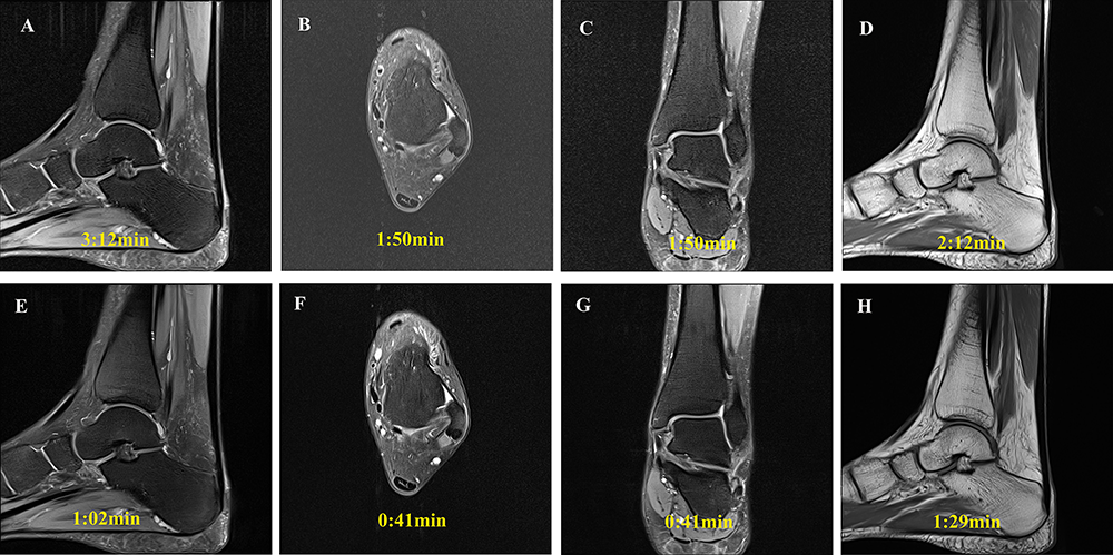
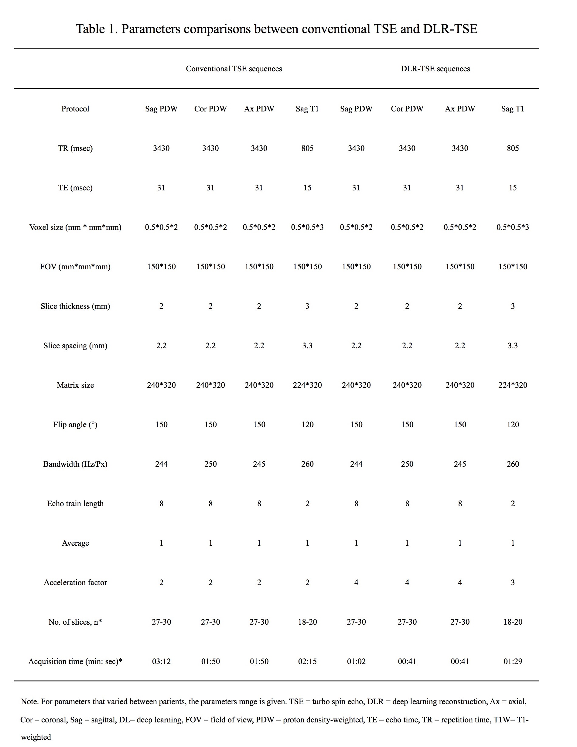
Between March and May 2023, 56 patients with an indication for ankle MRI were prospectively enrolled. Each patient underwent the conventional TSE sequences with GRAPPA acceleration factor of 2 and the research DLR-TSE sequences with an acceleration factor of 4 or 3. Each sequence was performed for sagittal T1-weighted imaging, as well as sagittal, coronal, and transverse proton density (PD)-weighted imaging with spectral fat suppression (Figure 1). Detailed parameters are shown in Table 1. All MRI examinations were performed using a 3-T MRI system (MAGNETOM Prisma, Siemens Healthineers, Erlangen, Germany) with patients in the supine position (feet first), using a dedicated 16-channel foot-ankle coil (Foot/Ankle 16, Siemens Healthineers). Four board-certified, subspecialized musculoskeletal radiologists (with 5 - 26 years of subspecialty experience) conducted independent assessments of the images. The images were presented in a random order of image quality and structural abnormalities. Image quality was assessed using a 5-point Likert scale for overall image quality, differentiation of anatomic details, diagnostic confidence, artifacts, and noise. Interchangeability analysis was performed to describe the ability of DLR-TSE to replace conventional TSE based on two readers’ assessment of the same patient in the detection of 18 pathological abnormalities.Results
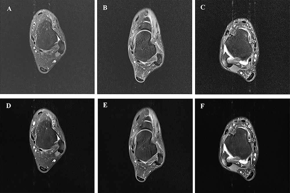
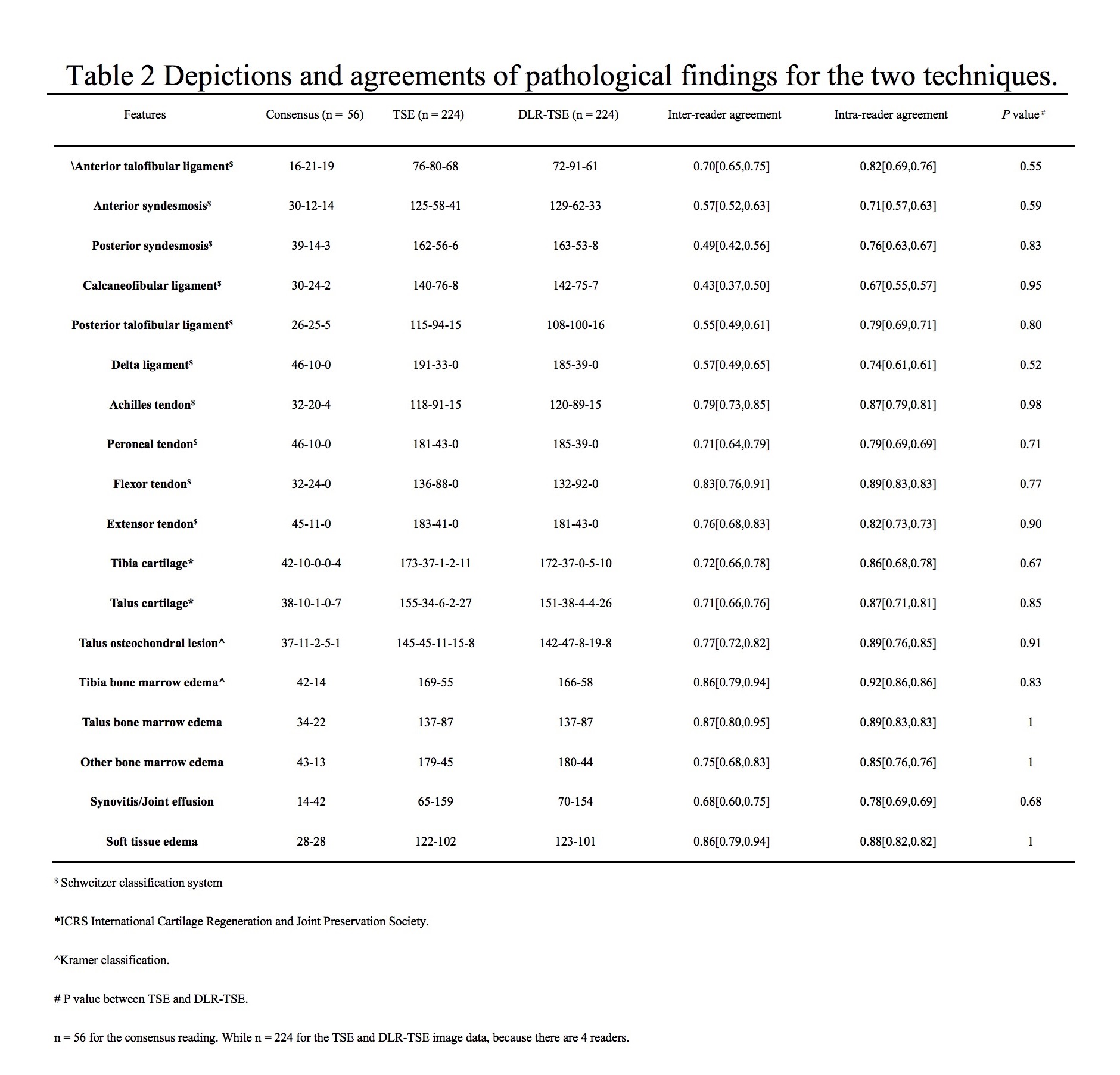
The DLR-TSE protocols, completed in 233 seconds, led to a 57.4% reduction in total acquisition time compared with the conventional TSE protocols (547 seconds). The average ratings from the four readers indicated that DLR-TSE images had superior overall image quality, reduced noise, and fewer artifacts with all comparisons showing statistical significance (all P < 0.001) compared with conventional TSE imaging. In conventional TSE, main artifacts observed were vascular pulsatile and motion artifacts, both of which were improved in DLR-TSE (36/56 vs. 14/56 for vascular pulsatile artifacts, as depicted in Figure 2, P = 0.02; 12/56 vs. 6/56 for motion artifacts, P = 0.3). Moreover, the differentiation of anatomic details and diagnostic confidence were rated as excellent or good, without no significant differences between the two imaging techniques (P > 0.05). In addition, there were no significant differences in the detection of pathological abnormalities between the two techniques (all P ≥ 0.52, as depicted in Table 2). A comparison of diagnostic evaluations between the two sequences revealed moderate to excellent inter-reader and intra-reader agreement (kappa value = 0.43 to 0.92, P < 0.001; as outlined in Table 2).Discussion and Conclusions
In a multi-reader study involving radiologists with various levels of experience in ankle MRI, this research compared DLR-TSE with conventional TSE. The results showed that DLR-TSE not only exhibited superior image quality but also demonstrated similar lesion detectability compared with conventional TSE, as well as a substantially reduced acquisition time.Acknowledgements
No acknowledgement found.References
1. Almansour H, Herrmann J, Gassenmaier S, et al. Deep Learning Reconstruction for Accelerated Spine MRI: Prospective Analysis of Interchangeability. Radiology. 2023;306(3):e212922.
2. Johnson PM, Lin DJ, Zbontar J, et al. Deep Learning Reconstruction Enables Prospectively Accelerated Clinical Knee MRI. Radiology. 2023;307(2):e220425.
3. Kaniewska M, Deininger-Czermak E, Getzmann JM, et al. Application of deep learning-based image reconstruction in MR imaging of the shoulder joint to improve image quality and reduce scan time. Eur Radiol. 2022.
4. Kijowski R, Fritz J. Emerging Technology in Musculoskeletal MRI and CT. Radiology. 2023;306(1):6-19.
5. Liu J, Li W, Li Z, et al. Magnetic resonance shoulder imaging using deep learning-based algorithm. Eur Radiol. 2023.
6. Siouras A, Moustakidis S, Giannakidis A, et al. Knee Injury Detection Using Deep Learning on MRI Studies: A Systematic Review. Diagnostics (Basel). 2022;12(2).
Figures