2273
Deep Learning Accelerated 3D SPACE DIXON for Improved Fat Suppression in Musculoskeletal MRI1Department of Radiology, NYU Langone School of Medicine, New York, NY, United States, 2Department of Radiology, University Hospital Basel, Basel, Switzerland, 3MR R&D Collaborations, Siemens Medical Solutions USA, Malvern, PA, United States, 4Siemens Healthcare GmbH, MR Application Predevelopment, Erlangen, Germany
Synopsis
Keywords: Whole Joint, Joints
Motivation: The application of 3D SPACE-DIXON techniques for musculoskeletal MRI has advantages in terms of fat suppression capacities, but drawbacks with regard to substantially longer acquisition times compared with SPAIR fat suppressed 3D SPACE.
Goal(s): To reduce the acquisition time of 3D SPACE-DIXON to that of 3D SPACE-SPAIR while maintaining superior fat suppression capabilities.
Approach: Six-fold CAIPIRINHA acceleration was combined with a deep learning-based image reconstruction algorithm to decrease the acquisition time and maintain image quality.
Results: Image quality parameters in volunteer scans were rated equivalent to the conventional 3D SPACE-DIXON sequence and superior to the 3D SPACE-SPAIR sequence.
Impact: We present a 3D SPACE-DIXON technique with improved fat suppression performance and similar acquisition time compared with 3D SPACE-SPAIR for musculoskeletal 3D MRI. Preliminary in vivo results indicate the clinical utility of this technique for proton density-weighted 3D MRI.
Introduction
In musculoskeletal MRI, chemical shift and inversion recovery-based fat suppression techniques are employed to improve the conspicuity of various acute and chronic abnormalities[1]. However, both techniques are affected by B0 and B1 field variations, which result in inhomogeneous fat suppression[2].Multi-echo DIXON techniques acquire data at different echo times (TE) to encode the chemical shift between fat and water, which is more robust to B0 heterogeneities and permits more uniform fat suppression[3,4]. Recently, we presented a 3D SPACE-DIXON technique for fat suppression in the knee[5]. While the SPACE-DIXON scheme provides homogeneous fat suppression compared to chemical shift techniques, it’s use has been limited due to the increased scan times.
In this work, we present a six-fold CAIPIRINHA-accelerated 3D SPACE-based 2-Point DIXON technique with deep learning-based image reconstruction (SPACE-DIXON-DL) and an improved fat-water separation algorithm for efficient fat suppression and generation of both fat-suppressed and non-fat suppressed images from a single acquisition. We compare the clinical utility to existing 3D SPACE-based techniques in volunteers.
Methods
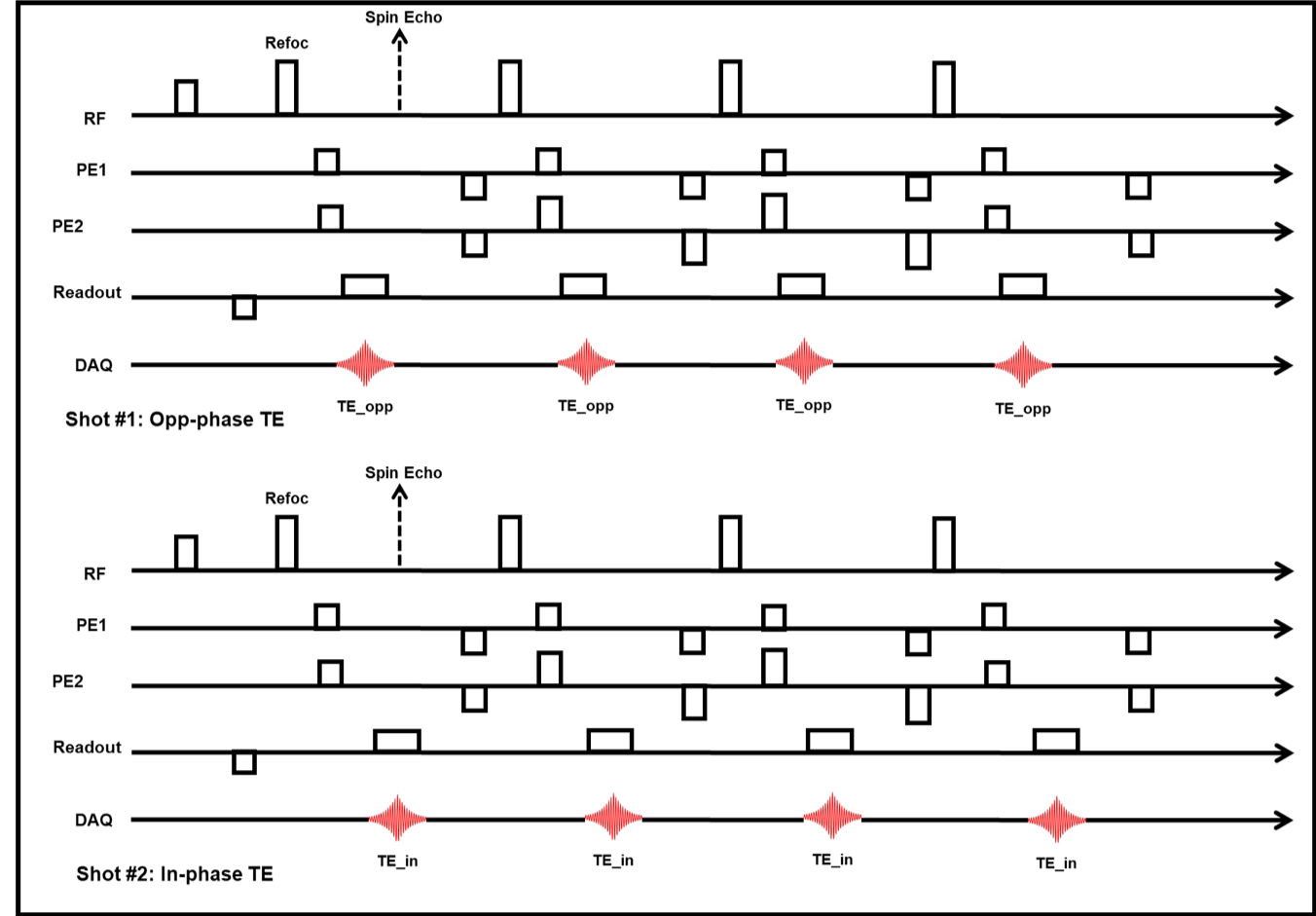
Data Acquisition and ReconstructionA prototype SPACE-DIXON sequence was developed to acquire data at the opposed-phase and in-phase echo times in separate shots, interleaved across TRs to minimize motion effects (Figure 1). The in-phase echo was acquired at the spin echo, corresponding to the conventional non-fat suppressed SPACE acquisition. This DIXON scheme is was integrated with the SPACE variable refocusing flip angle approach to enable SAR efficient sampling .The deep learning-based image reconstruction involved two sequential, independent processing steps. In the first step, images are reconstructed from k-space data on the acquired resolution using a network architecture inspired by variational networks[6]. A supervised training performed using about 5000 training pairs derived from about 500 fully sampled 3D datasets acquired from healthy volunteers on 1.5 and 3T scanners (MAGNETOM scanners, Siemens Healthcare, Erlangen, Germany) in head, abdomen and pelvis. The second processing step interpolates the acquired images using a deep learning-based superresolution algorithm[7,8].
Phantom Imaging
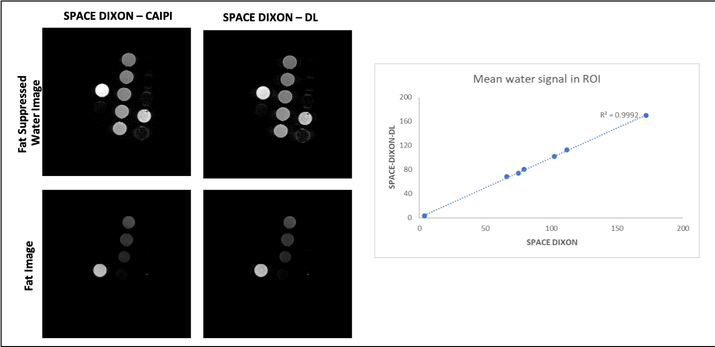
Fat suppression efficiency of the proposed SPACE-DIXON-DL technique was evaluated using a set of commercial PDFF phantoms (Calimetrix, Madison, WI USA) with PDFF values of 0%, 5%, 10%, 20%, 30%, 40%, and 100% on a 3T MRI system (MAGNETOM Vida, Siemens Healthcare, Erlangen, Germany). Data was acquired using both CAIPI SPACE-DIXON and the 6-fold accelerated SPACE-DIXON-DL techniques and the corresponding water fraction values were compared.
In-vivo MRI
Data from nine volunteers were acquired on a 3T MRI scanner (MAGNETOM Vida, Siemens Healthcare, Erlangen, Germany) after IRB approval and informed consent. Imaging consisted of three sequences: (1) a 3D PD SPACE dataset with the following parameters: resolution=0.68mm3, ETL=52, TE=26ms, TR=1150ms, acceleration factor = CAIPIRINHA 2x2, Acq Time=6min09sec, (2) a fat suppressed 3D T2 SPACE dataset with the following parameters: resolution=0.68mm3, ETL=42, TE=107ms, TR=1000ms, acceleration factor = CAIPIRINHA 2x2, Acq Time=6min36sec, (3) a 3D SPACE-DIXON dataset with matched parameters except ETL=52 and Acq Time=9min42sec, and (4) a 3D SPACE-DIXON-DL dataset with matched parameters except acceleration factor = CAIPIRINHA 3x2 and Acq Time=6min36sec.
Data Analysis
De-identified datasets were reviewed by one fellowship-trained musculoskeletal radiologist blinded to sequence type and parameters. Image quality was rated terms of (1) multiplanar reformation (MPR) capabilities, (2) edge sharpness, (3) soft tissue contrast, (4) artifacts, (5) fluid contrast, (6) image noise, and (7) fat suppression quality using a 5-point Likert scale (1=”very bad” to 5=”perfect” image quality). Medians with IQR were calculated for all parameters and compared between the different datasets.
Results
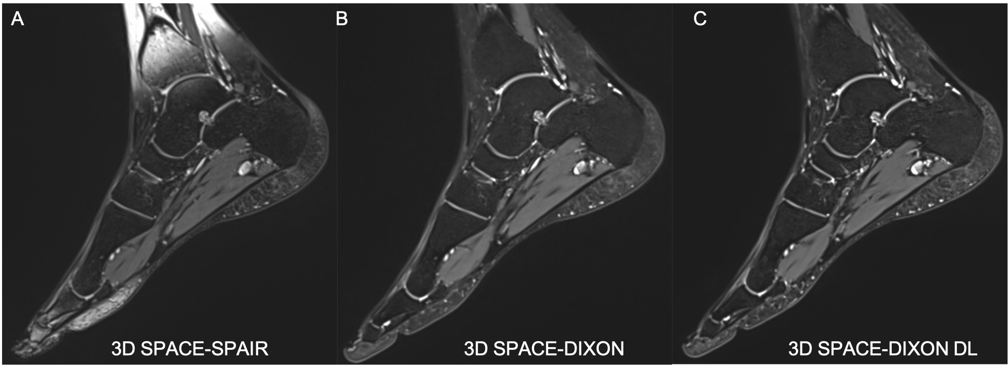
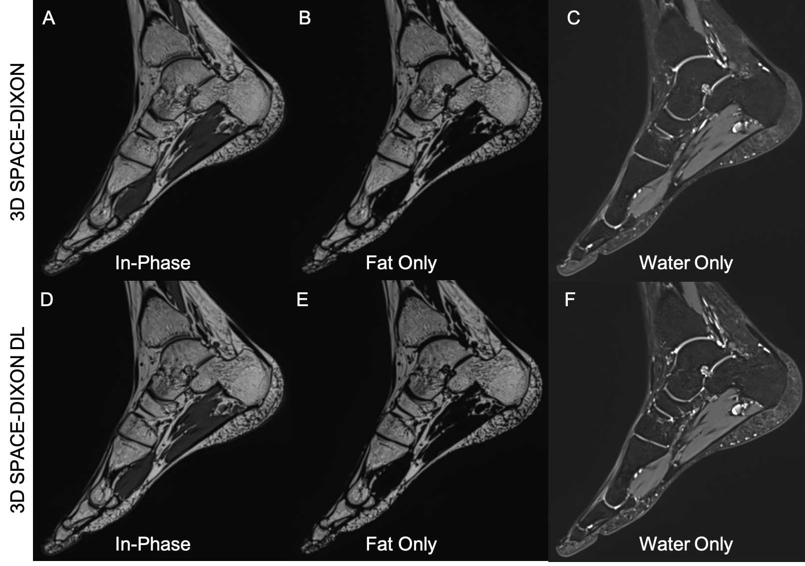
Phantom experiments demonstrated comparable fat suppression efficiency between the CAIPI SPACE-DIXON and accelerated SPACE-DIXON-DL techniques (Figure 2). Figure 3 compares representative whole foot images of the SPAIR fat suppressed SPACE sequence with the the water-only images from the SPACE-DIXON and SPACE-DIXON-DL acquisitions. It can be observed that the SPACE-DIXON water-only images have more uniform fat suppression compared to fat suppressed SPACE. The isotropic resolution of the SPACE-DIXON-DL allows multi-planar reformatting. Further, the in-phase and fat-only images can serve as surrogates for non-fat suppressed and T1-weighted contrasts respectively (Figure 4).The accelerated SPACE-DIXON-DL sequence was rated superior to the SPACE-SPAIR sequence for soft tissue contrast (5 [5,5] vs. 4 [4,4],p<.01), fluid contrast (5 [5,5] vs. 4 [4,4],p<.01), and fat suppression quality (5 [5,5] vs. 4 [3,4],p<.01). All other parameters were rated equivalent (Figure 5).
Compared with the conventional SPACE-DIXON sequence, the SPACE-DIXON-DL was rated equivalent for all parameters.
Conclusion
3D SPACE-DIXON-DL is feasible and provides improved fat suppression performance, soft tissue contrast and fluid contrast, while maintaining similar acquisition times than 3D SPACE-SPAIR for musculoskeletal MRI. Image acceleration does not negatively impact image quality compared with conventional 3D SPACE-DIXON.Acknowledgements
No acknowledgement found.References
1. Del Grande F, Santini F, Herzka DA, et al. Fat-suppression techniques for 3-T MR imaging of the musculoskeletal system. Radiographics. 2014;34(1):217-233. doi:10.1148/rg.341135130
2. Glover, Gary H. "Multipoint Dixon technique for water and fat proton and susceptibility imaging." Journal of Magnetic Resonance Imaging 1.5 (1991): 521-530.
3. Glover GH, Schneider E. Three-point Dixon technique for true water/fat decomposition with B0 inhomogeneity correction. Magn Reson Med 1991; 18: 371–383.
4. Glover GH. Multipoint Dixon technique for water and fat proton and susceptibility imaging. J Magn Reson Imaging 1991; 1: 521–530.
5. Mahesh Keerthivasan, Xiaodong Zhong, Mary Bruno, Iman Khodarahmi, Jan Fritz. A 3D SPACE-DIXON Technique for Fat Suppression in Knee Imaging. Proceedings of Annual Meeting of ISMRM 2022.
6. Hammernik, K., Klatzer, T., Kobler, E., Recht, M.P., Sodickson, D.K., Pock, T. and Knoll, F. (2018), Learning a variational network for reconstruction of accelerated MRI data. Magn. Reson. Med., 79: 3055-3071
7. Afat S, Wessling D, Afat C et al (2022) Analysis of a Deep Learning-Based Superresolution Algorithm Tailored to Partial Fourier Gradient Echo Sequences of the Abdomen at 1.5 T: Reduction of Breath-Hold Time and Improvement of Image Quality. Invest Radiol 57:157-162
8. Wessling D, Herrmann J, Afat S et al (2022) Application of a Deep Learning Algorithm for Combined Super-Resolution and Partial Fourier Reconstruction Including Time Reduction in T1-Weighted Precontrast and Postcontrast Gradient Echo Imaging of Abdominopelvic MR Imaging. Diagnostics (Basel) 12
Figures